Let's Provide You Detailed Information About Angiography with Our Expert Team.
Fill Out the Form Now
Let's Contact You Immediately
n
What is CT Coronary Angiography?
n
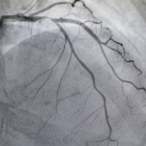
It is the painless visualization of the heart vessels using next-generation Computed Tomography devices in a very short time of 10-12 seconds. Today, CT devices with 40 and 64 detectors can obtain high-quality images. Approximately 300-500 slices are obtained from each patient, and the data contains more volumetric information than slices. Advances in information technology, computer-aided diagnostic programs, and advanced fast computers allow the obtained data to be processed with high speed and accuracy, yielding diagnostic images that can be nearly equivalent to catheter angiography performed via the femoral artery. In comparative studies with catheter angiography, which is considered the gold standard and performed via the femoral artery, the accuracy rates for coronary stenoses, sensitivity, and specificity values are between 90-100% for 40-64 detector devices. Particularly, the negative predictive value, known as the normal evaluation of coronary arteries in CT angiography, has been found to be 100% in many comparative studies.
n
n
Who is it performed on?
n
Conditions where CT coronary angiography can be applied:
n
n
- n t
- Detection of atherosclerotic plaques causing stenosis in heart vessels and evaluation of the degree of stenosis,
- Calcium scoring,
- Monitoring asymptomatic but at-risk patients,
- Control of vessels used during bypass surgeries, those opened with balloons, or those with stents,
- Examination of coronary artery anatomy to show abnormalities and normally accepted anatomical variations,
- Evaluation of coronary arteries in patients at high risk for catheter angiography,
- Obtaining complementary information in cases where a definitive decision cannot be made or where catheter angiography has failed.
n t
n t
n t
n t
n t
n t
n
n
n
CT coronary angiography is a preferred advanced imaging method that should primarily be used in situations where catheter angiography is insufficient or risky, and it is believed that it will not require catheter angiography for diagnostic purposes in the near future. In the last year, CT coronary angiography has begun to be accepted as the gold standard for evaluating the patency of vessels and stents used during bypass. This examination technique has been accepted by the Cardiology and Radiology Associations and Societies in Europe and America, and it is covered by insurance companies in these countries. Particularly, patients in the mild and moderate risk group with symptoms, congenital vascular abnormalities, stenoses, and plaques can be evaluated.
n
n
How is it performed?
n
Technically, CT coronary angiography is performed while the patient is awake and holding their breath for 10-12 seconds. Images are obtained using iodinated contrast material administered through veins in the arms or hands. There is a short preparation period for the procedure. During this period, the patient is informed about how the procedure will be performed, possible risks, and precautions to be taken against risks. After a brief preparation before the procedure, imaging can be performed, and patients can return to their normal lives within a few minutes after the imaging. CT angiography is performed by Radiology Department teams; the obtained data is processed with various computer programs and converted into different images, including three-dimensional ones, and then evaluated by Radiology Specialists, after which the necessary medical and interventional treatments are decided by Cardiology and Cardiovascular Surgery Specialists.
n
n
What are the benefits?
n
CT coronary angiography allows for treatment opportunities without experiencing a heart attack through early diagnosis. With the widespread use and development of the method, many silent coronary artery disease patients will have the opportunity for early diagnosis and treatment. With coronary CT angiography, not only the lumen (the channel where blood is located) of the coronary arteries but also the vessel wall structure, plaque characteristics, the anatomy of the heart, and the major arteries of the heart and lungs, as well as structural abnormalities can be examined, and functional studies (related to the heart’s work and function) can be evaluated with nearly real-time moving images of the heart chambers’ movements and heart valve functions during normal operation.
n
n
What are the future expectations?
n
Although not yet in routine use, studies conducted with 256 detector devices are being published. In the near future, the concept of the number of detectors will be eliminated, and images with much higher spatial and temporal resolution will be obtained using flat panel systems (high-resolution detectors in a plane beyond numbers), and catheter angiography procedures performed for diagnostic purposes will be replaced by this dizzying technology.
n