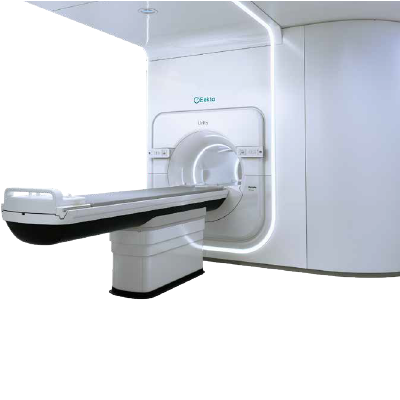
Elekta Unity 1.5 Tesla MR-LINAC
MR-LINAC is a “smart radiotherapy” method that destroys the tumor with rays by targeting it with special images and measurements.
MR-LINAC, which has more advanced equipment than conventional radiotherapy, provides new and important gains to the patient and radiation oncologist thanks to the MR (Magnetic Resonance) added to radiotherapy.
Thanks to the MR-LINAC method, even tumors in mobile organs can be irradiated and destroyed with pinpoint accuracy during treatment. While the tumor is irradiated, the surrounding healthy tissues are protected with great precision. By identifying the most active and aggressive parts of the tumor, higher doses of radiation can be delivered to this area.
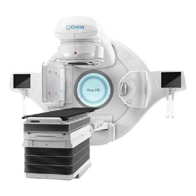
Versa Hd Linac
A modern linear accelerator (Linac) is a device that generates high-energy X-rays and electron beams for the treatment of cancer patients. Radiation therapy is based on the interaction between matter and radiation. Thanks to the Symmetry feature in the Elekta Versa HD device, errors that may occur during treatment can be prevented by monitoring tumor and healthy organ movements. Up to 1 mm. can treat brain tumors with special apparatus.
Thanks to Surface Tracking System (SIGRT):
- Reliable treatment
- Preventing the risk of wrong patient and wrong site
- Possibility to monitor patient movements at any time
- No need for marking with a marker
- Possibility of maskless treatment for claustrophobic patients
- No radiation exposure
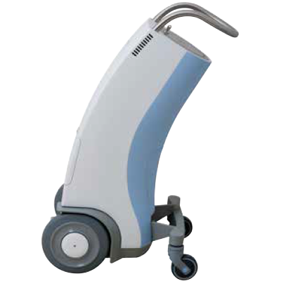
Targeted Irradiation in Cancer Treatment: Brachytherapy
Brachytherapy is a method used to increase the local control of the disease by increasing the dose received by the tumor before or after external radiotherapy.
Brachytherapy is a type of radiotherapy used to treat cancer patients. This type of treatment uses radiation to kill cancer cells. Brachytherapy, which biologically targets cancerous cells, is especially applied to cancerous areas in the body such as the breast, prostate, lung, brain, liver, and stomach. It is often used successfully in the treatment of gynecological cancers (uterus, cervix, vagina), lung cancer, and skin cancers.
Robotic and Fully Automated Drug Preparation
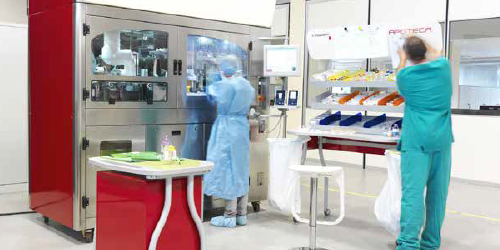
In the chemotherapy unit, which is allocated on a separate floor of the hospital, the treatment of patients is carried out in a comfortable environment, while at the same time, the medicines prepared for the patients are prepared with fully automatic and robotic systems, untouched, sterile, in a way to protect employee and patient safety.
Patient and environmentally sustainable anesthesia device
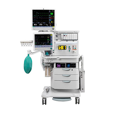
In our hospital, with our anesthesia devices that are economically, and environmentally sustainable and offer patient comfort, low-flow anesthesia is performed with the experience of our entire anesthesia team, especially our specialist physicians.
The fresh gas flow given to the patient in one minute is reduced from 4-5 liters to 0.4-0.5 liters. It is aimed to prevent the discomfort that may occur in the lungs of the patients, especially in long-term cases.
High precision rate in cancer diagnosis and treatment – PET-CT
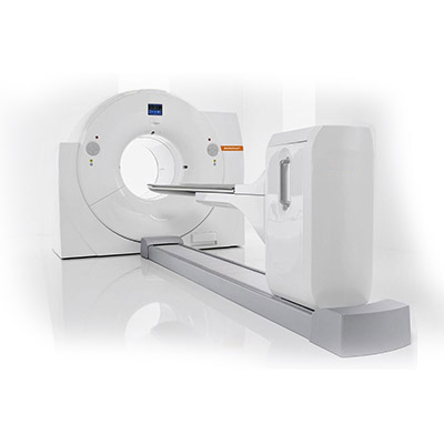
PET-CT device is an advanced technological imaging method formed by the combination of positron emission tomography and computerized tomography. It allows obtaining 3D images of changes in the body and tracking cancerous cells. With its high quality lesion detectability and spatial resolution features, detection accuracy is ensured in the detection of the smallest cancer lesions. PET-CT is used especially in detecting the location of incipient metastases and the stage of cancer.
C-arm X-ray shortening the operation time
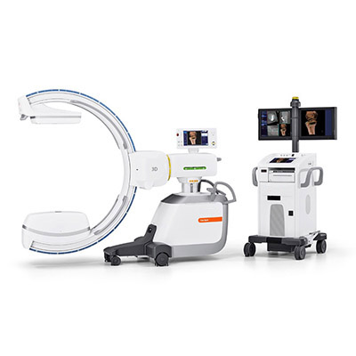
With the C-arm X-ray device, 3D images are obtained in dense tissues and in severe patients, thanks to the sensitive imaging opportunity with a volume of 16 cm. The ease of automatic location and wide patient intake depth provide convenience to our physicians in routine surgical operations and provide a more comfortable experience to our patients. On the other hand, it increases the diagnostic accuracy with its high X-Ray transmittance, especially in overweight patients.
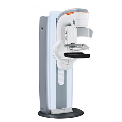
Mammography allowing simultaneous biopsy
With our new generation mammography device, effective images and successful results can be obtained in breast controls. Thanks to high mAs values, new technology compression improvements, software and hardware, the average dose taken during the shooting is reduced as well as keeping the patients at a minimum stress level. Stereotactic Biopsy performed during mammography, which is one of the most sought-after features of patients, is possible with our mammography device. On the other hand, with advanced technology breast tomosynthesis and 3D imaging feature, even the smallest lesions are successfully detected and the chance of early diagnosis of breast cancer increases.
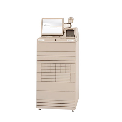
Computerized medicine system working with fingerprint
Our hospital aims to maximize patient experience and safety with the latest technological equipment. The drug management system is the method of taking the drugs prescribed to our inpatients through a closed system, in the desired amount, on behalf of the patient.
In the system, which is accessed in a controlled and secured with the personal password and fingerprint of the user nurse, it can be seen by whom, when and in what quantity the medicine was taken. Thus, the system minimizes medication errors.
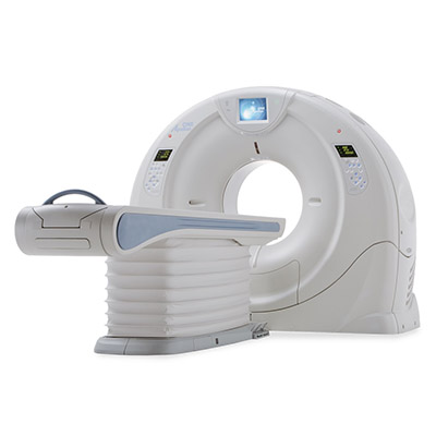
640 section tomography system with fast scanning and high diagnostic value
The new high-end Computed Tomography system using X-ray plays an active role in the early diagnosis and treatment process. By scanning 640 sections and an area of 16 cm in a single rotation, the system significantly reduces the radiation dose given to our patients, thanks to its fast shooting technology. In addition to being the most powerful scanner in the world, 3D examinations are offered with the highest diagnostic value and quality with the new CT System. Moreover, coronary CT angiography can be performed for 24 hours for our emergency patients with the tomography system.
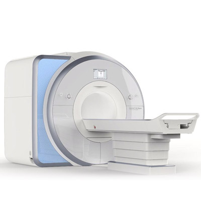
3 Tesla MRI with a larger and comfortable design
3 Tesla MRI, which is used in the early diagnosis and treatment of diseases, is one of the most advanced imaging methods. In addition to its high image quality that allows small lesions to be detected, the aim is the patient comfort. For many, MRI can be a dark, noisy and frightening process. 3 Tesla MRI offers patients a more satisfying experience with its wider design and Cinema Vision MRI Video System. Thanks to the opportunity to watch 3D movies and television, the MRI process can be completed without the need for sedation.
High precision in neurosurgery operations – surgical microscope
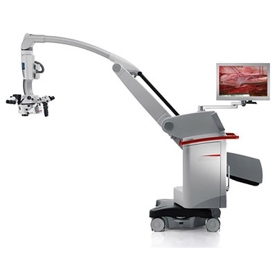
The Surgical Microscope aims to prevent damage to healthy cells by protecting the patient from the risk of stroke, thanks to its sharp focus, in sensitive brain and neurosurgery surgery. It gives the best results, especially in tumor surgery, identifying the tumor area and providing safe surgery in tumor-focused work. With its exclusive FusionOptics technology, advanced optics and three integrated fluorescent modes, the device allows to focus on every critical detail, giving the best possible support to ensure precise movement during surgery.
Next generation endoscopy – high resolution endoscopy system
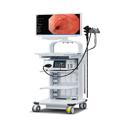
Non-surgical advanced endoscopic techniques are used in the treatment of early stage esophageal, stomach and colon cancers and polyps with the high-resolution endoscopy system. With HD quality imaging technology, it allows the removal of tumors in the digestive system with the ESD method without any incision in the body and the clear removal of early-stage polyps in the digestive system. Moreover, it is used as an endoscopic treatment method in rare diseases with diffuse esophageal spasm and excessive contraction of the esophagus, as well as achalasia.
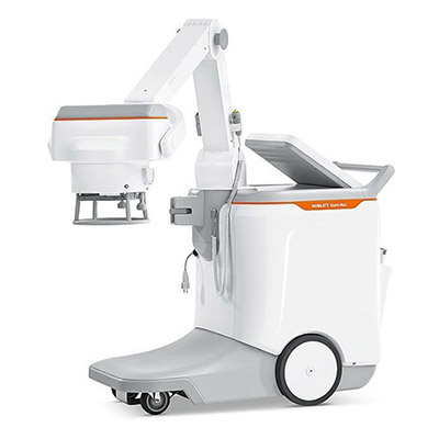
Minimum waiting time for X-Ray – mobile X-Ray
Mobil X-Ray enables to obtain images that will facilitate the diagnosis and treatment process, thanks to its high image enhancing feature. With its powerful battery system, motorized driving feature and positioning flexibility, it can be moved to the desired department in the hospital quickly and easily. In this way, the waiting time for X-rays of our patients is minimized.
