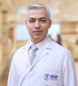
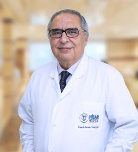
Hisar Intercontinental Hospital Radiology Department is an advanced diagnosis and treatment unit that owns modern hardware with cutting-edge technology and is able to perform any kind of radiology operations twenty-four-seven by its experienced specialist physicians and technicians. Our Radiology Department relies on the idea that correct diagnosis can be achieved by carefully selecting and performing the least risky, fastest and most suitable method for each patient.
Medical imaging devices that we use in the Radiology Department for the diagnosis and monitoring of the diseases are as follows:
- Multi Detector computed tomography (MDBT)
- 5 Tesla Magnetic Resonance (MR)
- Digital Angiography (DSA)
- Digital Fluoroscopy
- Digital X-Ray
- Digital Mammography
- Ultrasonography
- Color Doppler Ultrasonography
- Bone Densitometry
- CR mobile X-Ray device for inpatients
- Digital Panoramic X-Ray (Dental X-Ray)
Images of all the examinations performed by using these devices are transferred to PACS (Picture Archiving and Communication System) system, evaluated by the radiology specialists and stored. The fact that the system is integrated with the hospital management system facilitates instant access to the results from all the polyclinics and clinics, and performing consultation if required.
Interventional Radiology
In our interventional department, operations are performed by using special medical tools for the diagnosis and treatment of various diseases under the guidance of imaging systems. The aim of interventional radiology is to create an alternative to the surgical treatment in many cases, to provide a treatment opportunity in cases without a surgery possibility or to facilitate the surgery by preparing the patients for surgical treatment.
Monitoring Guided Diagnostic Interventions
Main method used in diagnostic practices is the needle biopsy administered percutaneous directly to various parts of the body. Most common regions of use are liver, pancreas, abdominal cavity and peritoneum, renal and suprarenal glands, lower abdominal and genital region, chest, all the bones and neck region. This enables the establishment of a final diagnosis in the cases that diagnosis such as malignant-nonmalignant tumors, abscess can’t be established by clinical data and imaging methods. Sampling from the formations that are considered to be inflammatory or dense effusions for the purpose of diagnosis and culture is performed as well as draining operation if required.
Therapeutic Intervention Radiology Operations
Operations are administered on two groups; vascular and extravascular. Operations are performed for the purposes of aneurysm obstructing the vein, vascular structural abnormalities, stopping the bleeding, tumor reduction. Interventional Radiology is used in the frequent tumor cases and disseminations, for the purposes of occluding the vein that supplies the tumor, increasing the effects and minimizing the side effects of chemotherapy. Regarding the extravascular operations, abscess and cysts formed in Kidneys, Urinary Tract, Bile Ducts and various areas can be drained percutaneous and treatment can be applied, if required, to prevent recurrence. In our Interventional Radiology Department, treatment of strictures and small kidney stones, opening of the bile duct obstruction with balloon and stent, treatment of the inflammation of the occluded system, removing the gallstones, treatment of the Hydatic Cysts by intervention, that are frequent in our country and may form in the liver and all body, are provided.


