

Nuclear Medicine
Nuclear medicine is the medical branch that enables safe, painless and affordable imaging of any part of the body and the treatment of diseases. Unlike diagnostic radiology, which provides only anatomical information, nuclear medicine methods provide information about both the structure and the function of any organ. Nuclear medicine methods make it possible to diagnose, treat and monitor serious illnesses.
About the Department
The scope, approach and highlighted services of the department.
Nuclear medicine is the branch of medicine that enables safe, painless and cost-effective imaging of any part of the body as well as the treatment of diseases. Unlike diagnostic radiology, which provides only anatomical information, nuclear medicine methods provide information about both the structure and the function of any organ. Nuclear medicine methods make it possible to diagnose, treat and monitor serious conditions. Diseases can usually be detected with nuclear medicine methods at an early stage, before they become detectable by other radiological methods and before clinical symptoms develop. This allows the disease to be treated more successfully before it progresses further. Very small amounts of radioactive material and radiopharmaceuticals are used for nuclear imaging. Pharmaceuticals are substances that, when administered to the body, bind specifically to a particular organ, bone or tissue. When bound to a radioactive substance, they are called radiopharmaceuticals; they travel to and bind to the organ to be imaged and, thanks to the radioactivity they carry, emit gamma rays that allow that organ to be imaged with gamma or PET cameras. These cameras are connected to computers, so that data and information about the imaged part of the body are obtained. The radiation dose the body receives during nuclear medicine procedures is approximately the same as with radiological X-ray diagnostic methods. The techniques frequently used in nuclear medicine are as follows. Planar imaging provides a two-dimensional image of the organ and information about its function. SPECT provides a three-dimensional image of the organ and information about its function. PET provides information about function in a specific organ, tumour or any metabolically active area while also providing three-dimensional imaging.
Although nuclear medicine is generally used for diagnostic purposes, it also plays an important role in the treatment of certain diseases. Nuclear isotopes are most commonly used in treatment for thyroid and prostate cancers, hyperthyroidism, polycythaemia vera (an abnormal increase in red blood cells) and bone cancer pain. In our Nuclear Medicine Department, diagnostic methods are performed for a wide range of diseases involving the cardiovascular, musculoskeletal, pulmonary, endocrine, neurological, genitourinary and gastrointestinal systems. Scintigraphic scans are performed with a dual-head Philips gamma camera.
Nuclear Medicine Applications
In Endocrinology
Thyroid Scintigraphy: Diagnosis and treatment follow-up of goitre, underactive or overactive thyroid gland, evaluation of nodules, post-operative follow-up.
Parathyroid Scintigraphy: Investigation of the causes of elevated blood calcium, detection of lesions (adenoma).
In Infection
Labelled White Blood Cell Study
Labelled Immunoglobulins (Human Polyclonal Immunoglobulins)
Gastrointestinal System
Liver-Spleen Scintigraphy: Performed to investigate abdominal pain, functional disorders, inflammatory conditions, post-operative complications and existing lesions.
Gastroesophageal Reflux Scintigraphy
Oesophageal Transit Study
Gastric Emptying Study (liquid and solid)
Hepatobiliary Scintigraphy: functional disorders; inflammatory conditions
Gastrointestinal Bleeding Study
Meckel's Diverticulum Scintigraphy
Salivary Gland Scintigraphy
Genitourinary System
Renal Cortical Scintigraphy (Static Renal Scintigraphy)
Dynamic Renal Scintigraphy (Renogram)
Diuretic Renal Scintigraphy (Diuretic Renogram)
Captopril Renal Scintigraphy (Captopril Scintigraphy)
Cardiovascular System
Myocardial Perfusion Scintigraphy: Determination of infarction and ischaemic areas, ejection fraction, myocardial viability, heart failure,
MUGA: A test that detects global or regional wall motion abnormalities of the heart. It is recommended before and after starting treatments that may damage the heart (particularly performed in cancer patients after chemotherapy to examine the regional and total EF of the heart).
Musculoskeletal System
Bone Scintigraphy:Investigation of the extent of primary or metastatic cancer spread to the bones or of the causes of pain, differential diagnosis of sports injuries, early detection of fractures
Bone Marrow Scintigraphy
In the Pulmonary System
Lung Perfusion Scintigraphy: Obstruction of blood flow and/or the airways, determination of probability in the diagnosis of pulmonary embolism. It is also performed to evaluate lung function before lung cancer surgery
Lung Perfusion/Ventilation Scintigraphy: Obstruction of blood flow and/or the airways, determination of probability in the diagnosis of pulmonary embolism
Quantitative Lung Perfusion Scintigraphy: It is also performed to evaluate lung function before lung cancer surgery
In the Central Nervous System
Brain Perfusion Scintigraphy (Perfusion SPECT): The most sensitive test for establishing a diagnosis even before clinical symptoms develop in epilepsy, tumours, circulatory insufficiency and especially in cases at risk of Alzheimer's disease.
Other Services
Lacrimal Duct Scintigraphy: The most sensitive and painless test for diagnosing blockage of the tear ducts.
Lymphoscintigraphy: Performed to investigate lymphatic drainage and the causes of blockage in the lymphatic vessels, and also to detect and mark the sentinel lymph node in diseases such as breast and prostate cancer.
Testicular Scintigraphy: Assists in the differential diagnosis when evaluating a painful testicle.
Breast cancer: Pre-operative lymphoscintigraphy that supports surgery by showing lymphatic drainage through an injection into the affected breast before the operation.
Thallium-201 Whole-Body Scan.
Whole-body tumour scan with Sesta MIBI
Radioactive Iodine Therapy for Thyrotoxicosis
Phosphorus-32 Therapy
Radionuclide Synovectomy.
Radionuclide therapy for pain palliation in patients with severe pain due to bone metastases
Nuclear medicine is the branch of medicine that enables safe, painless and cost-effective imaging of any part of the body as well as the treatment of diseases. Unlike diagnostic radiology, which provides only anatomical information, nuclear medicine methods provide information about both the structure and the function of any organ. Nuclear medicine methods make it possible to diagnose, treat and monitor serious conditions. Diseases can usually be detected with nuclear medicine methods at an early stage, before they become detectable by other radiological methods and before clinical symptoms develop. This allows the disease to be treated more successfully before it progresses further. Very small amounts of radioactive material and radiopharmaceuticals are used for nuclear imaging. Pharmaceuticals are substances that, when administered to the body, bind specifically to a particular organ, bone or tissue. When bound to a radioactive substance, they are called radiopharmaceuticals; they travel to and bind to the organ to be imaged and, thanks to the radioactivity they carry, emit gamma rays that allow that organ to be imaged with gamma or PET cameras. These cameras are connected to computers, so that data and information about the imaged part of the body are obtained. The radiation dose the body receives during nuclear medicine procedures is approximately the same as with radiological X-ray diagnostic methods. The techniques frequently used in nuclear medicine are as follows. Planar imaging provides a two-dimensional image of the organ and information about its function. SPECT provides a three-dimensional image of the organ and information about its function. PET provides information about function in a specific organ, tumour or any metabolically active area while also providing three-dimensional imaging.
Although nuclear medicine is generally used for diagnostic purposes, it also plays an important role in the treatment of certain diseases. Nuclear isotopes are most commonly used in treatment for thyroid and prostate cancers, hyperthyroidism, polycythaemia vera (an abnormal increase in red blood cells) and bone cancer pain. In our Nuclear Medicine Department, diagnostic methods are performed for a wide range of diseases involving the cardiovascular, musculoskeletal, pulmonary, endocrine, neurological, genitourinary and gastrointestinal systems. Scintigraphic scans are performed with a dual-head Philips gamma camera.
Sub-departments & Units
Clinics, centers and units within this department.
History and General Fields of Application of Nuclear Medicine
Nuclear medicine, in its simplest definition, is the use of radioactive substances in the diagnosis and treatment of diseases. Currently, in our country, nuclear medicine departments serving at universities, state and social security hospitals in major provinces, and some private centers perform functional imaging for nearly every organ system.
Frequently Asked Questions
What is nuclear medicine? It is a field of medicine in which very small amounts of radioactive agents are used in the diagnosis and treatment of various diseases. After the substances are administered into the body…
PET-CT
PET-CT, an advanced imaging method in which two separate devices are combined into a single unit, plays an active role in cardiac and neurological conditions, and especially in the field of oncology. Thanks to its ability to scan the entire body at low radiation and high speed, it is used in the diagnosis of cancer, treatment planning, and post-treatment evaluation.
Treatment Methods
The main treatments and procedures performed in our department.
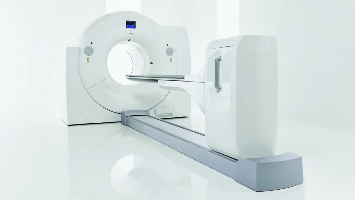
What Is PET-CT? In Which Diseases Is It Used for Diagnosis and Treatment?
Nuclear Medicine

What is Lutetium and How is it Treated?
Nuclear Medicine
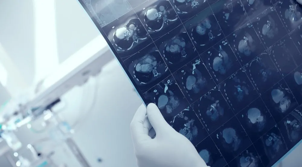
Gallium 68 Dota PET: What Is It? Which Diseases Is It Used For?
Nuclear Medicine

What Is PSMA PET? Which Diseases Is It Used For?
Nuclear Medicine

What is Actinium Treatment? In Which Diseases is it Used?
Nuclear Medicine
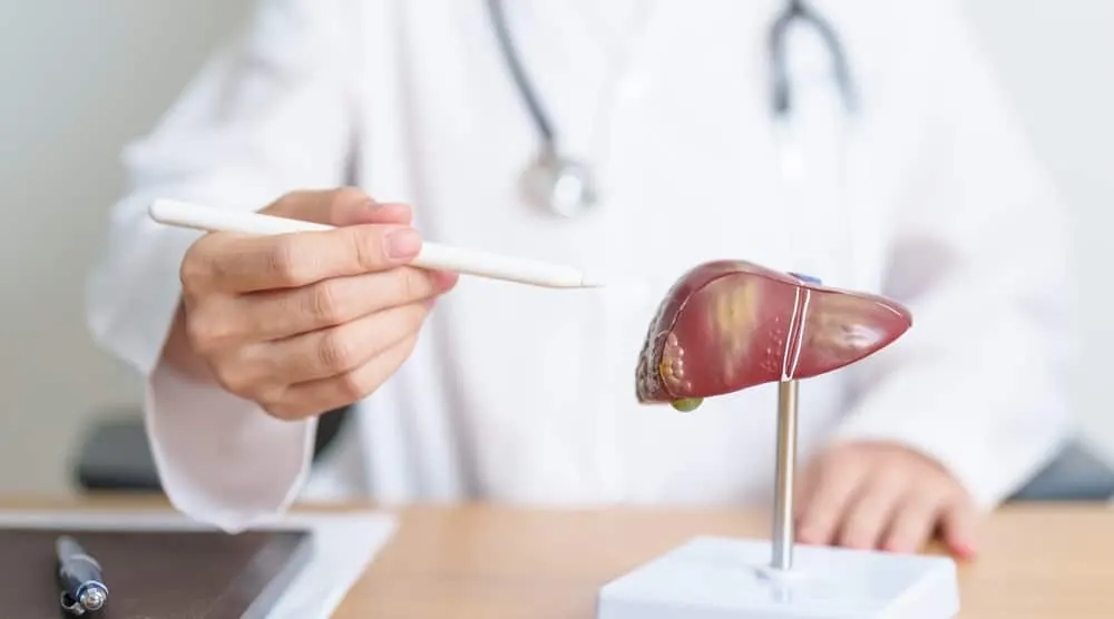
What Is TARE? Why Is It Performed?
Nuclear Medicine
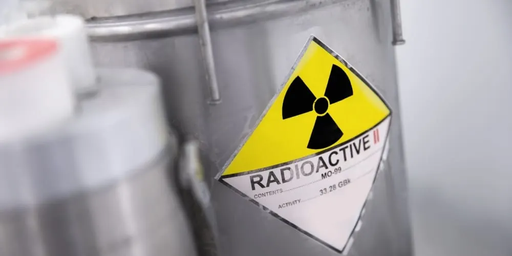
What Is Radium Therapy? Which Diseases Is It Used For?
Nuclear Medicine

What Is Nuclear Medicine?
Nuclear Medicine
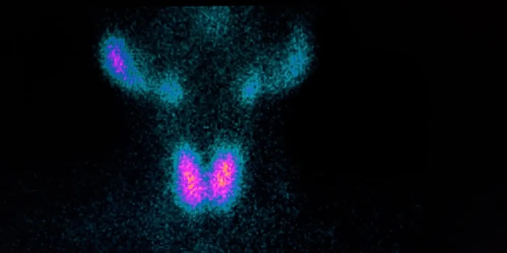
What Is Thyroid Scintigraphy?
Nuclear Medicine
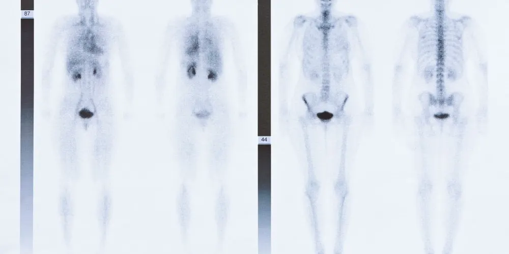
Bone Scintigraphy
Nuclear Medicine
What Is FAPI PET?
Nuclear Medicine
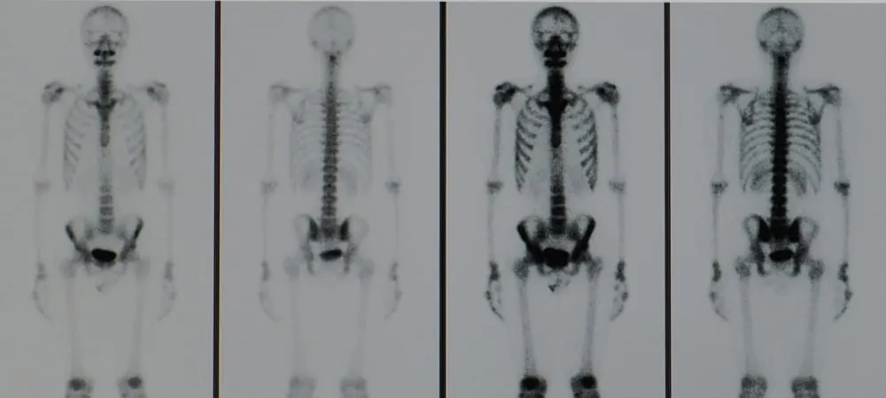
What is Myocardial Perfusion Scintigraphy (MPS)? How Is It Performed, Its Risks and Results
Nuclear Medicine
Department Doctors
Our specialist team working in the Nuclear Medicine department.
Related Videos
Informative videos from our specialists related to the Nuclear Medicine department.


Healthy Living Guide
Featured content related to the department.
nuclear-medicine
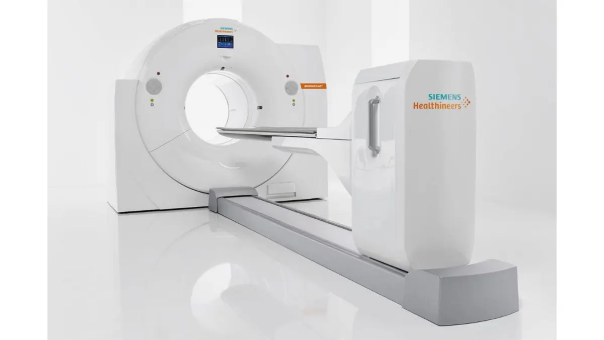
What is PET-CT?
PET-CT is an advanced imaging method that combines PET and CT technologies, playing a crucial role in the diagnosis and treatment planning of various diseases, especially in oncology.
nuclear-medicine
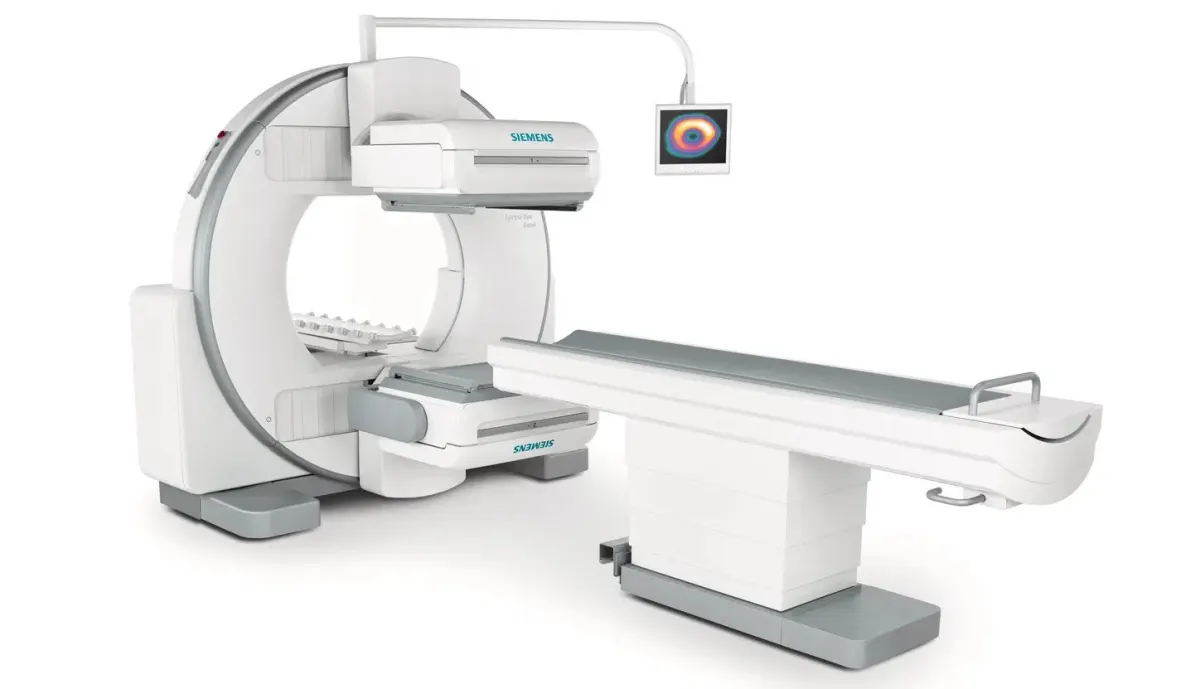
What is Scintigraphy and How is Scintigraphy Used in Oncology?
Scintigraphy, commonly used in the field of Nuclear Medicine, is one of the preferred methods for diagnosing and monitoring many diseases. Scintigraphy provides a detailed examination of organs and tissues at the molecular level and physiol
nuclear-medicine
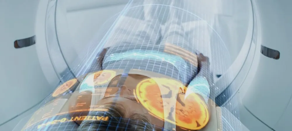
Brain PET Scan Uses, Procedure, and Frequently Asked Questions
A brain PET scan is a crucial imaging method for diagnosing and monitoring neurological diseases such as Alzheimer's, Parkinson's, and epilepsy. It provides insights into functional changes and aids in treatment planning.
Locations
At Which Hospitals?
The Nuclear Medicine department is available at the following locations.

Author
Web and Medical Editorial BoardReviewed by a specialist
 Assoc. Prof. Dr. Tamer AksoyNuclear MedicineReview date: 31 October 2026
Contact
Nuclear Medicine Department — Get in Touch
Fill out the form with your questions; our team will call you back as soon as possible.
The Hisar Hospital Web and Medical Editorial Board contributed to the development of this content. All content on this website is provided for general informational purposes only. Please consult a physician for the evaluation, diagnosis and treatment of your condition.
Last Updated: