Congenital anomalies, also known as malformations or birth defects, are structural or functional disorders that occur during the development of the fetus. These disorders, which can affect different organs and systems, can arise from many causes, ranging from genetic factors to environmental influences. Some congenital anomalies can be serious enough to threaten a person's life. Congenital heart diseases, neural tube defects, and chromosomal anomalies such as Down syndrome are the most common congenital anomalies. Today, thanks to advancing healthcare technologies, signs of anomalies in babies can be largely detected at an early stage through fetal anomaly screenings performed during the prenatal period or newborn screenings.
What is an Anomaly?
An anomaly is a general term that describes structural or functional disorders present at birth in the human body. Anomalies can seriously affect a person's life and lead to long-term disability. According to World Health Organization data, an estimated 240,000 newborns worldwide die within the first 28 days after birth, and 170,000 children between the ages of 1 month and 5 years die due to congenital disorders. Congenital anomalies are not always unavoidable. Adherence to vaccination programs and methods such as adequate intake of folic acid and iodine before and during pregnancy can help prevent some congenital anomalies.
Birth defects can occur due to the influence of one or more genetic, infectious, nutritional, or environmental factors. However, it is often difficult to determine the exact causes of congenital anomalies. The main causes of congenital anomalies are as follows:
Genetic factors: Chromosomal anomalies account for approximately 20% of congenital anomalies. Chromosomal anomalies such as Down syndrome (Trisomy 21) or single-gene defects such as cystic fibrosis can be given as examples of genetic causes of congenital anomalies. For this reason, the risk of congenital anomalies may be higher in geographies where consanguineous marriages are common.
Socioeconomic and demographic factors: Research shows that congenital disorders are more common in low-income families and countries due to factors such as pregnant women not having access to adequate food, being more exposed to infections and harmful substances, or having limited access to healthcare services and screenings. It is estimated that approximately 94% of serious congenital disorders occur in low- and middle-income countries. Additionally, maternal age is also an important factor that increases the risk of chromosomal abnormalities, especially Down syndrome.
Environmental factors: Environmental factors also play an important role in the emergence of congenital anomalies. The fact that congenital anomalies are more common in certain parts of the world where people use dangerous pesticides and herbicides is one of the most important proofs of this. Furthermore, the expectant mother's exposure to infectious diseases such as syphilis or rubella, radiation, or certain chemical substances during pregnancy, vitamin and mineral deficiencies such as iodine and folate deficiency, or chronic health problems such as diabetes can be effective in the emergence of congenital anomalies. In addition, the expectant mother's consumption of alcohol or use of various medications during pregnancy can also increase the risk of congenital anomalies.
In addition to those listed, the causes of some congenital anomalies are not known with certainty. Experts believe that genetic and environmental factors have a combined effect on the emergence of anomalies such as congenital heart diseases, cleft lip or palate, and clubfoot.
What Are the Symptoms of an Anomaly?
Birth defects can manifest with various symptoms before, during, or after birth. Symptoms of an anomaly in a baby can be mild or life-threatening, depending on the baby's age, the affected organs or systems, and the type of anomaly. Screening tests performed during pregnancy allow for the early detection of some anomalies. For example, abnormal fluid accumulation at the back of the fetus's neck can be detected in ultrasonographic examinations, or structural defects in the heart structure can be detected via fetal echocardiography. Deviations of protein levels from expected values in blood tests can also be an indicator of an anomaly.
In the postnatal period, anomalies can often become more apparent. Common symptoms observed in babies and young children include abnormal heart rhythms, respiratory difficulties, feeding difficulties, and developmental problems. For example, a child's failure to show age-appropriate development, inability to follow objects with their eyes, or lack of response to auditory stimuli may indicate a congenital anomaly. In addition to these symptoms, noticeable differences in the child's facial, head, eye, and ear structures can also be an indicator of an anomaly.
What Are the Types of Anomalies?
Congenital anomalies are basically divided into two main categories: structural and functional (developmental). Among the structural anomalies that develop as a result of a specific body part being missing or malformed, congenital heart diseases, cleft lip or palate (division of the upper lip or roof of the mouth), spina bifida (split spine disease), and pes equinovarus (inward turning of the feet) are the most common disorders. Accordingly, the main types of anomalies are as follows:
Congenital heart diseases: Heart defects are disorders seen as a result of the heart wall, valves, vessels, or heart chambers not developing as they should. As a result of the heart's anatomy not being as it should be, serious complications such as heart failure and rhythm disorders can occur. Congenital heart diseases, which are among the most common congenital anomalies, are usually noticed shortly after birth and may require surgical intervention.
Cleft lip and palate: Cleft lip or palate are structural anomalies that occur as a result of the upper lip or roof of the mouth not closing completely. Cleft lip and/or palate can affect the baby's basic functions such as feeding, speech, and dental development. For this reason, surgical treatment of structural anomalies in the lip or palate is as important from a functional perspective as it is from an aesthetic one.
Spina bifida: Also known as split spine disease, spina bifida is a type of congenital anomaly characterized by the neural tube, which should normally be closed, not closing completely and leaving an opening in the spine. Split spine disease can be seen in 1 out of every 1000 births worldwide. Depending on the degree of the opening in the spine, spina bifida can cause serious health problems such as paralysis. With early diagnosis and surgical intervention, the quality of life of patients can be significantly improved.
Clubfoot: Clubfoot, known in medical literature as pes equinovarus, is a structural anomaly characterized by the inward turning of one or both feet, resulting from the bones, muscles, and tendons of the foot not developing as they should. If clubfoot is not treated early with methods such as casting, the use of special shoes, or surgery, the child may encounter walking difficulties when they start to walk.
Functional (developmental) congenital anomalies are anomalies that cause a part or system of the body to not work properly, leading to intellectual or developmental problems. The most common functional or developmental birth defects are as follows:
Down syndrome: Under normal conditions, humans have 46 chromosomes. In people with Down syndrome, there are 3 copies of the 21st chromosome instead of 2. In other words, babies born with Down syndrome have 47 chromosomes. This condition causes intellectual and physical developmental delays in the child. Down syndrome is examined in 3 classes: Trisomy 21, mosaicism, and translocation. In Trisomy 21, the most common type of Down syndrome, there is an extra chromosome in every cell. In mosaicism, there is an extra chromosome in only some cells. In translocation, although the total number of chromosomes is 46, one of them contains an extra piece of the 21st chromosome. In addition to their developmental problems, people with Down syndrome may be more sensitive to other health problems such as heart diseases, hearing loss, and hypothyroidism. Thanks to developing pregnancy screenings, Down syndrome can be diagnosed while the baby is still in the womb.
Sickle cell anemia: Sickle cell anemia is a genetic blood disease that causes red blood cells to lose their normal round structure and take on a sickle or half-moon shape. Thanks to their normally disk-shaped structures, red blood cells, also known as erythrocytes, whose primary duty is to carry oxygen to tissues and organs, can easily pass through small vessels. However, in the case of being sickle-shaped, these cells can get stuck in the vessels and prevent blood from being transported to different parts of the body. As a result, severe pain, organ or tissue damage can occur. Symptoms of sickle cell anemia can usually manifest in infancy with symptoms such as fatigue, swelling of hands and feet, frequent infections, and anemia.
Cystic fibrosis: Cystic fibrosis is an autosomal recessive (inherited from both mother and father) genetic congenital anomaly characterized by the loss of water in exocrine glands and the thickening of body secretions due to a mutation in the gene that produces the cystic fibrosis transmembrane conductance regulator (CFTR) protein. Cystic fibrosis causes the accumulation of thick, sticky mucus in various organs, primarily the lungs, pancreas, liver, and intestines. Symptoms of cystic fibrosis such as recurrent lung infections, chronic cough, and constipation can appear in early infancy, but in some cases, no symptoms may be seen until adolescence. However, cystic fibrosis can be diagnosed at an early stage through screenings performed in the newborn period. There is no definitive cure for the disease. However, symptoms can be controlled with the help of mucus-thinning medications or antibiotics.
In addition to those listed, there are many other types of congenital anomalies such as kidney anomalies, uterine anomalies, and developmental venous anomalies.
How is an Anomaly Diagnosed?
Early diagnosis of birth defects is important for both the baby's and the mother's health. Today, experts can diagnose anomalies with the help of fetal anomaly screenings during pregnancy or some tests and examinations they perform after birth. The main anomaly diagnostic methods frequently used by experts are as follows:
Ultrasonography: With the help of ultrasound, experts can check whether there is abnormal fluid accumulation at the back of the fetus's neck. Abnormal fluid accumulation at the back of the child's neck may indicate congenital heart diseases or chromosomal anomalies.
Blood tests: Experts can measure protein levels in the expectant mother's blood or circulating cell-free fetal DNA with the help of blood tests they perform in the first trimester of pregnancy, i.e., the first 3 months. In case of abnormal results, the presence of chromosomal disorders in the baby may be suspected. Blood tests performed in the second trimester of pregnancy can help diagnose health problems such as spina bifida.
Amniocentesis: Amniocentesis is the process of taking a sample of amniotic fluid, also known as the baby's water, which surrounds the fetus during pregnancy. Amniocentesis can help confirm the diagnosis in cases where chromosomal anomalies are suspected or diagnose whether there are infections such as cytomegalovirus (CMV) in the fetus.
Chorionic villus sampling (CVS): Especially in high-risk pregnancies, CVS, which is performed by taking a small piece from the placenta in the early stages of pregnancy, can help detect chromosomal anomalies.
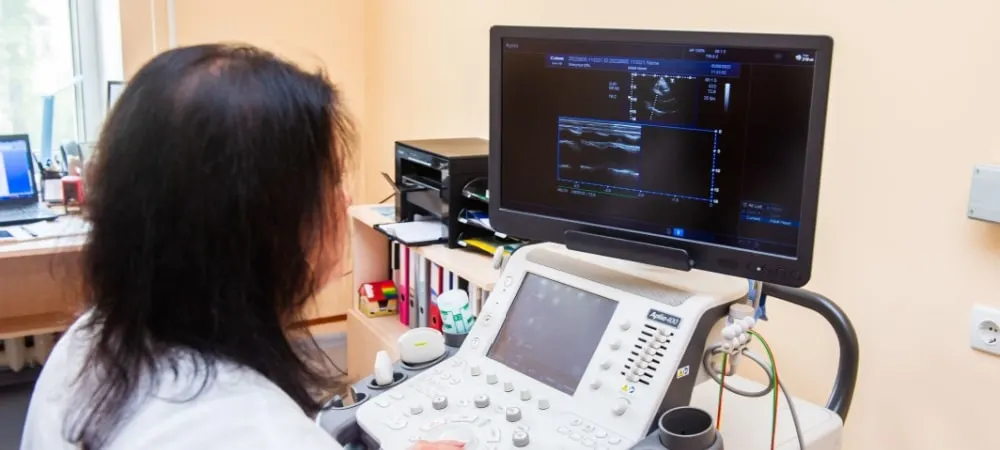
Fetal echocardiography: Fetal echocardiography can help detect abnormalities in the fetus's heart. However, it may not be possible to diagnose all heart diseases before birth with the help of ECHO.
Fetal magnetic resonance imaging: Fetal MRI helps to examine the fetus's brain and nervous system and to obtain information that cannot be obtained with ultrasound.
In addition to those listed, anomalies such as cystic fibrosis can be diagnosed with the help of newborn screenings performed after birth.
How is Anomaly Treatment Performed?
Anomaly treatment varies according to the type and severity of the birth defect. In the treatment process, the main goal of experts is to alleviate symptoms, correct structural abnormalities, and prevent possible complications. In the treatment of congenital anomalies such as cleft lip or congenital heart diseases, experts may resort to surgical methods. In addition, medication may be used to treat birth defects or reduce the risk of complications. In some cases, certain medications given to mothers during pregnancy can help correct abnormalities detected in the fetus.
In addition to those listed, physical therapy and rehabilitation in children born with congenital anomalies that affect motor skills, especially cerebral palsy, can significantly increase the child's quality of life by improving their mobility.
Frequently Asked Questions
Babies born with structural or functional disorders that occur during the developmental process in the womb are called babies with anomalies. Sex-linked chromosomal anomalies are genetic disorders that arise as a result of structural or numerical changes in the chromosomes that play an important role in the determination of sex and reproductive functions. For example, Turner syndrome can cause growth retardation and delayed puberty in girls due to a missing or structurally defective X chromosome. Klinefelter syndrome, on the other hand, manifests in boys with symptoms such as less muscle development and a decrease in sperm production during puberty due to the presence of an extra X chromosome. Such chromosomal anomalies can sometimes cause serious health problems such as infertility. Anomaly ultrasound is an important screening test performed between the 18th and 21st weeks of pregnancy. During the anomaly ultrasound, experts carefully examine the brain, spine, heart, kidneys, and other organs. In addition, the baby's arms, legs, face, and the condition of the placenta are evaluated during the screening. In this way, the baby's development can be examined in detail and structural anomalies can be diagnosed at an early stage. An anomaly in a baby can be determined by fetal anomaly screening performed before birth, or it can be understood by newborn screenings performed after birth. It may not always be possible to prevent a congenital anomaly. However, the risk of anomalies due to environmental influences can be reduced with precautions such as avoiding the use of medications not prescribed by a doctor and avoiding alcohol consumption. Additionally, paying attention to folic acid intake before pregnancy in planned pregnancies can also help prevent anomalies. Chromosomal anomalies are usually caused by errors that occur during cell division or genetic mutations. There are several important risk factors in the formation of chromosomal anomalies. The risk of chromosomal anomalies may increase due to genetic predisposition in people with a history of chromosomal anomalies in their family. Maternal age is also an important factor. Research shows that the risk of chromosomal anomalies may increase in mothers aged 35 and over. Additionally, environmental factors such as exposure to radiation and certain chemicals or some infectious diseases can also increase the risk of anomalies. During some blood tests performed during pregnancy, abnormal protein levels in the expectant mother's blood or circulating cell-free fetal DNA may suggest a chromosomal disorder to experts. Additionally, the accumulation of extra fluid at the back of the baby's neck during ultrasound imaging may also indicate chromosomal anomalies such as Down syndrome.
Although it is not always possible to prevent or treat congenital anomalies, the risk of complications can be minimized through early diagnosis. Regular check-ups, screenings, and expert support before and during pregnancy are of great importance in this process. Hisar Hospital Department of Obstetrics and Gynecology provides comprehensive healthcare services to women who are planning a pregnancy or are in the process of pregnancy. We are with you at all stages from pre-birth to post-birth with our experienced physicians such as Op. Dr. Elif Erol from our expert staff.
Commencez par le bon bilan pour Causes, Symptoms, Types, and Diagnostic Methods of Congenital Anomalies
Partagez vos symptômes avec nos médecins spécialistes ; définissons ensemble le diagnostic et le plan de traitement adaptés.
Attention
Si vos symptômes sont apparus soudainement, évoluent rapidement ou limitent nettement votre vie quotidienne, consultez un médecin sans tarder.
Planifiez votre parcours Causes, Symptoms, Types, and Diagnostic Methods of Congenital Anomalies avec nos spécialistes.
Remplissez le formulaire ou prenez rendez-vous pour connaître les options de traitement, le déroulement et la préparation.