Symptoms of a patellar dislocation include sudden swelling in the knee, deformity, and severe pain. When your kneecap dislocates, your movement is restricted and you have difficulty walking. In fact, 45% of people who experience a patellar dislocation may experience this problem again. In this article, we will cover what a patellar dislocation is, its symptoms, and its diagnosis. Moreover, we will examine patellar dislocation treatment options and the recovery process in detail.
What Is a Patellar Dislocation?
Anatomy of the Kneecap
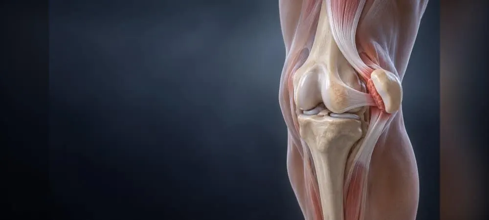
The kneecap is a triangular structure located between the thigh bone and the shin bone. Normally, its back surface glides within the groove of the thigh bone. This bony structure sits in front of the knee joint and serves a protective function.
The attachment of the patella to the knee joint by multiple ligaments provides its stability. Embedded within the tendon formed by a muscle group, this structure reduces friction during joint movement thanks to its cartilage. It also acts as a lever, increasing the strength of the muscle and allowing effective movement with less energy.
For the kneecap to stay in place, the ligament tissues around it must be strong. Ligament structures such as the medial patellofemoral ligament (MPFL) reduce the risk of injury by preventing the kneecap from sliding sideways. Weakness or tearing of these ligaments can cause the patella to dislocate.
Definition of a Patellar Dislocation
A patellar dislocation is a condition in which the kneecap slides out of its natural position. The kneecap bone leaving the track in which it moves is called a patellar dislocation. This usually occurs toward the outer side of the knee.
For the kneecap to dislocate, the ligaments that hold it in place must tear. These dislocations occur at a rate of 6 per 100,000 in the population. They are more common in girls and in young people under 17 years of age. In 15% of people with a patellar dislocation, another family member has also had a dislocation.
After a patellar dislocation in one knee, the risk of a dislocation in the other knee is quite high. For this reason, early diagnosis and the right treatment approach are important.
Types of Patellar Dislocation
Acute patellar dislocation occurs traumatically. It develops as a result of a blow to the side of the knee or an abnormal strain during twisting. In this type of dislocation, there is tearing of the ligaments and sudden displacement of the joint.
Recurrent patellar dislocations occur when the knee structures injured during the first dislocation do not heal completely. Kneecap dislocations seen in the adult age group tend to recur continually. Although a severe strain is required during the first dislocation, in untreated dislocations the soft tissues stretch as they heal, so recurrent dislocations can occur after much simpler movements.
Congenital patellar dislocation is the rarest form. In some children, the patella is already partially dislocated from birth or due to development. In these children, a complete dislocation can occur during a simple twisting movement. In the rarest cases, the patella is located on the outer side of the knee rather than at the front, and it cannot be put back in place without surgery.
Symptoms of a Patellar Dislocation
The moment the kneecap dislocates, your body notices it immediately. You feel severe pain and may hear a "pop" sound from the knee. These symptoms are usually sudden and very noticeable.
Sudden Pain and Swelling
The first symptom of a patellar dislocation is sudden, severe, stabbing pain. Together with the deformity of the knee, this pain is a typical finding. Especially in dislocations caused by trauma, pain and swelling may be more pronounced because the likelihood of cartilage damage is higher.
Within a short time, swelling occurs in the knee due to bleeding inside the joint. This condition is called hemarthrosis and causes major swelling in the knee. The swelling may be tender to the touch. You may also feel pain behind or around the knee.
Visible Deformity
The most obvious and striking symptom of a kneecap dislocation is the abnormal appearance in the knee area. The patella usually slides toward the outside of the knee. As a result, swelling and a deformed appearance develop on the outer part of the knee.
After a sudden twisting movement in the knee, people feel the patella bone slide toward the outside of the knee. You have a distinct sensation that the kneecap has slipped out of place. You can see the kneecap appearing as a bulge on the outer side of the knee.
Restricted Movement and Difficulty Walking
The knee's range of motion is significantly restricted. The normal stability of the knee decreases. You may not be able to fully straighten and bend your leg. The knee usually stays locked in a bent position.
You cannot put weight on the affected leg or walk. The knee's weight-bearing ability is lost. A patient with a dislocated kneecap cannot bear weight on the affected side and feels fear. In addition, pain and swelling prevent a passive assessment.
Feeling of Instability in the Knee
The knee may suddenly give way, creating a feeling of insecurity. You experience a sensation of slipping or twisting in the knee. You may also feel weakness in the knee. Patients say that the knee gave way and that they heard a "popping" sensation.
In recurrent dislocations, the most important and bothersome symptom is a feeling of instability. Especially during movements such as going down stairs, making sudden turns, or squatting, you fear that the kneecap will dislocate again at any moment. You generally experience a feeling of the knee giving way or weakness. This fear leads you to restrict your movements.
Causes and Risk Factors of a Patellar Dislocation
There are several different causes of a kneecap dislocation. The most common cause is direct or indirect trauma. Your kneecap can dislocate while playing sports, during an accident, or with a sudden movement.
Sudden Movements and Trauma
Trauma is the most common cause of patellar dislocation. Sudden changes of direction during sports can trigger this problem. The risk is quite high in sports such as football, basketball, and volleyball.
Unwanted accidents such as falling onto the knee also lead to dislocation. Twisting or spraining of the knee can displace the patella. Direct blows to the side of the knee cause the kneecap to be pushed out of the groove.
The most common cause is a tear of the ligament that holds the kneecap in place, which we call the MPFL. This usually develops as a result of trauma. It occurs when the body rotates while the foot is planted.
Muscle and Ligament Weakness
Weakness of the ligaments that hold the kneecap in place is among the common causes of dislocation. Muscle imbalances also make it difficult for the patella to stay in its normal position.
When the muscles at the front of the thigh are weak, the kneecap tends to slide outward. This risk increases especially in people who sit for long periods, do not exercise, or have muscle imbalances. Imbalances between the muscles on the inner and outer sides of the thigh can lead to movement problems in the kneecap.
Anatomical Structural Abnormalities
Anatomical and genetic factors also play an important role. A patella positioned higher than normal is one of these risk factors. A shallow groove on the femur makes it easier for the patella to slide outward.
Looseness of the ligaments around the knee reduces stability. Knock knees (X-shaped legs) push the kneecap outward and disrupt the load balance. Such structural problems increase the risk of dislocation, especially in young people aged 10-17.
In some individuals, structural differences can trigger a kneecap dislocation. Conditions such as bowlegs increase the risk of kneecap dislocation. Hypermobile joints and anatomically weak ligaments can also be among the causes of patellar dislocation.
Genetic Predisposition and Congenital Factors
Certain genetic characteristics may increase the predisposition to kneecap dislocation. In 15% of cases, there is a history of dislocation in another family member. In 15% of people with a patellar dislocation, another family member has had a dislocation.
After the first dislocation, the risk of another dislocation is between 40% and 70%. After a second dislocation occurs, the risk of recurrence approaches 100%. The reason is that the already flexible soft tissues become even looser after each dislocation.
The risk of recurrence is higher in people with an underlying structural abnormality. A previous kneecap dislocation also makes recurrence easier. This is because after the first dislocation, the ligaments loosen and joint stability decreases.
Diagnosis of a Patellar Dislocation
To make an accurate diagnosis of a patellar dislocation, the patient's history, physical examination findings, and imaging methods are evaluated together. Your orthopedic or emergency physician asks detailed questions about how the injury occurred and about the symptoms of the patellar dislocation. Especially in children, difficulties in diagnosis may arise because they cannot describe the event well, and the injury may mistakenly be assessed as a meniscus or ligament injury.
Physical Examination
First, your doctor carefully observes your knee to look for swelling, bruising, and muscle wasting. Tenderness on the outer part of your knee, and pain at the medial edge of the patella and the lateral femoral condyle, are frequently detected. When there is bleeding inside the joint, this area becomes especially tender.
In addition, your knee's range of motion, flexibility, and stability are tested. Your doctor may ask you to straighten and bend your knee. To see how the patella sits within the groove, the area is carefully palpated and the alignment of the bones is checked.
One of the most important tests performed during the diagnosis of a patellar dislocation is the "apprehension" test. During this test, when the doctor gently pushes your kneecap outward, becoming anxious, feeling fear, or involuntarily contracting your muscles to prevent this movement is a strong sign of instability. Tests such as the J sign, which shows whether the knee follows an abnormal path during movement, may also be performed.
Imaging Methods
Next, imaging tests are used to support the physical examination. The diagnosis of a patellar dislocation is confirmed with a two-view X-ray evaluation of the knee. X-ray films show the position of the patella, the depth of the bony groove, the height of the kneecap, and possible bone fractures. Special X-rays taken from different angles reveal underlying structural problems such as patellofemoral dysplasia.
Magnetic resonance imaging is the gold standard in the diagnosis of a patellar dislocation. MRI shows soft tissues and cartilage in remarkable detail. Whether the MPFL ligament is torn, and where the tear is and how severe it is, are understood most clearly with MRI. In addition, it detects whether there is damage to the joint cartilage and whether there are loose fragments floating within the joint.
Computed tomography is used especially when precise measurements of bony alignment are needed. Measurement of the TT-TG distance and of femoral and tibial rotation is performed with CT. These measurements are critically important for predicting the risk of recurrence of the dislocation and for guiding surgical planning.
Detection of Additional Injuries
During a patellar dislocation, the underside of the patella strikes the outer part of the femur, and this impact can cause cartilage damage and bone fractures. An osteochondral fracture is seen in 42% of cases. The presence of fat droplets in the blood collected in the joint indicates an osteochondral fracture.
Accordingly, loose fragments inside the joint can cause catching and locking symptoms during knee movements after the pain subsides. These loose fragments are detected with imaging methods, and the treatment plan is shaped accordingly.
Treatment of a Patellar Dislocation and the Recovery Process
Treatment of a patellar dislocation is determined by the severity of the dislocation and the damage to the knee structures. Treatment usually begins with putting the kneecap back in place. If the ligament and muscle tissues around the knee are intact, non-surgical methods are preferred.
Reduction of the Dislocation
If the kneecap has not returned to its place at the time of the dislocation, your doctor performs a manual reduction. With this procedure, the kneecap is carefully brought back to its normal position. In some patients, the knee may settle back into place on its own when the leg is straightened.
After this procedure, you usually feel immediate relief. Then, the condition of the dislocation and whether there is additional damage are checked with imaging. After the dislocation has been reduced, the knee is immobilized for several weeks.
The immobilization period generally ranges from 2 to 4 weeks. For this purpose, knee braces, splints, or special stabilizing devices are used. During this period, the knee should not be overstrained.
Medication Treatment and Ice Application
Ice can be applied to reduce swelling and pain. Applying ice for 15-20 minutes several times a day is recommended. Cold application helps reduce edema and inflammation by constricting the blood vessels in inflamed tissues.
Painkillers are effective in controlling pain and inflammation, especially in the first days. Anti-inflammatory medications are used as recommended by your doctor. The RICE protocol (rest, ice, compression, and elevation) may be applied.
Physical Therapy and Exercises
One of the most important stages of the recovery process is physical therapy. The muscles that stabilize the knee, especially the quadriceps muscle, are strengthened. Muscle-strengthening exercises increase the stability of the knee.
In addition, flexibility exercises, balance work, and coordination training are applied. This process helps prevent recurrent dislocations. Exercises for the hip region and core area are also included in the program.
Regular physical therapy increases the chance of success. The rehabilitation process lasts about 1 month, and during this time muscle strengthening and joint range of motion are gradually increased.
Need for Surgical Intervention
If the kneecap dislocation recurs, if there is a tear in the ligaments, or if the cartilage structures are damaged, surgery may be required. Patients who have had recurrent patellar dislocations are treated surgically.
The surgeries performed include repair of the ligaments that hold the patella in place (MPFL reconstruction). Realignment of the kneecap or repair of the joint surfaces may be performed. If there are loose cartilage fragments in the joint, surgery is essential.
Recovery After Treatment and Precautions
In patients treated without surgery, the recovery process is generally shorter. Full recovery may take an average of 6 weeks. In cases where surgical intervention is performed, the recovery time varies between 3 and 6 months.
After treatment, the patient is called in for check-ups at certain intervals. During this process, the stability of the knee, muscle strength, and joint range of motion are evaluated. Especially during the first 2-3 weeks, weight should not be placed on the knee.
Full recovery is generally achieved in patients who adhere regularly to treatment. Thanks to the right rehabilitation program, you can fully regain knee function.
Frequently Asked Questions About Patellar Dislocation
Patients ask many questions about patellar dislocation. These questions focus on topics such as recovery time, risk of recurrence, and return to daily life. Below you can find answers to the most frequently asked questions.
How long does a patellar dislocation take to heal?
The recovery time varies depending on your condition and the treatment applied. In first-time dislocations, noticeable improvement is seen within 2-4 weeks. However, in recurrent dislocations, this period may extend to 6-8 weeks.
In patients treated without surgery, full recovery takes about 6 weeks. In addition, recovery after surgery varies between 3 and 6 months. You need to keep the knee immobilized for the first few weeks.
From the third month onward, you can return to your daily activities. Athletes in particular should undergo a specialist evaluation before starting physical activities. You can start sports such as walking, cycling, and swimming after the 6th week. However, you should start high-impact sports that involve running and squatting after the 6th month.
Does a patellar dislocation recur?
After the first dislocation, the risk of another dislocation is between 40% and 70%. Therefore, the likelihood of recurrence is quite high. After a second dislocation occurs, the risk of recurrence approaches almost 100%.
The reason is that the soft tissues become looser after each dislocation. The risk of recurrence is higher in people with an underlying structural abnormality. If recurrent dislocations are not treated, they can lead to irreversible cartilage damage in the knee joint.
In addition, painful osteoarthritis (wear and tear of the joint) may develop at an early age. Undergoing regular physical therapy during the recovery process reduces the risk of another dislocation.
Does a patellar dislocation go back into place on its own?
It generally needs to be put back in place by an orthopedic specialist. Checking the cartilage and ligament structures is important. In some patients, the kneecap settles back into place when the leg is straightened.
Spontaneous recovery occurs only in the pediatric age group and if it is the first kneecap dislocation. The kneecap may settle back into place while the patient is being transported to the emergency room. However, dislocations in the adult age group and recurrent dislocations do not heal on their own.
Can you walk with a patellar dislocation?
A patient with a dislocated kneecap cannot bear weight on the affected side and cannot walk. You feel fear and severe knee pain. The knee's weight-bearing ability is lost. After the dislocation has been reduced and the pain has decreased, you can gradually begin to walk.
Patellar Dislocation in Summary
Although a patellar dislocation is a serious injury, full recovery is possible with the right diagnosis and treatment. Undergoing regular physical therapy after the first dislocation is especially important. Otherwise, the risk of recurrence ranges between 40% and 70%. Therefore, you should not neglect muscle-strengthening exercises.
When you notice sudden pain, swelling, or deformity in your knee, get a specialist evaluation immediately. Early intervention prevents permanent cartilage damage and speeds up the recovery process. If you follow your treatment plan, you can return to your normal activities with confidence.
Fill Out the Form for Detailed Information and Contact!
Start with the right assessment for What Is a Patellar Dislocation? What Are Its Symptoms? How Is It Diagnosed and Treated?
Share your symptoms with our specialist physicians; let us determine the right diagnosis and treatment plan together.
Warning
If your symptoms started suddenly, are progressing rapidly, or significantly restrict your daily life, consult a physician without delay.
Plan your What Is a Patellar Dislocation? What Are Its Symptoms? How Is It Diagnosed and Treated? journey with our specialists.
Fill in the form or book an appointment to learn about treatment options, the process and preparation.