Gonarthrosis is one of the most common causes of knee pain in adults and is quite prevalent in the community. Living with a worn knee joint is both painful and uncomfortable. Knee osteoarthritis symptoms usually start insidiously and increase over time. In this article, you will find clear and understandable answers to questions such as what gonarthrosis is, what its symptoms are, how knee osteoarthritis is diagnosed, and what treatment options are available for knee osteoarthritis.
What is Knee Osteoarthritis (Gonarthrosis)?
What Does Gonarthrosis Mean?
Knee osteoarthritis is medically referred to as gonarthrosis. Gonarthrosis is a condition that occurs with the deterioration of structures inside the knee joint. It develops as a result of chronic and progressive damage to the cartilage tissues.
Gonarthrosis can occur in two different ways. Primary gonarthrosis is the spontaneous degeneration of the articular cartilage. In this type of osteoarthritis, there is fundamentally no single factor causing the degeneration. However, the vast majority of gonarthrosis cases are secondary gonarthrosis. In this case, it occurs as a result of post-traumatic injury to the articular cartilage, natural wear and tear with age, or rheumatic diseases.
What Happens in the Knee Joint?
During the gonarthrosis process, many structural changes occur in the knee joint. First, the joint space narrows and the cartilage structure begins to soften. The cartilage thins over time and may disappear in places.
Cartilage tissue is a slippery structure covering the bones in the joint. It ensures that movements are performed smoothly. However, when gonarthrosis begins, this protective tissue starts to wear down. The cartilage surface becomes rough and irregular. The joint no longer moves smoothly, catching and grating.
Additionally, the structure of the synovial cells that secrete synovial fluid is disrupted. Consequently, these cells cease to function properly. Similarly, tears may occur in the menisci. This entire clinical picture is called knee osteoarthritis.
In advanced stages, increased friction leads to painful inflammation in the knee and fluid accumulation in the joint capsule. Bones form spurs to keep the joint in balance, but these harden the joint.
Stages of Gonarthrosis
The disease is examined in four stages. Each stage is classified according to the degree of cartilage loss:
Stage 1 (Mild): There are invisible signs of wear in the articular cartilage. The cartilage becomes less flexible compared to healthy tissue. Initial knee pain may occur during heavy loading. The joint space has narrowed by less than 50%.
Stage 2 (Moderate): The cartilage tissue thins and its surface becomes rough. Bones begin to form initial spurs. The joint space has narrowed by more than 50%, but subchondral bone is not yet exposed. In this stage, you can slow down the course of the disease with exercises and weight control.
Stage 3 (Advanced): Cartilage is partially lost and bones rub against each other in some places. X-rays show prominent bone spurs and narrowing of the joint space. Significant movement restrictions and pronounced knee pain emerge.
Stage 4 (Severe): The joint space is significantly narrowed and cartilage is almost gone. Bones suffer damage, the joint becomes deformed, and frequently inflames. The fourth stage is the final phase of the disease.
What Are the Symptoms of Knee Osteoarthritis?
Symptoms of knee osteoarthritis usually develop slowly and begin insidiously. These symptoms, which are difficult to notice in the early stages, can reach a level that affects daily life over time.
Early Stage Symptoms
The most common early symptom is pain. Initially, it is felt especially after standing for a long time or following a walk. After taking a few steps, the pain usually decreases or disappears.
A feeling of stiffness is experienced in the knees in the mornings. Joint stiffness occurs after night sleep. However, this stiffness decreases as you move during the day.
Knee pain becomes prominent after resting or after being inactive for a long time. These pains are usually relieved by rest and the use of painkillers.
Slight crackling or grinding sounds are heard when moving the knees. During joint movements, a rustling sound called crepitation occurs. The crunching sounds coming from the knee increase depending on the frequency of movement.
Complaints Arising as the Disease Progresses
Over time, pain may continue even during rest. If necessary precautions are not taken, pain is felt even while resting. Especially when the knee joint is strained during the day, night pain can be severe. Knee-area pains that wake the person from sleep occur.
Going down and up stairs, squatting, and long walks are the movements that increase the pain the most. Difficulty is experienced in squatting down and getting up, and in climbing stairs. Sitting with bent knees becomes difficult.
Swelling develops in the knee joint. From time to time, swelling occurs due to fluid accumulation. The feeling of stiffness in the joint area increases.
Movement restriction becomes pronounced. The knee becomes unable to fully straighten or bend. Bending and straightening the knee causes pain. Catching and locking are observed in the joints.
The problem of not being able to stand for a long time begins. Difficulty is experienced when sitting down and getting up. Restrictions arise in daily activities.
Weakness occurs in the thigh muscles. In advanced stages, deformities may occur in the knee. Legs mostly take an O-shape (bowlegs).
Findings Seen in Physical Examination
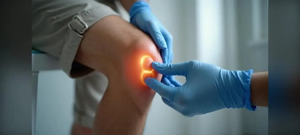
During the physical examination, tenderness, pain, and inflammation conditions in the joint are evaluated. The location of the pain and the range of joint motion are checked.
The presence of catching and locking is examined. It is evaluated whether there is pain related to the kneecap bone.
Swelling around the joint is observed. Slight temperature increase may be detected. Redness on the skin and warm skin findings can be seen.
As the disease progresses, complaints continue to increase. Symptom severity varies from person to person.
How is Knee Osteoarthritis Diagnosed?
The diagnosis of knee osteoarthritis is made by using clinical evaluation and imaging methods together. The diagnostic process begins with listening to the patient's complaints and continues with a systematic evaluation.
Physical Examination
The first step of the diagnostic process is to take a detailed patient history. It is questioned when and how the pain started, where it is located, and at what times of day it increases. What types of movements trigger the pain are learned.
A careful physical examination is then performed. During the examination, several evaluations are carried out. First, both knees are compared. It is checked whether there is swelling, deformity, redness, or muscle wasting.
Next, by touching the knee, temperature increase, tender points, and fluid accumulation within the joint are checked. By measuring how much the knee can bend and straighten, the restriction in the range of motion is determined. At this time, an attempt is made to feel a crackling sound while the joint is moved.
Special tests that provoke pain are applied. Gait analysis is performed and muscle strength is tested. Whether there is catching in the knee is examined. In clinical examination, pain is usually widespread in the knee, especially in the anterior and inner parts.
Radiological Examinations
Imaging methods are utilized to confirm the diagnosis and determine the degree of osteoarthritis. The most frequently resorted method is X-ray.
Standing weight-bearing knee radiographs are very important in diagnosis. In anteroposterior and lateral radiographs taken under load, a decrease in the joint space indicates cartilage loss. In patients planned for joint space evaluation with a preliminary diagnosis of gonarthrosis, a standing direct graph imaging is sufficient.
In X-ray films, you can clearly see the narrowing of the joint space, which is an indicator of cartilage loss, sharp projections at the bone edges, and advanced stage findings where bones begin to rub against each other. Changes such as bone spurs at the joint margin, whitening in the bone tissue under the joint, and cystic structures are observed.
The disease is radiologically divided into four stages according to the Kellgren-Lawrence classification. The fourth stage is an advanced stage of osteoarthritis where the knee joint is completely closed.
MRI and Advanced Imaging Methods
Magnetic resonance imaging is not an absolutely necessary diagnostic method. It is not required in ordinarily progressing gonarthrosis. It is not a routinely requested test.
However, it is resorted to when the diagnosis is uncertain or when another problem accompanying osteoarthritis is suspected beneath the pain. It is preferred in evaluating early-stage cartilage damage. It is useful in examining soft tissue structures such as menisci and ligaments.
It does not contain radiation and is an imaging modality where soft tissues can be evaluated in detail. Menisci, cartilage, and joint ligaments along with bone structures can be evaluated in detail.
MRI may be requested in case of increased pain after minimal trauma. Bone marrow edema can be detected and conditions such as osteonecrosis can be identified.
Blood tests do not provide a direct diagnosis. However, if it is confused with other rheumatic diseases such as rheumatoid arthritis, some blood tests may be requested for differential diagnosis. Routine blood test results are expected to be normal.
When there is prominent swelling in the knee joint, the knee joint can be evaluated with ultrasound, and the fluid inside the knee can be drained and sent to the laboratory.
How is Knee Osteoarthritis Treated?
The treatment plan is personalized according to the patient's age, the stage of the disease, the severity of the pain, and the level of daily activity. It starts with conservative methods, and when these are insufficient, more advanced treatments are progressed to.
Non-Surgical Treatment Methods
Lifestyle changes form the basis of treatment. Weight control reduces the load on the knee, relieves pain, and slows the progression of the disease. Losing weight is one of the most effective methods in overweight patients.
Activity regulation is extremely important. You need to avoid movements that increase pain. You should limit activities such as standing for a long time, running, and jumping. Instead, you can turn to low-impact movements such as swimming, cycling, and water exercises.
Physical therapy and exercise programs are very valuable. Exercises performed under the supervision of expert physiotherapists strengthen the muscles around the knee. Specifically, strengthening the quadriceps, hamstring, and hip abductor muscles is targeted. Preserving joint range of motion and ensuring flexibility are also among the goals of these programs.
Paracetamol can be used as a first choice in drug treatment. Non-steroidal anti-inflammatory drugs such as ibuprofen, naproxen, and diclofenac reduce both pain and inflammation. However, since they may have side effects on the stomach, kidneys, and heart, they should be used under doctor supervision.
Assistive devices such as canes and walkers reduce the load on the knee joint. Specially designed knee braces can also alleviate pain by supporting the joint.
Intra-Articular Injection Treatments
When adequate responses cannot be obtained from conservative treatments, intra-articular injections are applied. Corticosteroid injections are powerful anti-inflammatory drugs. When injected into the joint, they rapidly reduce pain and swelling. Their effects are temporary and usually last from a few weeks to a few months. Since they may have negative effects on the cartilage, they are not administered more than three or four times a year.
Hyaluronic acid injections are a component of joint fluid. They are used to lubricate the joint and increase smoothness. They can be administered as one to five injections spaced a few weeks apart. Its effect starts later than cortisone but can last longer. It is effective in patients with early and intermediate stage osteoarthritis.
PRP injections are platelet-rich plasma obtained from the patient's own blood. It has the potential to stimulate healing and reduce inflammation. Scientific evidence is still evolving and results vary.
Surgical Treatment Options
If pain persists despite trying all other treatment methods and daily life activities are severely restricted, surgical treatment is considered.
Arthroscopy is not the primary treatment for knee osteoarthritis. It can only be useful for mechanical symptoms accompanying osteoarthritis, such as a meniscus tear or joint mice.
Osteotomy is generally applied to patients under 60 years of age, active, and having alignment disorders. The bone is cut to correct the angle, and body weight is transferred from the damaged area to the healthy area. It can delay the need for a prosthesis.
Partial knee replacement is applied only when there is osteoarthritis in one of the inner, outer, or patellofemoral compartments of the knee. It offers a smaller incision and faster recovery compared to total replacement.
Knee Replacement Surgery
Total knee replacement is the gold standard surgical method in the treatment of advanced stage gonarthrosis. If physical therapy or medications are ineffective, the only solution is knee replacement surgery. Worn femur, tibia, and sometimes patella joint surfaces are removed and replaced with a prosthesis made of metal and polyethylene materials.
It is more frequently preferred in patients over 60 years of age. Thanks to modern technology, prostheses can be used from the age of 50 onwards. With today's prosthesis designs, lifespan has extended up to 15-20 years.
Prostheses are not applied to young patients except in mandatory situations. Other methods protecting the joint and cartilage are preferred for them.
Surgery is usually performed under general anesthesia. However, when general anesthesia is not possible, regional anesthesia below the waist can be used. Damaged joint surfaces are cleaned and the prosthesis is placed. At the end of the surgery, an 8-12 cm long scar remains in the operated area.
Postoperative Recovery Process
The length of hospital stay is an average of two to three days. Early mobilization after surgery is of vital importance. Walking usually begins the day after surgery.
With an intensive physical therapy program, knee range of motion, muscle strength, and walking function are regained. This process continues for months. Full recovery and reaching maximum function can take six months to one year.
Sutures are removed 21 days after surgery. After six weeks, the hospital is visited again and an examination is performed. Regular exercises should be performed, and blood-thinning medications should be used correctly. Water should not touch the operated area until the first check-up.
Frequently Asked Questions About Knee Osteoarthritis
Patients ask many questions about knee osteoarthritis. Below you can find clear and understandable answers to the most frequently wondered topics.
Why does knee osteoarthritis occur?
Knee osteoarthritis occurs with the wear or damage of the cartilage tissue in the knee joint. Many factors play a role in this process.
With advancing age, the rate of damage in joints increases. Especially at age 50 and after, the frequency of gonarthrosis increases significantly. Cartilage tissue naturally loses its capacity for regeneration.
Excess weight is one of the most important causes. Every kilogram gained adds about 3 to 4 kilograms of weight to the knees. Having a body mass index of 30 and above can lead to osteoarthritis. Obesity also accelerates the progression of the disease by increasing inflammation in the joint.
Genetic predisposition is an important factor. The risk is higher in individuals with a family history of osteoarthritis. Some individuals may genetically have weaker cartilage structure.
Movements that apply pressure to the knee and the frequent repetition of these can damage the cartilage. Athletes and heavy laborers are at risk in this respect. Past knee injuries, meniscus tears, and ligament injuries accelerate osteoarthritis.
Individuals with autoimmune diseases such as rheumatoid arthritis carry the risk of knee osteoarthritis. Additionally, deformities such as bowlegs and knock-knees put more pressure on specific parts of the joint.
Can knee osteoarthritis be prevented?
Gonarthrosis is not a fully preventable condition, but its onset can be delayed. Maintaining a healthy weight is the most effective method. Studies show that every 1 kg of weight loss reduces the load on the knee joint by 4 kg.
Doing regular exercises protects knee health. Low-impact activities such as swimming, cycling, and walking reduce the pressure on the joints. These exercises strengthen the muscles around the knee.
A balanced diet supports cartilage health. A diet containing Omega-3 fatty acids, vegetables, and fruits is effective in protecting joint tissue health.
It is important to avoid knee traumas. Individuals who suffer an anterior cruciate ligament tear or meniscus tear at a young age may encounter gonarthrosis at an early age if they are not treated.
What movements should I avoid in knee osteoarthritis?
You should avoid exercises involving running and jumping. Sports such as basketball, volleyball, and artificial turf football place shock loads on the prosthesis.
Long-distance running, climbing hills, squat exercises, and weight lifting are not recommended. It is very important not to kneel on the knees. You should also avoid bending forward movements.
You should mobilize the knee region approximately every half hour. You should not remain in the same position for long periods. You should take frequent breaks during long walks.
It is necessary to avoid or limit jobs that require standing for a long time.
How long does a knee replacement last?
The lifespan of knee replacements applied today has generally extended up to 20-25 years. 95% of modern knee replacements are still functioning smoothly 15 years after surgery.
Several factors affect prosthesis lifespan. The experience of the physician performing the surgery, the quality and durability of the prosthesis used are important. The patient's way of using the prosthesis is also decisive.
In overweight patients and individuals living a very active lifestyle, prosthesis lifespan may shorten. Situations such as constantly jumping, skipping, and falling with the prosthesis accelerate wear.
The vast majority of patients do not need a second surgery throughout their lives. Revision surgery may be required in case of prosthesis loosening.
In Summary, Knee Osteoarthritis (Gonarthrosis)
Knee osteoarthritis can make your life difficult, but it is a manageable condition with the right approach. You can slow down the progression of the disease with early diagnosis and an appropriate treatment plan. Simple steps like weight control, regular exercise, and physical therapy make a noticeable difference. Painkillers and intra-articular injections relieve symptoms. In advanced stages, knee replacement surgery allows you to return to a pain-free life. Consult a specialist without delay when your complaints begin. Thanks to timely precautions, you can move more comfortably and preserve your quality of life.
Start with the right assessment for What is Knee Osteoarthritis (Gonarthrosis)? What are Its Symptoms? How is It Diagnosed and Treated?
Share your symptoms with our specialist physicians; let us determine the right diagnosis and treatment plan together.
Warning
If your symptoms started suddenly, are progressing rapidly, or significantly restrict your daily life, consult a physician without delay.
Plan your What is Knee Osteoarthritis (Gonarthrosis)? What are Its Symptoms? How is It Diagnosed and Treated? journey with our specialists.
Fill in the form or book an appointment to learn about treatment options, the process and preparation.