What is EUS?
Endoscopic ultrasonography, or EUS for short, is a diagnostic method aimed at obtaining high-resolution images of the digestive system and surrounding organs. This procedure is performed with the help of an ultrasound probe placed at the tip of a thin, flexible tube called an endoscope. Thanks to EUS, the internal structure of organs can be imaged in detail. This method yields much more precise results compared to classical endoscopy or external ultrasound. It is particularly effective in evaluating areas such as the stomach, pancreas, esophagus, bile ducts, and liver. EUS not only provides imaging but also allows for procedures such as biopsies when necessary.
What is an EUS (Endoscopic Ultrasound) Device?
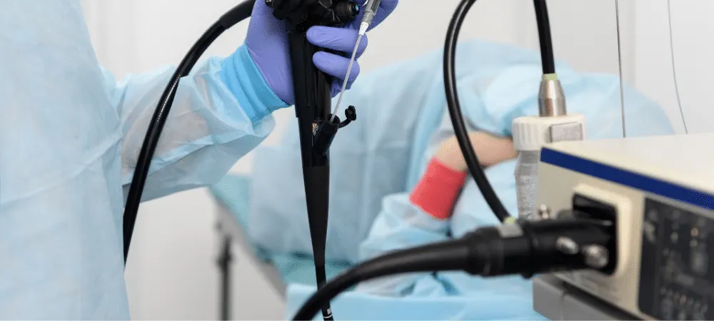
The EUS device is one of the advanced technologies in the field of modern medical imaging. This device makes the detailed examination of internal organs possible by combining endoscopy with ultrasound. A small ultrasound probe located at the tip of the device reaches the organs by producing high-frequency sound waves and creates an image using the reflected waves. Thus, structures such as the stomach, pancreas, liver, bile ducts, and intestines can be clearly visualized. The EUS device is available in hospitals with advanced technological infrastructure. It is especially preferred for cancer research, tumor evaluations, and detailed analysis of internal structures. Thanks to the sensitivity of this device, even small-sized formations that cannot be noticed by other methods can be clearly imaged. At the same time, the ability to perform both diagnosis and interventional procedures in the same session is an important feature that distinguishes this device from other imaging methods. Constantly updated technologically, this device plays an important role in achieving earlier and more accurate diagnoses in medicine.
What is EUS (Endoscopic Ultrasound) Used For?
EUS provides great convenience in reaching a diagnosis in many medical conditions. First and foremost, it is used in digestive system diseases. It is highly successful in staging tumors in regions such as the esophagus, stomach, duodenum, and rectum. Deeply seated lesions, such as pancreatic tumors, are better visualized with EUS. In addition, EUS is preferred when detailed examination of the bile ducts and liver is required. Not only imaging but also procedures such as biopsies can be performed with this method. Especially in the diagnosis of suspicious masses, tissue samples taken with EUS confirm the diagnosis. Pancreatic cysts, gallstones, and certain infectious conditions can also be detected through EUS.
What Are the Advantages of EUS (Endoscopic Ultrasound)?
EUS offers significant advantages in many aspects compared to conventional imaging methods. Providing high-resolution images allows small masses or lesions to be noticed at an early stage. The ability to examine the layers of organs individually makes it possible to detect details that cannot be noticed by other techniques. Administering sedation during the procedure increases patient comfort. Biopsies can be taken from suspicious areas without the need for surgery. Furthermore, since it works with sound waves, it does not contain X-rays and thus carries no radiation risk. EUS is generally an outpatient procedure; the patient can be discharged on the same day. Since results are obtained quickly, the diagnostic process is accelerated and the treatment planning of the disease can be made in a shorter time.
In the Detection of Which Diseases is EUS Used?
Endoscopic ultrasonography is an effective method in the diagnosis of various diseases. The staging of digestive system tumors is one of the most common areas where EUS is used. The degree of spread and lymph node involvement of cancers developing in the esophagus, stomach, pancreas, bile ducts, and rectum are evaluated with EUS. Structural problems such as pancreatic cysts or masses, gallbladder stones, bile duct strictures, and blockages can also be clearly examined using this method. The relationship of the liver with neighboring organs and lymph node enlargements can likewise be analyzed with EUS. In addition, EUS is quite successful in evaluating submucosal masses and inflammatory conditions developing in the walls of the gastrointestinal system. In this respect, it holds an important place in terms of both diagnosis and treatment planning.
How is the EUS (Endoscopic Ultrasound) Procedure Performed?
The EUS procedure begins with the patient preparation process. Patients are generally asked not to consume food and beverages for 6 to 8 hours prior to the procedure. This preparation is important in terms of ensuring the stomach and intestines are clean and obtaining clearer images. On the day of the procedure, the patient is taken to the endoscopy unit and sedated with medications administered intravenously. The procedure performed under sedation ensures that the patient does not feel pain or discomfort during the procedure. The endoscope is advanced through the mouth into the esophagus and then toward the stomach. The internal and peripheral structures of the organs are scanned in detail with the ultrasound probe at the tip of the device. Images are simultaneously transferred to a monitor, and the specialist physician evaluates these images. If there is a suspicious structure, a biopsy can be taken from that area with the help of a special needle. The procedure typically takes 20 to 40 minutes. Afterward, the patient is allowed to rest for a couple of hours and can be discharged on the same day.
Frequently Asked Questions
EUS imaging is performed under sedation. First, a sedative medication is administered intravenously to the patient. Then, the stomach and surrounding organs are examined using the endoscopic device advanced through the mouth. The patient does not feel any pain during the procedure and is kept under observation for a short time after the procedure is completed. The images obtained during the scan are evaluated by the specialist physician. If there is a suspicious finding, a tissue sample can be taken during the procedure. This entire process takes approximately 30 minutes. It is generally considered a safe procedure, but like any medical intervention, it carries some minor risks. There may be mild throat irritation or difficulty swallowing after the procedure. Although rare, conditions such as infection or bleeding may develop in the area where the biopsy was taken. Perforation, meaning damage to the wall of the digestive tract, is a very rare but potentially serious risk. These risks are minimized when applied by experienced healthcare teams. If only imaging was performed during the EUS procedure, the doctor can share the initial assessment immediately after the procedure, but if a biopsy was taken, the tissue samples sent to pathology need to be examined. This analysis is usually completed within 2-4 business days. When the results are ready, the patient is informed and a treatment plan is made for the next step. Throat irritation, nausea, or gas complaints may be observed after the procedure. These are generally mild and pass quickly. In cases where a biopsy is taken, there may be a risk of bleeding or infection. In rare cases, damage to the digestive tract may occur. All these complications have a very low probability of occurring and the procedure is safe when performed by experienced teams. Due to sedation, a feeling of sleepiness, dizziness, or grogginess may occur after the procedure. These effects are temporary and disappear within a few hours. Side effects such as a stinging sensation in the throat or temporary coughing may also be observed. These side effects are usually mild in intensity and do not require medical intervention. Although ERCP and EUS are applied in similar fields, they serve different purposes. ERCP is performed to visualize the bile ducts and pancreatic ducts and to intervene in these areas when necessary. During ERCP, X-rays are taken by administering a contrast agent, and therapeutic procedures such as stone removal and stent placement are usually performed. EUS, on the other hand, is mostly for diagnostic purposes. Internal organs are examined with sound waves, and detailed images are obtained without the need for surgery. EUS yields much more sensitive results, especially in detecting small masses. The special balloon placed around the ultrasound probe located at the tip of the EUS device and inflated with water is called the EUS balloon. Thanks to this balloon, the contact of the probe with the tissues is increased and the image clarity is enhanced. The balloon is used to improve image quality, particularly in hollow organs such as the stomach or rectum. Thus, a more sensitive and detailed examination can be performed. EUS biopsy is the procedure of taking a tissue sample from a suspicious area with the help of a thin needle during endoscopic ultrasound imaging. These samples are sent to the laboratory and examined under a microscope to reach a definitive diagnosis. It allows biopsies to be safely taken from suspicious areas in the pancreas, lymph nodes, or stomach wall. This procedure is of critical importance in diagnosing the disease and determining the treatment plan. EUS screening is a planned imaging process that provides a detailed evaluation of the organs belonging to the digestive system. This screening is applied for early diagnosis, especially in individuals carrying high risk. EUS screening may be recommended for those with a family history of pancreatic cancer, those experiencing unexplained abdominal pain, or individuals in whom suspicious findings were detected in previous tests. Thanks to this screening, small masses or formations can be noticed early and necessary interventions can be planned in a timely manner.
If you are experiencing symptoms such as abdominal pain, unexplained weight loss, jaundice, indigestion, or if a suspicious finding has been detected in your examinations, it is very important to consult our Gastroenterology Specialist without delaying the diagnostic process.
Thanks to the Endoscopic Ultrasound (EUS) screening applied at Hisar Hospital Intercontinental, early diagnosis of digestive system diseases is made possible; thus, the treatment process can be initiated in a timely and effective manner.
With our advanced technological infrastructure and expert physician staff, we perform the EUS procedure in a comfortable and safe manner. If you have any doubts about your health, please know that you are at the right address.
You can contact us by filling out the form below or create an application directly from our book an appointment page.