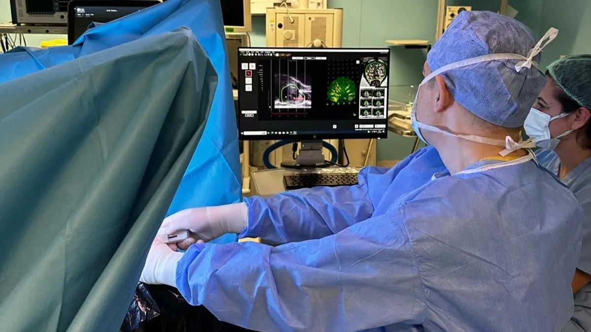
The prostate is a small gland located in men just below the bladder and in front of the rectum. Its main function is to produce the fluid that nourishes and transports sperm. Various problems can occasionally develop in this gland. Particularly with advancing age, prostate enlargement or prostate cancer may be encountered. Prostate cancer is one of the most common cancers affecting men worldwide. Early diagnosis is important for successful treatment outcomes, and advances in diagnostic techniques are of critical importance for increasing detection rates. MR fusion prostate biopsy can be described as a revolutionary innovation in the diagnosis of prostate cancer.
What Is an MR Fusion Prostate Biopsy?
MR fusion prostate biopsy is a state-of-the-art procedure that combines two powerful imaging technologies: magnetic resonance imaging (MRI) and ultrasound. This combination provides a more precise and targeted approach to prostate biopsy and significantly increases the accuracy of cancer detection. The integration of MRI and ultrasound not only increases the accuracy of targeting suspicious areas, but also helps detect tumors that may be missed by conventional biopsy methods. This means fewer false and more definitive diagnoses. As a result, it provides patients with better treatment decisions and outcomes.
Before the procedure, an MRI scan of the prostate is performed on the patient. This scan produces detailed images of the gland and marks any suspicious region that may indicate the presence of cancer. During the procedure, the MRI images are combined with real-time ultrasound images. In this way, it provides the urologist with a comprehensive roadmap for guiding the biopsy needle precisely to the exact location of the suspicious tissue.
Why Is an MR Fusion Prostate Biopsy Performed?
MR fusion prostate biopsy is generally used in cases where prostate cancer is suspected, based on elevated prostate-specific antigen (PSA) levels and abnormal findings on digital rectal examination (through the rectum):
Suspicious Spots on MRI: If magnetic resonance imaging of the prostate reveals suspicious lesions or areas of concern, MR fusion technology is used to confirm the presence of cancer and determine its aggressiveness.
Elevated PSA Despite Previous Negative Biopsies: This procedure can help identify prostate tumors that were missed in patients who have persistently high PSA levels despite previous negative results.
Active Surveillance and Disease Monitoring: MR fusion biopsy can accurately detect disease progression or recurrence in individuals at risk of prostate cancer or with a history of treated prostate cancer, thereby guiding treatment decisions.
Evaluation of Treatment Response: MR fusion biopsy is used to identify residual or recurrent tumors after treatments such as radiation therapy or radical prostatectomy.
Evaluation of Complex Cases: It can be used to clarify prostate abnormalities in cases where the clinical presentation or imaging findings are unclear or complex.
What Are Its Advantages Over Classic Biopsies?
Prostate cancer remains a significant health problem for men worldwide, and therefore accurate and precise diagnostic techniques are required for early detection and appropriate management. Compared with classic biopsies, MRI-TRUS fusion prostate biopsy has emerged as a revolutionary approach offering many advantages. These advantages are as follows:
Increased Accuracy: MR fusion biopsy combines the high-resolution imaging capabilities of magnetic resonance imaging technology with real-time ultrasound imaging. Combining these imaging modalities allows urologists to precisely target suspicious areas identified on the MRI scan.
Better Detection of Tumors: Unlike conventional biopsies, MR fusion prostate biopsy allows targeting of specific lesions identified on the MRI scan. This targeted approach increases the likelihood of detecting tumors while reducing the detection of low-grade, clinically insignificant lesions that do not require intervention.
Reduced Risk of Missed Diagnosis and Unnecessary Treatment: By accurately characterizing suspicious lesions, MR fusion biopsy reduces the risk of a missed diagnosis that could be overlooked by standard biopsies. It also helps prevent unnecessary treatment for low-risk tumors, thereby reducing the risk of unnecessary treatment and associated side effects.
Improved Risk Stratification and Treatment Planning: By providing detailed information about the size, location and aggressiveness of tumors, MR fusion biopsy makes it possible to tailor treatment strategies to individual patients.
Reduced Need for Repeat Biopsy: In some patients, conventional biopsies may fail to detect cancer and may need to be repeated. MR fusion biopsy reduces the need for repeat procedures by improving the accuracy of initial results.
Reduced Risk of Infection in the Patient: This method causes less infection compared with conventional biopsies and offers a less invasive approach.
How Should You Prepare for a Prostate Biopsy?
First, you can schedule a consultation appointment with your urologist to talk in detail about the prostate biopsy. During this consultation, you can provide your urologist with your complete medical history, including past prostate procedures, allergies, current medications and existing medical conditions. Your urologist may order blood tests to check your prostate-specific antigen (PSA) levels and other relevant markers before the biopsy. Before the procedure, you will be asked to completely empty your bladder and bowels. This provides a clearer image during the MRI scan and makes the procedure more comfortable. Depending on your health care provider's instructions, you may need to fast for several hours to prevent possible complications related to anesthesia or sedation. If you have any concerns or questions about the procedure, you can ask your doctor.
What Is the Process After a Prostate Biopsy?
After the procedure, health care staff will take you to a recovery area to monitor your condition. This observation period is necessary to make sure you have recovered well after the procedure. This may take 2-4 hours. It is important to rest for the remainder of the day after the biopsy and avoid strenuous activities. Some people may experience mild discomfort such as mild pain, blood in the urine or semen. These symptoms usually resolve within a few days. Although rare, it is important to watch for signs of infection or other complications after the procedure. Your doctor will schedule a follow-up appointment to discuss the results and determine the next steps. At this appointment, the biopsy findings will be reviewed, treatment options will be discussed if necessary, and any questions or concerns will be addressed.
Frequently Asked Questions
An MR fusion biopsy takes approximately 45 to 60 minutes. This time includes the time required for preparation and the procedure. However, the exact duration may vary depending on the individual's anatomy, the complexity of the condition and the protocols of the medical facility where the procedure is performed. Fusion biopsy samples are taken specifically from the prostate gland via the perineal region (between the anus and scrotum). During the procedure, targeted areas within the prostate identified by MRI imaging are sampled to obtain tissue for analysis. Yes, fusion MR biopsy is generally performed under general anesthesia or sedation. The reason for this is to keep the patient still during the procedure, thereby increasing the accuracy of the biopsy. Patients are evaluated by anesthesiologists beforehand. However, for patients who cannot receive anesthesia, local anesthesia is also applied to numb the area around the prostate and aims to minimize discomfort during the procedure. After an MR fusion biopsy, most people can return to their daily activities within one or two days, although this may vary depending on how the person feels. To allow the body to fully recover, it is recommended to avoid heavy lifting or strenuous activities for several days. It is also important to follow your doctor's instructions for complete recovery. MR fusion biopsy results are available within one to two weeks after the procedure. The exact timeline may vary depending on factors such as the laboratory's workload, the complexity of the cases and the specific protocols followed by the facility. No pain is felt during an MR fusion biopsy. However, patients who receive local anesthesia to numb the area around the prostate may feel mild discomfort or a sense of pressure during the procedure, although the use of anesthesia significantly helps relieve pain. In addition, health care professionals make efforts to ensure the patient's comfort during the procedure. If you have concerns about pain or discomfort, you should discuss them with your health care provider in advance. The margin of error of MR fusion biopsy is quite low compared with standard biopsy techniques. Although it provides highly accurate results in detecting and diagnosing prostate cancer, there is a small risk of false-positive or false-negative findings. Factors such as the quality of imaging, the expertise of health care professionals and tumor characteristics affect accuracy. Collaboration among radiologists, urologists and pathologists helps reduce errors and increase diagnostic precision.
If you are experiencing prostate enlargement or suspect prostate cancer, you may undergo an MR fusion biopsy on your doctor's advice for more effective treatment.