What Is Capsule Endoscopy and How Is It Performed?
Capsule endoscopy is a major innovation in medical diagnosis for both patients and physicians. With this new technique, pathological conditions in the small intestine, once a difficult area to reach, can now be visualized and diagnosed more easily. Capsule endoscopy combines state-of-the-art technology with minimally invasive methods to offer patients a more comfortable alternative.
What Is Capsule Endoscopy?
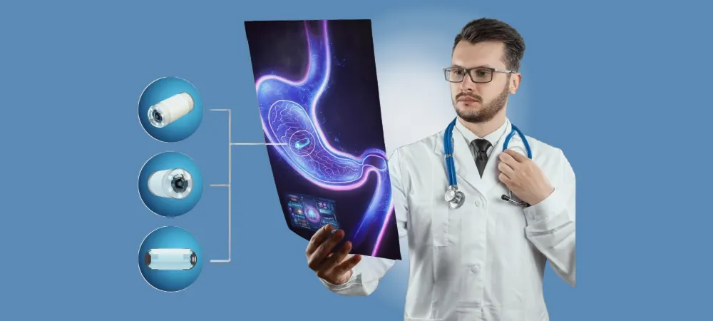
Capsule endoscopy, also known as wireless capsule endoscopy or video capsule endoscopy, is a medical procedure that allows healthcare professionals to examine the inside of the gastrointestinal tract in a non-invasive way. The technique involves the use of a small, pill-sized camera that the patient swallows. As the capsule travels through the digestive system, it captures thousands of images, allowing the small intestine to be examined in detail. Although the entire large intestine can be examined by colonoscopy, capsule endoscopy makes it possible to view the large intestine in patients who cannot tolerate colonoscopy due to sedation or other reasons.
The capsule itself is about the size of a large vitamin pill. Inside it are a small camera, light-emitting diodes for illumination, a battery and a radio transmitter. The thousands of images captured as the capsule passes through the gastrointestinal tract are sent to a recorder worn on a belt around the waist. Once recording is complete, the recorded images are reviewed by a physician on a computer using a special program to determine whether there is any abnormality in the small or large intestine. One of the most important advantages of capsule endoscopy is its ability to visualize parts of the small intestine that are difficult or impossible to reach with conventional endoscopic methods. This is especially valuable in diagnosing conditions affecting the small intestine, such as Crohn's disease, celiac disease and obscure gastrointestinal bleeding.
The procedure is generally well tolerated by patients, as it requires no sedation and no tube to be placed in the body. It is a good option for people who are concerned about more invasive procedures or who have anatomical problems that make conventional endoscopy difficult. Although capsule endoscopy is a very important innovation and advance in the diagnosis of diseases of the gastrointestinal tract, it is important to note that it has some limitations. Once swallowed, the capsule cannot be steered or controlled. In addition, the procedure may not be suitable for patients who have known or suspected gastrointestinal strictures or obstructions, because the capsule has the potential to become lodged at the site of a narrowing in the digestive system.
Why Is Capsule Endoscopy Performed?
Capsule endoscopy is performed for various reasons, primarily to investigate and diagnose conditions affecting the gastrointestinal system, especially the small intestine. By providing insight into areas of the digestive system that were previously difficult to examine thoroughly, this procedure has become an important tool in gastroenterology. Capsule endoscopy may be performed in the following situations:
Gastrointestinal Bleeding: One of the most common reasons for capsule endoscopy is the investigation of “obscure gastrointestinal bleeding,” in which the source of the bleeding cannot be identified. When the source of bleeding cannot be determined by conventional endoscopic methods or other imaging techniques, this method provides a comprehensive view of the small intestine. In this way, it may be possible to identify small lesions, ulcers or vascular abnormalities that could be the cause of the bleeding.
Inflammatory Bowel Diseases: Capsule endoscopy is frequently used in the diagnosis and management of inflammatory bowel diseases, especially Crohn's disease. It can help visualize areas of inflammation, ulceration or narrowing in the small intestine that cannot be seen with other diagnostic methods. This information is essential for determining the extent and severity of the disease, guiding treatment decisions and monitoring the response to treatment.
Peutz-Jeghers Syndrome: It can be used to monitor and evaluate hereditary polyposis syndromes such as Peutz-Jeghers syndrome. In these cases, it can help detect and monitor polyps in the small intestine.
Celiac Disease: In patients with suspected celiac disease, capsule endoscopy can be valuable for assessing the extent of intestinal damage or identifying complications such as lymphoma.
Intestinal Tumors: It is used to detect both benign and malignant tumors of the small (and large) intestine. Existing tumors can be difficult to detect with other imaging methods, but the high-resolution images provided by capsule endoscopy can reveal small lesions or masses.
Unexplained Conditions: When other diagnostic tests have not yielded results, capsule endoscopy may be used to provide detailed images for the evaluation of patients with unexplained abdominal pain or chronic diarrhea. It may be possible to identify small intestinal ulcers, areas of inflammation or other abnormalities that are not apparent by other means and may be the cause of these symptoms.
How Is Capsule Endoscopy Performed?
The capsule endoscopy process is simple and minimally invasive, which makes it a comfortable experience for most patients. The procedure begins with some preparations to ensure the best possible visualization of the gastrointestinal tract:
Before the procedure, patients are generally instructed to fast for about 12 hours. This helps keep the digestive system as clean as possible and provides better image quality. In some cases, particularly when examination of the lower part of the small intestine or the colon is important, bowel preparation is recommended to obtain clear images.
On the day of the procedure, the patient arrives at the healthcare facility and a belt or strap containing the receiver that will collect data from the capsule is fitted. The capsule itself is then activated and given to the patient, who swallows it with water. The capsule is about the size of a large vitamin pill, which makes it easy for most people to swallow.
After swallowing, the capsule begins its journey through the digestive system, moved along by the natural muscle contractions of the gastrointestinal tract. While traveling, the capsule captures images at a rate of several frames per second and transmits them wirelessly to the receiver worn by the patient.
During the procedure, patients can continue their normal daily activities with some restrictions. They are generally advised to avoid strenuous physical activity and to stay within the range of the receiver. Patients are also given guidance on when they can resume eating and drinking, usually a few hours after swallowing the capsule.
Throughout the day, the capsule continues to transmit images as it moves through the stomach, small intestine and large intestine. The duration of the journey may vary between individuals and is affected by factors such as bowel motility and the presence of any gastrointestinal condition.
The recording device can be removed 8 hours after the start of the procedure or after the capsule is seen in the stool, whichever occurs first. At the end of the procedure, the patient returns to the healthcare facility so the receiver can be removed. The capsule itself is designed to pass out with a natural bowel movement one or two days after the procedure. Patients are instructed to monitor their stools to confirm that the capsule has been passed.
The data collected by the receiver is then downloaded and processed, and a video of the capsule's journey through the digestive system is created. A gastroenterologist or a specially trained healthcare professional reviews this video, which may contain up to 50,000 images, looking for any abnormality or sign of disease.
The endoscopy capsule is generally considered safe; however, there are risks associated with capsule endoscopy, particularly in patients with known strictures or obstructions in the gastrointestinal system. In such cases, additional precautions may be taken or alternative diagnostic methods may be considered.
What Should Be Considered After Capsule Endoscopy?
There are several important points to be aware of in order to complete the capsule endoscopy procedure in the best possible way and minimize potential complications:
First, patients should follow the specific instructions on diet and activities given by their healthcare providers. While many patients can return to their normal diet and routines shortly after swallowing the capsule, some may be advised to limit themselves to liquids for a period of time.
It is very important for patients to be aware of any symptoms that may indicate a complication. Although rare, capsule retention may occur, especially in patients with undiagnosed strictures or obstructions in the gastrointestinal system. Symptoms such as persistent or worsening abdominal pain, nausea, vomiting or bloating should be reported to a healthcare provider immediately.
Patients should be aware of the need to confirm that the capsule has passed from their body. This usually occurs with a natural bowel movement one or two days after the procedure. It is appropriate for patients to collect or examine their stools to visually confirm the passage of the capsule. If there is any doubt about whether the capsule has passed, the patient may need an X-ray to confirm the position of the capsule.
In the days following the procedure, patients should avoid any magnetic resonance imaging (MRI) examinations until they are sure that the capsule has passed. The strong magnetic fields used in MRI can potentially interact with the capsule and cause harm if it is still present in the body.
Frequently Asked Questions
Capsule endoscopy is applied to patients who have symptoms related to small intestinal pathology that is difficult to diagnose by other methods, or in whom colonoscopy cannot be performed for patient-related reasons. It is an important diagnostic tool, especially in individuals with unexplained gastrointestinal bleeding, suspected Crohn's disease, celiac disease, small intestinal tumors, polyps and hereditary polyposis syndromes. It is also used for monitoring patients who have undergone small intestine surgery or who have known bowel abnormalities. The capsule endoscopy procedure itself takes only a few minutes, as it involves only swallowing the capsule. However, the capsule captures images and transmits them to the recorder while moving through the digestive system for approximately 8 hours. Patients carry the data recorder during this time, and most patients can continue their normal activities. The capsule is usually passed within 24 to 72 hours. Capsule endoscopy is performed by a gastroenterologist who specializes in diseases of the gastrointestinal system. The procedure is generally carried out in the gastroenterology department of a hospital or in a specialized outpatient clinic. The images obtained with the capsule are analyzed by a gastroenterologist to diagnose and evaluate any abnormality in the small intestine.
If you are also experiencing unexplained digestive complaints, you can apply to our Hisar Hospital Gastroenterology Clinic for a detailed evaluation and appropriate diagnostic methods. Our expert physicians carefully analyze your health with advanced diagnostic methods such as capsule endoscopy.