Patellar tendinitis is a condition that occurs in the tendon below your kneecap, causing pain in the front of the knee. Also known as jumper's knee, this problem was first defined in 1963 as a knee issue resulting from jumping. It is particularly seen in athletes aged 16-40 and is more common in men. It is prevalent in sports that require jumping, such as volleyball, basketball, and soccer.
In this article, you will find what you need to know about patellar tendinitis symptoms, diagnostic methods, patellar tendinitis treatment, and patellar tendinitis exercises.
What is Patellar Tendinitis (Jumper's Knee)?
The patellar tendon, which connects the kneecap to the shinbone, is one of the strongest tendons in your body. Thanks to this tendon, you can straighten your knee, walk, run, and jump. The quadriceps muscle from above and the patellar tendon from below work together to enable the movement of your knee.
Patellar tendinitis is a degenerative injury characterized by microscopic tears and wear in the tendon fibers resulting from overuse of this tendon. Repetitive movements and excessive load on the tendon lead to deterioration in the internal structure of the tendon. Contrary to traditional belief, there is no inflammation in the tendons in this condition.
The Function and Importance of the Patellar Tendon
The patellar tendon is a critical part of the knee's extensor mechanism. It transmits the power of your quadriceps muscles to the shinbone, making it possible to extend your leg. Therefore, the basic movements in your daily life depend on the health of this tendon.
Activities such as standing up from a sitting position, climbing stairs, and kicking are performed thanks to the patellar tendon and the knee's extensor mechanism. The tendon acts as a lever, strengthening the power of the quadriceps muscle.
Other Names for the Disease
Patellar tendinitis is known by different names in the literature and among the public. The most commonly used name is "jumper's knee." This name stems from the fact that the disease is frequently seen in jumping sports.
It is also called runner's knee. In some sources, the term patellar tendinopathy is used. All these names describe the same condition: wear of the kneecap tendon due to overuse.
Who Does It Affect?
Patellar tendinitis is generally seen in the 16-40 age range, but it can also occur in adolescent athletes and athletes over 30. The incidence rate in male athletes is significantly higher than in women: 25.3% in men and 13.1% in women.
The prevalence is higher, especially in sports where the thigh extensor muscle group is used intensively:
- Volleyball: The prevalence reaches up to 40-50% in elite volleyball players
- Basketball: It is frequently seen due to the intensity of jumping movements
- Soccer: Running and kicking movements pose a risk
- Athletics: It is common in long jump and high jump disciplines
Athletes involved in professional-level jumping sports are at higher risk. Individuals with long training durations and those who play on hard surfaces are also in the risk group. Being overweight is a significant risk factor for the disease.
What Are the Symptoms of Patellar Tendinitis?
Patellar tendinitis symptoms usually start slowly and worsen over time. In the first stage, the pain is only felt after intense activity, but if left untreated, it becomes something that affects daily life.
Pain in the Front of the Knee
The most prominent finding of the disease is pain felt in the front part of the knee. The pain is localized specifically under the kneecap, on the patellar tendon. Although some patients have difficulty finding the exact location of the pain, it is mostly concentrated in the area near the kneecap.
Pain increases with activity. It intensifies while performing jumping, running, or leaping sports. Pain occurs on the tendon during movements that strain the knee, such as climbing stairs and standing up from a chair. Pain may start when you begin physical activity or immediately after intense training.
In the early stage, the pain that appears after activity manifests itself when starting sports as the disease progresses. In more advanced stages, pain is felt even at rest or while sitting. Therefore, the timing and intensity of the pain indicate the stage of the disease.
Swelling and Tenderness
Swelling is seen under the kneecap and in the front of the knee. Swelling occurs on the tendon as a result of fluid accumulation in the sheath of the patellar tendon. Mild or moderate edema may occur in the area.
Tenderness is felt when the tendon is touched. There is tenderness to touch along the tendon under the kneecap. Pain occurs when the inflamed area is pressed lightly. Additionally, an increase in temperature and burning may be seen in the knee.
Stages of the Disease
Patellar tendinitis is classified into 4 stages. This classification determines the severity of the disease and the treatment approach.
Stage 1: Pain is felt only after sports activity. In this stage, the pain goes away with rest, but it returns when starting sports.
Stage 2: Pain starts at the beginning of sports activity, decreases or disappears after warming up. Pain reappears with fatigue.
Stage 3: There is pain during sports activity and at rest. The pain does not go away with rest and is felt even when going up and down stairs. It is intense enough that sports cannot be continued.
Stage 4: A rupture of the patellar tendon is in question. In advanced levels, pain occurs even when something touches the tendon.
Difficulty in Daily Activities
As the disease progresses, difficulty is experienced in daily movements. Pain is felt on the tendon during simple activities such as climbing stairs or standing up from a chair. It becomes difficult to fully bend and straighten the knee.
Weakness and loss of power are seen, especially in straightening the knee. Movement restriction occurs and loss of strength is experienced. Pain increases during movements where the knee is used intensively, such as squatting. If the pain is ignored and training is continued, it can cause larger tears in the patellar tendon.
Causes and Risk Factors of Patellar Tendinitis
Multiple factors contribute to the development of patellar tendinitis. Usually, not a single cause but a combination of various factors lies at the root of the disease. The most basic mechanism is loading the tendon more or more frequently than it can handle.
Overuse and Training Errors
Repetitive movements lead to excessive stress on the tendon, causing inflammation. Activities such as running and jumping are the most common triggering factors. Especially during sports activities, excessive straining of the tendons is common.
Increasing training intensity in a short time damages the tendon. Inadequate warm-up and stretching movements also pose a risk. Sudden increases in activity intensity, training pace, and weekly loads cause the development of tendinitis.
Insufficient rest periods prevent the tendon from repairing itself. As the tears in the tendon multiply, your body struggles to repair them. Playing on hard surfaces or engaging in high-impact sports without proper warm-up increases the risk.
Incorrect posture positions and techniques while exercising or during work can damage the tendon. Not using the proper technique leads to tendinitis. Even changing sports shoes involves risk.
Muscle Imbalance and Stiffness
Excessive tension in the thigh muscles increases the tension on the patellar tendon. Stiffness and shortening in both the front (quadriceps) and back (hamstring) muscles of the thigh put extra stress on the patellar tendon. Muscles shorten over time as you train.
If some muscles in your legs are much stronger than others, the stronger muscles pull your patellar tendon more. This uneven pulling causes tendinitis. Excessively strong quadriceps and hamstring muscles also create problems.
If your hip muscles are not strong enough or if you have a restriction in your ankle, your knee takes on the load that these areas should handle. This faulty alignment increases the pressure on the structures inside the knee.
Anatomical Factors
Leg length discrepancy or gait abnormalities strain the tendon. Structural problems such as a high Q angle and pelvic tilt pose a risk. Flat feet or hip-knee-ankle alignment problems can cause pain in the knee.
Obesity is a physical risk factor. Excess weight and obesity weaken the tendons and muscles around the knee. In overweight individuals, the knee joint struggles to carry body weight.
Chronic Diseases
Chronic diseases such as rheumatoid arthritis, diabetes, and gout increase the risk of tendon inflammation. Some diseases impair blood flow in the tendons, which weakens the tendon.
Chronic kidney failure, autoimmune diseases such as rheumatoid arthritis, and metabolic diseases such as diabetes and high blood lipids constitute risk factors. Lupus can also be given as an example.
Tendons lose their elasticity as age progresses. As people age, their tendons become less flexible, which makes them easier to injure. In old age, tendons withstand less stress.
How Is Patellar Tendinitis Diagnosed?
Your doctor first listens to your complaints and performs a physical evaluation to diagnose patellar tendinitis. They take detailed information about when the pain started, which activities increase it, and your past knee injuries. Clinical evaluation, physical examination, and imaging methods are used together in the diagnostic process.
Physical Examination
During the physical examination, your doctor identifies the tender points in your knee, checks for swelling, and evaluates your range of motion. Tenderness is sought by touch on the patellar tendon under the kneecap.
The VISA score is used to determine the severity of the disease. This scoring system measures the severity of the disease according to your symptoms.
The Royal London Hospital test is quite useful in the diagnosis of patellar tendinitis. This test has an 88% sensitivity and 98% specificity rate. The test is performed as follows: your doctor checks for tenderness by pressing the patellar tendon from top to bottom while your knee is in a full straight position. Then, they bend your knee 90 degrees and press the same area again. If the pain decreases or disappears in the bent position, the test is considered positive.
Imaging Methods (MRI and Ultrasound)
Ultrasound and MRI are the best tools in the diagnosis of patellar tendinitis. These imaging methods are valuable for confirming changes in the tendon.
Ultrasound examination: Shows thickening in the tendon and irregular fiber structure. Hypoechoic areas, i.e., regions that appear dark on ultrasound, are detected. These lesions are usually observed in the posterior part of the patellar tendon, near the lower pole of the kneecap. When Doppler ultrasound is used, neovascularization, i.e., new vessel formation and increased blood flow, can be shown in symptomatic cases. According to research, if the tendon thickness is over 6.67 mm, the disease can be diagnosed with 100% sensitivity and 92% specificity.
MRI imaging: Characterized by thickened tendon structure and increased signal intensity. It shows the changes in the tendon and surrounding structures in detail. Signal changes can be seen at the lower pole of the kneecap and in the infrapatellar fat pad. With MRI, a tendon thickness over 7.08 mm shows 100% sensitivity and 89.4% specificity. MRI is the gold standard method in differential diagnosis.
X-ray: Can be taken to rule out fractures or bone-related problems.
Differential Diagnosis
It is necessary to rule out other conditions that show similar symptoms. Patellofemoral pain syndrome, quadriceps tendinitis, and tendon tears should be considered in the differential diagnosis.
Osgood-Schlatter disease and Sinding-Larsen-Johansson syndrome should be kept in mind in adolescent athletes. Infrapatellar bursitis can also cause similar pain. Suprapatellar fat pad irritation and plica syndrome are also evaluated. MRI is the gold standard method in distinguishing these lesions.
How Is Patellar Tendinitis Treated?
Patellar tendinitis treatment is largely based on conservative methods. The treatment strategy is personalized according to the severity, duration of symptoms, and your functional expectations. Most cases heal without the need for surgery.
Rest and Activity Modification
The first step of treatment is to reduce or stop the activity that causes the symptoms. Especially high tempo and squats are prohibited. In professional athletes, the training program should be changed or activities where the legs are used should be avoided.
In the first stage, knee braces, ice application, and painkillers are used. Cold packs relieve pain and reduce swelling. However, long-term use of painkillers is not recommended. These drugs may suppress the proliferation of tendon cells.
Bandage applications and special knee braces reduce the pressure on the tendon. Kinesiology taping can be used to relieve pain and support the tendon.
Physical Therapy and Patellar Tendinitis Exercises
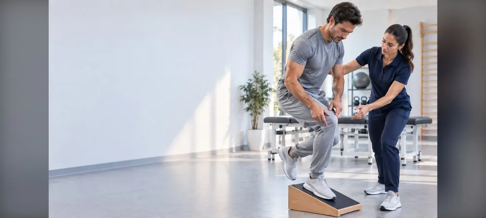
Physical therapy aims to increase the strength and flexibility of the knee extensor mechanism. Exercise programs applied with a physiotherapist form the basis of treatment. Especially eccentric exercises are accepted as the gold standard.
Stretching exercises are performed on the quadriceps and hamstring muscles. By focusing on eccentric strengthening, muscle length is increased. These exercises simulate the working of muscles during landing. Therefore, it is an accurate choice for the healing of the injury and preventing its recurrence.
It starts with open kinetic chain exercises. Movements such as straight leg raises strengthen the quadriceps without provoking the tendon. After sufficient progress is made, it moves on to closed kinetic chain exercises such as squats and lunges. Gluteal muscles and abdominal muscles should also not be neglected.
PRP and Biological Injections
In cases where no response is obtained from conservative methods, 2-3 sessions of PRP treatment can be performed. PRP is the concentration of platelets taken from your own blood and given to the damaged tissue. The growth factors it contains increase vascularization and trigger collagen production.
Precise PRP applications performed under ultrasound guidance provide a reduction in tissue thickness and structural improvement. However, the success of the treatment depends on the application technique and rehabilitation compliance. Eccentric exercise programs applied following the injection are factors that multiply success.
Biological repair starts from the 2nd week and reaches its peak at the 6th week. In tendinitis cases, functional improvement is seen in 3-6 weeks. Cortisol injections have no place in the treatment of patellar tendinitis.
When Is Surgery Necessary?
Surgical treatment comes to the agenda in patients who do not respond to all non-surgical treatments for at least 6 months and whose functional limitation continues. If the damage to the tendon in the MRI imaging is severe, surgery is planned.
Surgically, debridement and revascularization surgery are performed. The dead part where the tendon is damaged is removed. Fibrotic areas are cleaned and mucoid tissues are taken. The remaining tendon pieces are sutured to each other and re-fixed to the kneecap.
Return to Sports After Treatment
Return to sports can be between 5-6 months after surgery. Criteria for return to sports are painless full joint range of motion, symmetrical muscle strength, and successful completion of functional tests. It has been reported that the vast majority of athletes can return to their former performance level.
Before starting to run, there should be no pain in daily life. Routines such as climbing stairs and walking uphill should be painless. The first run should not exceed 10 minutes and if it is painless, the duration should be increased gradually.
Frequently Asked Questions
Questions frequently wondered by patients and their answers:
How long does patellar tendinitis take to heal?
The healing time depends on the severity of the disease. In the early stages, a decrease in symptoms can be seen within 6-12 weeks. However, full recovery can take 3-6 months. In advanced stages, the treatment period is longer.
Which exercises should be done for patellar tendinitis?
Eccentric exercises are the most effective patellar tendinitis exercises. It starts with straight leg raises, then moves on to movements such as squats and lunges. Exercises should be done under the supervision of a physiotherapist. You should avoid movements that cause pain.
When can I start running?
You can start running when there is no pain left in your daily activities. Climbing stairs and walking uphill should be painless. The first run should not exceed 10 minutes and should be increased gradually.
Is wearing a knee brace useful?
Knee brace and bandage applications reduce the pressure on the tendon. Especially during activity, supportive knee braces can be used. Therefore, it helps to relieve pain.
What is the recovery time after surgery?
Return to sports after surgery takes place between 5-6 months. Full joint range of motion must be gained and muscle strength must be symmetrical. After successfully passing functional tests, one can return to sports.
Does patellar tendinitis recur?
If appropriate treatment and rehabilitation are applied, the risk of recurrence decreases. Also, the regulation of your training program is important. You should avoid sudden load increases. Regular stretching exercises are protective.
What precautions can I take at home?
You can apply ice when there is pain. It is essential to avoid activities that increase pain. You can do regular stretching movements. However, you must determine your treatment plan with your doctor.
In Summary, Patellar Tendinitis
Patellar tendinitis is a treatable condition. With early intervention, most cases heal without surgery. Therefore, it is important to take action immediately when you feel pain in the front of the knee.
The basis of treatment is rest, physical therapy, and eccentric exercises. Especially strengthening programs performed with a physiotherapist increase the success rate. Also, you should increase your training intensity gradually and avoid sudden loads.
In general, you can return to your former performance with appropriate treatment and regular rehabilitation. If you are involved in jumping sports, do not neglect protective measures. Consulting your doctor instead of ignoring the pain in your knee prevents permanent damage.
Fill Out the Form for Detailed Information and Contact!
Start with the right assessment for What is Patellar Tendinitis (Jumper's Knee)? What Are Its Symptoms? How Is It Diagnosed and Treated?
Share your symptoms with our specialist physicians; let us determine the right diagnosis and treatment plan together.
Warning
If your symptoms started suddenly, are progressing rapidly, or significantly restrict your daily life, consult a physician without delay.
Plan your What is Patellar Tendinitis (Jumper's Knee)? What Are Its Symptoms? How Is It Diagnosed and Treated? journey with our specialists.
Fill in the form or book an appointment to learn about treatment options, the process and preparation.