Osteochondritis dissecans is a joint disorder observed particularly among children in their growth period and young athletes. It occurs when the bone and cartilage tissue on the joint surface weakens and separates due to impaired blood supply. It is most frequently seen in the knee joint and is more common in boys.
In this article, we will answer questions such as what osteochondritis dissecans is, what causes it, and how its symptoms manifest. In addition, we will discuss in detail how osteochondritis dissecans is diagnosed, what the treatment options are, and when surgery for osteochondritis dissecans is necessary.
What is Osteochondritis Dissecans and How Does It Develop in Joints?
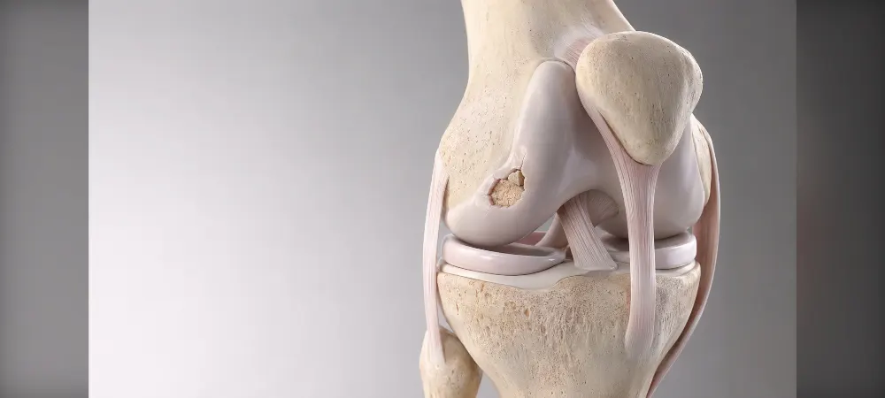
When the blood supply to the bone tissue on the joint surface is disrupted, a nutritional problem arises in a localized area. This condition causes the bone to weaken and, over time, separate from the underlying healthy bone bed. Because the cartilage covering the detached fragment is nourished by the joint fluid, it maintains its viability, but no living cells remain in the underlying bone tissue.
Although connection to the main bone may continue in the early period, if healing does not occur, the fragment may completely detach. The loose fragment begins to circulate freely in the joint cavity and becomes an intra-articular foreign body.
The disease usually begins before adolescence. The majority of cases seen in adults are old lesions that originated in childhood but have only recently begun to produce symptoms. The incidence in males is higher compared to females. Bilateral involvement is seen in 15-30% of cases, and findings on both sides may not follow the same course.
Changes Occurring in Joint Structure
Decreased blood circulation in the subchondral bone leads to the development of necrosis in the bone tissue. The retraction or early cessation of blood vessels during growth creates small-scale dead areas in the growth cartilage. Over time, these areas cause the normal ossification process to falter and turn the cartilage into a weak, unsupported region.
If the vessels nourishing the cartilage cannot perform their function fully, that region cannot be adequately nourished. The weak area fails to harden during the period when it should ossify, leaving a thin cartilage island unsupported from underneath. The articular cartilage on top loses its support and begins to separate over time.
Strains such as sports activities, jumping, running, or spraining can cause this weak area to crack easily. A subchondral stress fracture may develop due to repetitive microtraumas, which progresses over time and leads to separation in the fragment.
Most Frequently Affected Joints
Any cartilaginous joint in the body can theoretically be affected by this disease, but anatomical structure and the distribution of loads exposed to in daily life make certain joints targets. The knee joint is the most frequently involved region, and in the knee, the medial femoral condyle is particularly affected. The capitellum in the elbow and the talus region in the ankle are the points where the disease settles. Rarely, it can also be seen in the hip and shoulder joints.
The incidence of osteochondritis dissecans in children is 9.5-29 per 100,000 in knees, 2.2 in elbows, and 2-4.6 in ankles. In adults, this rate is approximately 3.4 per 100,000. The risk increases significantly in children aged 12 and older.
Causes and Risk Factors of Osteochondritis Dissecans
Although the exact cause is unknown, osteochondritis dissecans occurs through the combination of multiple factors. Research shows that a single cause is not responsible. Biological, mechanical, and hereditary factors play a role in the development of the disease.
Blood Circulation Disorders
There are extremely thin vessels feeding the bone tissue immediately beneath the cartilage. If the blood flow in these microscopic capillaries slows down or stops completely for various reasons, the bone cells in that region remain oxygen-free and unnourished. This vascular problem comes to the fore especially in cases without a clear trauma history.
During growth, some vessels complete their function and regress. However, if this regression is premature or interrupted, unnourished areas form in the cartilage tissue. Bone tissue with impaired blood supply weakens, and the cartilage on it collapses. Animal studies have revealed that most of these vascular interruptions heal spontaneously, but permanent damage develops when trauma is added.
Repetitive Microtraumas and Sports Activities
It is the result of a much more complex process than a sudden, simple fall or sprain event. Mechanical strains and repetitive microtraumas spread over years trigger the disease. Repetitive stress on the joint causes the weakening of cartilage and bone tissue.
Certain sports activities particularly increase the risk:
- Basketball and football (sudden stopping, changing direction)
- Gymnastics (weight bearing, repetitive loading)
- Baseball and volleyball (throwing motions)
- Wrestling (pressure on the joint)
Constantly straining the same joint can compress vessels and impair the nutrition of the cartilage. Anatomical differences and joint alignment also lead to greater strain on certain areas. The dominant side joint is mostly affected.
Growth Period and Genetic Predisposition
In children of growing age, the processes of longitudinal bone growth and maturation continue rapidly. At this critical stage, delays or fusion defects sometimes occur in the centers where the cartilage tissue at the joint ends transforms into bone. When the ossification process does not proceed flawlessly, the risk of developing the disease increases.
Genetic factors also play a role. Individuals with a family history of similar joint problems have a higher probability of occurrence. Its appearance in multiple joints in some families underscores a hereditary predisposition. If there is a history in close relatives, the risk may increase.
Age and Gender Factors
It most frequently occurs between the ages of 13 and 21. Patients' ages generally range from 11 to 23. The typical patient profile is males aged 15-20.
The incidence in males is twice as high as in females. The risk is significantly high in boys. It is generally unilateral, but bilateral involvement can be seen in some cases. The occurrence of the disease at progressively younger ages may be related to children starting sports at an early age and the increase in childhood obesity.
Symptoms of Osteochondritis Dissecans
Early Stage Symptoms
At the onset of the disease, symptoms are vague and a sensation of pain that cannot be precisely defined dominates. Aching that occasionally appears in the joint cannot be clearly expressed by patients. Sometimes there may be no complaints at all, while sometimes intermittent pain and swelling are observed.
Pain is usually felt during or after activity. Playing sports, walking, or movements related to the joint increase the pain. It eases during resting periods. It appears as a deep ache and is expected to worsen over time.
Mild swelling can be noticed in the affected joint. Limping becomes prominent during painful periods. A feeling of fatigue and temporary joint stiffness may be experienced. Especially in the knee, these symptoms concentrate in the front of the knee.
Advanced Stage Symptoms
When the cartilage fragment begins to detach from its bed, the clinical picture changes completely. When a loose body circulating within the joint forms, patients describe that an object is roaming inside during movement. In superficial joints, the patient can feel this fragment themselves.
Catching and locking occur when the free fragment gets caught between the joint. Straightening and bending the knee joint becomes difficult. The person can neither fully open nor fully close their joint. This condition is quite painful and causes intense swelling.
Clicking or popping sounds may come from the joint. Movement restriction becomes prominent, and a decrease in joint strength is observed. Throbbing-type pain, swelling, and clicking complaints in the ankle may be added. Difficulty when climbing stairs or squatting is frequently reported.
Physical Examination Findings
During the examination, the affected joint area is painful when pressed with the hand. This finding is known as the Axhausen sign. Fluid accumulation in the joint at varying degrees can be detected.
The Wilson test is frequently used in the knee. While straightening the knee between 30 and 90 degrees, the lower leg is rotated inward. The patient feeling pain and the pain decreasing when the lower leg is rotated outward indicates that the test is positive. However, this test is only valid for lesions in the classic location.
The radiocapitellar compression test is applied in the elbow. If the wrist is rotated while the arm is fully straight, the pain reappears. These tests may not yield definitive results, and if positive, advanced imaging is required.
How is Osteochondritis Dissecans Diagnosed?
The diagnostic process is supported by imaging methods following physical examination findings. The orthopedics specialist first requests a plain radiograph, and more detailed examinations may be required afterward.
X-Ray Imaging
X-ray imaging is the first imaging method applied. It is sufficient to make a diagnosis in the majority of patients. In addition to anteroposterior and lateral radiographs, tunnel view images taken when the knee is bent 30-50 degrees are used. This angle clearly shows the condyle regions on the back side of the femur bone.
On the X-ray, the lesion appears as a slightly whitened bone island separated from the underlying healthy bone by a thin clear line. However, X-rays only show hard bone tissue. They cannot show cartilage, ligaments, or the amount of fluid inside the joint. In the very early periods of the disease, cell death in the bone may be too small to reflect on the X-ray, and the image may appear completely normal.
The most common location in the knee joint is the lateral side of the medial femoral condyle, near where the posterior cruciate ligament attaches in the intercondylar notch. This region is called the classic location and constitutes 50-70% of cases.
Magnetic Resonance Imaging (MRI)
MRI is the gold standard in diagnosing osteochondritis dissecans. It provides three-dimensional images of not only the bone, but also the thickness and quality of the cartilage, edema within the bone, and the condition of the damaged fragment. It is indispensable for determining the stage of the disease and deciding whether surgery is necessary.
MRI images can largely predict whether the fragment will detach from its bed. On T-2 weighted slices, seeing a high-intensity line indicating the presence of fluid behind the fragment, this line being associated with a crack or defect in the articular cartilage, and the lack of increased intensity on contrast-enhanced images taken with gadolinium are indicators that the fragment tends to detach from its bed.
Arthroscopy is one of the most valuable methods for diagnosis and evaluating the stability of the lesion. The interior of the joint is examined using a camera, and the fragment's dimensions, location, and connection to the underlying bed are evaluated.
Disease Stages and Classification Systems
A 4-stage staging system is generally used to classify the severity of the disease. The International Cartilage Repair Society (ICRS) classification is the most useful:
Stage 1: Softened on palpation, stable, cartilage continuity not disrupted
Stage 2: Partial detachment, stable on palpation
Stage 3: Completely detached, but resting in its bed
Stage 4: Empty crater, free body (crater smaller or larger than 10 mm)
In all classifications, the severity of the disease increases from Stage 1 to 4. A lesion that is stable in its bed in the early stage separates in advanced stages and becomes a free body.
Differential Diagnosis
Other joint disorders showing similar symptoms must be distinguished. Meniscal tear, ligament injuries, osteonecrosis, chondromalacia patellae, and other osteochondral lesions should be considered in the differential diagnosis.
Osteochondritis Dissecans Treatment Options
Treatment planning is made individually according to the patient's age, the stage of the lesion, and joint stability. Non-surgical methods can be successful in young patients whose growth plates are open and in stable lesions. In advanced stages, surgical intervention may be required.
Conservative (Non-Surgical) Treatment Methods
Non-surgical treatment is primarily applied in children whose skeletal maturation is not completed and in stable lesions. This process is not a magical solution; it requires serious discipline and patience. It usually lasts between 6 months and 1 year.
The most important rule is to completely remove the mechanical pressure on the sensitive bone region trying to heal. If body weight continues to load onto that area, healing cells cannot do their work. Therefore, all sports involving running, jumping, and impact must be stopped immediately.
The treatment process includes rest, crutch use, partial weight-bearing, special braces, and physical therapy. It is essential to maintain joint movement and necessary to reduce muscle strength loss. Healing is monitored with regular MRI follow-ups.
It has been reported that juvenile osteochondritis dissecans cases heal with non-surgical treatment at a rate of 50-95%. Osteochondritis dissecans detected before puberty and not separated from the underlying living bone bed can heal without surgery in more than half of children.
Conditions Requiring Surgical Treatment
In some cases, non-surgical treatments are just a waste of time and lead to permanent damage in the joint. If the patient is an adult and bone growth has stopped, the body's probability of repairing that region on its own is very low.
If the fragment has completely broken away from its place (Stage 4) or is about to break away (Stage 3), it is impossible for it to fuse back on its own. Despite a 6-month non-surgical treatment process where rules are strictly followed, if pains do not pass and no healing is detected in imaging, surgical intervention now becomes inevitable.
Surgery is required in unstable lesions, the presence of a free body, or situations unresponsive to conservative treatment. Surgery is considered in cases of cartilage integrity disruption, mechanical symptoms, and large symptomatic lesions.
Osteochondritis Dissecans Surgery Techniques
In situations where the cartilage surface is intact but the underlying bone has not fused, the drilling technique is applied. Surgeons enter the joint via arthroscopy and open microscopic-diameter tunnels into the dead bone region. Thanks to these holes, healthy blood and stem cells deep in the bone seep to the surface.
If the bone-cartilage fragment has separated from the main bed but maintains its integrity, gluing it back into its own socket is the best option. The socket beneath the fragment is cleaned, fresh bleeding is created, and the fragment is placed precisely back into its place. Various materials are used for fixation: titanium screws, steel screws, bioabsorbable nails, melting pins, and bone grafts.
Mosaicplasty is applied for fragments that are too thin or deformed to allow fixation. Cartilage and bone cylinders taken from the non-weight-bearing part of the joint are transplanted to the damaged region. Autologous chondrocyte implantation is planned for large lesions.
Post-Surgery Recovery and Rehabilitation Process
Performing orthopedic surgery flawlessly is only half of the process; the other half is entirely related to the patient's compliance with the rehabilitation program. The first 6-week period is entirely directed at protecting the tissue. The patient is strictly forbidden from giving weight to the leg, but moving the joint with special devices to prevent stiffness is mandatory.
Around the second month, controlled weight-bearing with crutches begins, and muscle strengthening exercises come into play. After the third month, full weight-bearing is given, and activities such as swimming and cycling are transitioned to. True running and returning to professional sports are possible between the 6th and 12th months when biological fusion takes place.
Frequently Asked Questions
Does osteochondritis dissecans heal on its own?
In children whose growth period continues and if the fragment has not separated from its place, healing is possible with rest and activity restriction. However, the probability of healing on its own is low in adults and advanced stages. If diagnosed early, the chance of non-surgical treatment increases.
Which sports cause this disease?
Sports that place repetitive pressure on joints, such as basketball, football, gymnastics, and wrestling, increase the risk. Activities involving sudden stopping, changing direction, and jumping movements can particularly cause the problem. Beyond a single sport, constantly straining the same joint creates the real problem.
When can I return to sports after surgery?
The complete healing process varies between 6 months and 12 months. No weight is given for the first 6 weeks, controlled activities start in the 3rd month. Running and returning to professional sports are possible at the earliest in the 6th month. Each patient's healing speed is different, and doctor follow-up is mandatory.
Are both joints affected at once?
Unilateral involvement is seen in the majority of cases. The rate of bilateral occurrence is low, and the disease on both sides may not progress in the same way. If there is a problem in one knee, it is beneficial to have the other knee checked as well.
What happens if it is not treated?
Fragments roaming within the joint form, and these fragments continuously damage the joint surface. Over time, cartilage wear accelerates, and degenerative joint disease may develop at an early age. Movement restriction and chronic pain reduce quality of life.
Is an MRI scan mandatory, won't an X-ray suffice?
X-rays are used for initial evaluation, but they cannot show the stage of the disease and the condition of the cartilage. MRI is necessary to determine the condition of the fragment, the chance of healing, and whether surgery is required. Treatment planning is made according to MRI results.
Is the chance of treatment higher in children?
In children whose growth plates are open, the body can work its own repair mechanisms better. The rate of healing with non-surgical treatment is significantly higher compared to adults. Full recovery is possible with early diagnosis and appropriate treatment.
In Summary, Osteochondritis Dissecans
Osteochondritis dissecans is a serious joint problem encountered especially in athlete children. When diagnosed early, the chance of recovery is quite high. If you feel pain, swelling, or locking in your joint, consult an orthopedics specialist immediately.
Non-surgical treatment mostly yields successful results in patients whose growth period continues. In advanced stages, surgical intervention may be required. In any case, it is mandatory to strictly comply with the treatment plan recommended by your doctor.
Taking a break from sports may feel difficult, but your joint health is of critical importance for your future performance. Be patient and take the rehabilitation process seriously.
Fill out the Form for Detailed Information and Contact!
Start with the right assessment for What is Osteochondritis Dissecans? What Are Its Symptoms? How Is It Diagnosed and Treated?
Share your symptoms with our specialist physicians; let us determine the right diagnosis and treatment plan together.
Warning
If your symptoms started suddenly, are progressing rapidly, or significantly restrict your daily life, consult a physician without delay.
Plan your What is Osteochondritis Dissecans? What Are Its Symptoms? How Is It Diagnosed and Treated? journey with our specialists.
Fill in the form or book an appointment to learn about treatment options, the process and preparation.