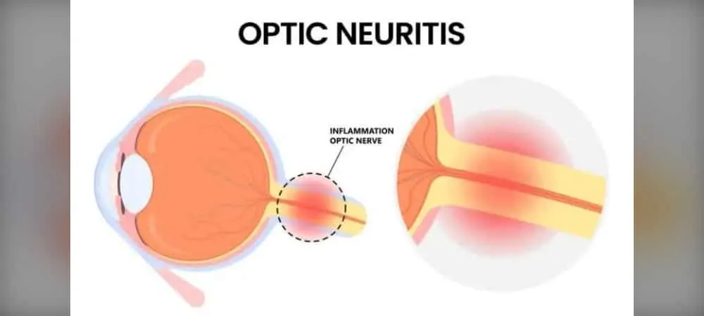
Optic neuritis is an inflammation of the optic nerve that directly affects your visual functions. Symptoms of optic neuritis, such as sudden vision loss, pain with eye movement, and faded color vision, usually manifest within days. In this article, you will find everything you need to know about the causes, diagnostic methods, and treatment of optic neuritis.
What is Optic Neuritis?
The Optic Nerve and Its Function
The optic nerve is the primary structure that carries images formed on the retina to your brain. You can think of it as an electrical cable. Each wire inside it transmits a different part of the image. A single optic nerve exits from each of your eyes, and this nerve delivers signals in the form of electrical impulses to the brainstem.
The optic nerve starts from the back of the eye and extends to the visual center in the brain. Thanks to this structure, everything you see becomes meaningful and visual information is processed.
What Happens in Optic Neuritis?
Optic neuritis is the inflammation of the optic nerve. This inflammation can affect the entire nerve or just a portion of it. Sometimes the nerve sheath is involved, and sometimes the section where it enters the eye is affected.
When inflammation occurs, edema develops in the optic nerve. When there is inflammation in the whole or a part of the nerve, the function is not fully realized, and you experience problems with vision. The disease usually affects one eye and is rarely bilateral.
Optic neuritis can occur at any time between the ages of 18 and 45. It is seen more frequently in the 30s. It is more common in women than in men. The disease starts very suddenly. Therefore, it is important to clarify the cause.
Differences Between Typical and Atypical Optic Neuritis
Optic neuritis is divided into two forms for clinical reasons: typical and atypical.
Typical optic neuritis is usually seen in young adults between the ages of 20-45 and mostly affects one eye. More than 90% of patients experience pain that increases especially with eye movements. This form often appears as the first symptom of multiple sclerosis. Vision usually improves significantly on its own or with treatment within weeks or months.
Atypical optic neuritis, on the other hand, shows different characteristics from the typical presentation. It may involve both eyes, be painless, or not respond well to corticosteroid treatment. It can be accompanied by very severe and permanent vision loss, as well as significant swelling and hemorrhages at the optic nerve head. In atypical cases, the improvement in visual acuity may be poorer, and there is a higher risk of developing optic nerve atrophy.
What Are the Symptoms of Optic Neuritis?
Although symptoms vary from person to person, they generally follow a course that starts suddenly and progresses rapidly. Symptoms of optic neuritis appear within hours or days, and early recognition of the disease increases treatment success.
Vision Loss and Blurred Vision
Vision loss is the most important finding of optic neuritis. Most patients experience temporary vision reduction in one eye. Noticeable vision loss usually develops within hours or days. Vision may worsen within a few days, then usually improves partially or completely within weeks.
In most cases, visual functions return to near normal within 8-10 weeks. However, complete and permanent vision loss can also be seen in some patients. The severity of vision loss varies from patient to patient, and in some people, vision is not completely lost.
Blurred vision may increase after exercising or taking a hot shower. The temporary worsening of vision in situations where body temperature rises is known as the Uhthoff phenomenon.
Pain with Eye Movements
People who develop optic neuritis have eye pain. This pain occurs with eye movements and increases especially when looking right-left or up-down. The pain is usually felt as a dull ache behind the eye.
Pain that increases with eye movements begins when vision worsens and disappears when vision improves. Sometimes the pain can also be perceived as a dull ache behind the eye.
Impairment in Color Perception
Impairment in color vision is a characteristic finding for optic neuritis. The appearance of colors as faded or gray is particularly evident on the red-green axis. Bright colors begin to look dull, and colors appear less vivid than normal.
In particular, a decrease in the vividness of the color red is common. The perception of objects as less bright in the affected eye also points to a decreased brightness perception.
Changes in the Visual Field
Various typical findings can be observed in the visual field. Central scotoma is one of the most common findings, and a gap in central vision occurs when the macular fibers of the optic nerve are affected. A centrocecal scotoma is a loss covering the area between the center and the blind spot.
Dark spots or missing areas may appear in the visual field. Additionally, some patients experience light flashes or light flickers during eye movements. Flashes or light glares can be seen especially during eye movements.
When to See a Doctor?
If you have sudden onset vision changes and eye pain, you should consult an ophthalmologist without delay. If symptoms of pain or changes in vision in one eye are worsening within hours or days, apply as soon as possible.
If you have symptoms such as vision loss in both eyes, double vision, and numbness in one or more limbs, you should definitely consult an ophthalmology and neurology specialist.
Causes and Risk Factors of Optic Neuritis
Although the exact cause of optic neuritis cannot always be determined, it is thought to occur as a result of your immune system mistakenly targeting the optic nerve. Your body attacks its own tissues and damages the myelin sheath.
Relationship with Multiple Sclerosis (MS)
The most common known cause is multiple sclerosis. In 15-20% of patients diagnosed with MS, optic neuritis is seen as the first symptom. 15% of multiple sclerosis patients start with optic neuritis.
The lifetime risk of developing MS after having optic neuritis is approximately 50%. If there are lesions on brain MRI, this risk increases further. However, if there are no new lesions on brain MRI, the risk of developing MS in the long term is quite low.
In 50% of MS patients, an episode of optic neuritis is seen at some point in their lives. Autopsy findings have revealed that more than 90% of MS patients have optic nerve involvement.
Autoimmune Diseases
Neuromyelitis optica is similar to MS but has a more severe course. In this disease, both the spinal cord and the optic nerve become inflamed. Recovery after attacks is rarer than in MS.
Myelin oligodendrocyte glycoprotein (MOG) antibody disorder causes inflammation in the optic nerve, spinal cord, and brain. Recovery from MOG attacks is generally better than in neuromyelitis optica.
Sarcoidosis, systemic lupus erythematosus, and Behçet's disease can cause recurrent optic neuritis. These are autoimmune conditions where the body attacks its own tissues.
Infections and Medications
Viral infections such as measles, mumps, and influenza can lead to optic neuritis. The herpes virus is also among the triggering factors. A history of infection is present in 28.6% of cases.
Bacterial infections such as Lyme disease, syphilis, and cat-scratch disease can also be causes. Viral and bacterial infections usually trigger an autoimmune response.
The drug ethambutol, used in the treatment of tuberculosis, is the most well-known drug that causes optic neuritis. In addition, methanol found in antifreeze, paints, and solvents is associated with optic neuritis.
Radiotherapy applied to the head and neck region is a significant risk factor. Uncontrolled diabetes can lead to optic nerve inflammation, especially due to small vessel damage.
Age, Gender, and Genetic Factors
Optic neuritis can be seen at any age, but it often occurs in young adults between 18-45 years old. The average age is 30. It is most commonly seen between the ages of 20-40.
Women are at twice the risk compared to men. The gender difference is significant, and it is much more likely to develop in women than in men.
Optic neuritis is more common in the white race. The race factor plays a role in the frequency of the disease.
Some genetic mutations may create a predisposition to optic neuritis or MS. Genetic factors increase the risk of developing the disease.
How Is Optic Neuritis Diagnosed?
The diagnostic process requires a detailed evaluation and is supported by multiple tests. First, it is questioned when your complaints started, how they progressed, and whether you have any accompanying problems. Whether you have experienced similar situations before, your known diseases, and the medications you use are also important for diagnosis.
Eye Examination and Clinical Evaluation
The ophthalmologist performs a fundus examination to check for edema or pallor in the optic nerve. In some patients, no findings may be observed. When the fundus is examined with an ophthalmoscope, enlargement due to edema in the optic nerve is seen. The optic disc is edematous in one-third of patients.
During the pupillary light reflex test, a bright light is shone into your pupil. In pupils affected by optic neuritis, constriction does not occur as it does in a healthy eye. This finding, called Marcus Gunn pupil or RAPD, is an important sign indicating optic nerve dysfunction.
In the eye examination, edema at the optic nerve head, swelling due to edema, redness, and sometimes thin splinter-like hemorrhages can be seen. These structural changes are followed throughout the treatment.
Vision Tests (Acuity, Field, Color)
Visual acuity is measured with the Snellen chart. In most patients, there is a decrease in visual acuity in the first days. Visual acuity can follow a different course in each patient throughout the diagnosis and treatment process.
Visual field tests are performed with automated devices using static or dynamic methods. The patient's ability to perceive lights coming from the periphery while looking at a fixed point is measured. It is evaluated whether there is a loss in the visual field. These tests objectively reveal the impairment due to the disease.
Color vision tests evaluate your ability to distinguish between red and green tones. Your ability to distinguish the color red is specifically tested. Color vision and light reflexes are important functions of the optic nerve that are affected.
OCT and Visual Evoked Potentials
Optical coherence tomography (OCT) analyzes the retinal layers and the optic nerve head in detail. It serves to show a tissue cross-section by utilizing the optical properties of tissues in real-time. It works based on infrared rays. It evaluates the thickness of the optic nerve and the retinal structure.
The visual evoked potential test measures the time it takes for signals from the eye to reach the brain. The patient is shown patterns or flashing lights. Electrodes placed on the scalp record the electrical response of the brain to these stimuli. If there is a delay, it is understood that there is a conduction disorder in the optic nerve. Prolongation of P100 latency provides strong support for the diagnosis.
MRI Imaging and Blood Tests
Magnetic resonance imaging (MRI) shows whether there is inflammation or damage in the brain and optic nerve. When contrast material is used, inflammation in the optic nerve can be seen. Lesions specific to MS are investigated in the brain. Damaged areas in the brain are identified.
Blood tests are requested to determine the underlying cause. Measuring various markers may be necessary to detect autoimmune diseases or infections. The sedimentation test is performed to evaluate the relationship with temporal arteritis. In the examination of cerebrospinal fluid, the presence of cells, protein, and sugar levels can be checked.
How Is Optic Neuritis Treated?
The treatment approach is based on slowing the rate of vision loss, accelerating recovery, and managing the underlying cause. Each patient's treatment is personalized and determined according to the severity of the attack.
Steroid Treatment and Methods of Administration
High-dose corticosteroid treatment is the first option in the treatment of optic neuritis. Cortisone, called methylprednisolone, is usually given intravenously. Treatment is applied for about 3 to 10 days depending on the severity of the patient's vision loss.
After intravenous steroid treatment, the dose is gradually reduced and discontinued in oral tablet form. However, low-dose oral steroid treatment is not recommended alone because it may increase the risk of a new attack.
Steroid drugs help vision improve faster by reducing inflammation and swelling in the optic nerve. However, steroids do not change the final visual level in the long term much more than a placebo; they mainly accelerate recovery.
Side effects include weight gain, mood swings, facial flushing, stomach pain, and insomnia. In cases with very severe vision loss or those that do not respond well to steroid treatment, plasma exchange or intravenous immunoglobulin treatment may be considered.
Immunomodulatory Treatments
If there is a disease such as MS, NMOSD, or MOGAD, long-term treatment planning is required. These treatments are used to regulate the response of the immune system. In patients at risk of MS, immune-modulating treatment may be started after cortisone treatment in the first attack to delay the onset of MS.
Immunomodulatory drugs are given to slow the progression of the disease and reduce attacks. These treatments are usually managed by neurology specialists.
Duration of Treatment and Recovery Process
In the vast majority of patients who have typical optic neuritis, vision improves significantly within a few weeks to a few months. In more than 90% of patients, visual functions return to near normal within 8-10 weeks.
Even if visual acuity improves, some people may have difficulty seeing colors as vividly as before, difficulty distinguishing contrast, or mild defects in the visual field. Starting treatment early reduces the risk of permanent damage.
Management of Recurrent Attacks
Optic neuritis is a condition that can recur. The risk of recurrence depends on the underlying cause. In patients with MS, optic neuritis attacks may recur from time to time.
The use of appropriate and effective treatment reduces the risk of recurrence, especially in optic neuritis associated with MS. Regular follow-up is important because recurrent attacks increase the risk of permanent damage to the nerve.
Frequently Asked Questions
Does optic neuritis cause permanent vision loss?
In most patients, vision improves significantly within a few weeks. In more than 90% of patients, visual functions return to near normal. However, in some people, vision may not return completely to its previous level.
Impairment in color perception recovers more slowly than visual acuity. Contrast loss or defects in the visual field may be permanent. How early treatment is started, the severity of the inflammation, and the underlying cause affect the outcome.
Early treatment brings better visual results. In some patients, the loss of color perception may be permanent even after recovering from optic neuritis. Therefore, it is important to start treatment without delay.
Does every optic neuritis turn into MS?
No, not every optic neuritis turns into MS. If there are no new lesions on brain MRI, the risk of developing MS in the long term is quite low. Approximately 50% of patients who have optic neuritis for the first time are diagnosed with MS within 15 years.
In fact, it is necessary to know that not every optic neuritis is related to MS. MS is a disease that causes destruction in the myelin layer around the nerves and presents with optic neuritis in 20-30% of patients. However, optic neuritis can also remain as a single event.
What happens if it is not treated?
Leaving optic neuritis untreated poses a serious risk for permanent vision loss. Once nerve tissue is damaged, full recovery is rare. If the optic nerve is not treated in time, it can be damaged beyond repair.
The decrease in visual acuity may be permanent, and permanent damage in the form of islands of low vision may be seen in the visual field. Especially in some cases of optic neuritis that develop due to trauma, successful results may not be observed with any treatment.
Does the disease recur?
In some patients, optic neuritis can recur. Depending on the underlying cause, the disease can be recurrent. It can recur, especially in autoimmune-based diseases.
The use of appropriate and effective treatment reduces the risk of recurrence, especially in optic neuritis associated with MS. In the presence of recurrent optic neuritis, the risk of decrease in visual acuity and permanent damage to nerve fibers increases. In patients with MS, optic neuritis attacks may recur from time to time.
What should be paid attention to in daily life?
Regular check-ups are essential. You should not neglect vision changes and should have regular neurological and eye examinations. Regular eye examinations are important to monitor the risk of recurrence of optic neuritis and the development of other conditions such as MS.
It is important to protect against infections and maintain a lifestyle that balances immunity. Underlying diseases are investigated, and the optic nerve is followed up in the long term.
In Summary, Optic Neuritis
Although optic neuritis may seem serious, recovery is generally possible in most patients with early intervention. When you notice symptoms such as vision loss and eye pain, you should consult a doctor immediately. Timely started steroid treatment accelerates the recovery process and reduces the risk of permanent damage.
Regular follow-up is important, especially due to its connection with MS. However, not every optic neuritis turns into MS, so it is enough to be under specialist control without panicking. Your chance of treatment is high, and the vast majority of patients regain their visual functions within a few months. Do not neglect regular check-ups to protect your vision health.
Get comprehensive information about the symptoms, diagnostic methods, and treatment options for optic neuritis. Prevent vision loss with early diagnosis and treatment.
Start with the right assessment for What is Optic Neuritis? What Are the Symptoms? How Is It Diagnosed and Treated?
Share your symptoms with our specialist physicians; let us determine the right diagnosis and treatment plan together.
Warning
If your symptoms started suddenly, are progressing rapidly, or significantly restrict your daily life, consult a physician without delay.
Plan your What is Optic Neuritis? What Are the Symptoms? How Is It Diagnosed and Treated? journey with our specialists.
Fill in the form or book an appointment to learn about treatment options, the process and preparation.