Mediastinal diseases can occur in any age group, and approximately 50% of mediastinal masses are located in the anterior region. These diseases, which develop in the mediastinum—the area in the middle of your chest between your lungs—may be asymptomatic or present with serious symptoms. In this article, we will explain what mediastinal diseases are, their symptoms, causes, diagnosis, and treatment methods in clear language. Therefore, you will learn what you need to know about health problems in this region in an understandable way.
What Are Mediastinal Diseases?
Mediastinal Region and Anatomy
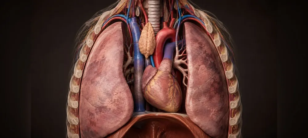
The mediastinum is the name of the anatomical region within the rib cage located between both lungs. This area begins at the thoracic inlet at the top and extends down to the diaphragm. It is bounded by the sternum (breastbone) in the front and the spine in the back.
This region houses vital organs. The heart, major blood vessels, trachea, esophagus, nerves, and lymph nodes are located within the mediastinum. Therefore, any disease that develops here can affect these structures.
For surgical planning and diagnostic purposes, the mediastinum is divided into three main compartments:
· Anterior mediastinum: Located just behind the sternum. The thymus gland, adipose tissue, major vessels, and lymph nodes are found in this section. In adults, the vast majority of mediastinal masses originate from here.
· Middle mediastinum: Contains the heart, pericardium, trachea, main bronchi, and major vessels. It is the most crowded area with the least amount of space.
· Posterior mediastinum: The narrow region in front of the spine. The esophagus, nerve structures, and the main lymphatic duct are located here.
Benign and Malignant Mediastinal Diseases
Mediastinal diseases can be benign or malignant. Benign structures usually grow slowly and do not spread to surrounding tissues. Cysts, some thymomas, and nerve sheath tumors are in this group.
Malignant diseases behave more aggressively. Lymphoma, thymic carcinoma, and some germ cell tumors show a tendency to spread to surrounding tissues.
In children with mediastinal masses, the probability of malignancy ranges from 27% to 77%. In adults, approximately 60% of these diseases present with symptoms.
Mediastinal Cysts
Cysts are fluid-filled structures and are mostly congenital. Bronchogenic cysts form as a result of faulty development of the respiratory tract while the baby is in the womb. They are filled with dense fluid.
Pericardial cysts result from developmental defects of the pericardium. They usually contain clear water and are harmless. Enteric cysts are developmental errors of the esophagus and may sometimes contain gastric cells.
Thymic cysts originate from the thymus gland. These cystic structures usually do not cause problems unless they become infected. However, when they grow, they can put pressure on surrounding organs.
Mediastinal Tumors
The type of tumor is closely related to the region where it is located. In the anterior mediastinum, thymoma (31%) and lymphoma (23%) are the most common tumors. Germ cell tumors usually appear at a young age.
Enlarged lymph nodes are frequently seen in the middle mediastinum. Tuberculosis, sarcoidosis, or metastatic diseases can cause lymph node enlargement in this region.
Neurogenic tumors are dominant in the posterior mediastinum. Benign nerve sheath tumors account for 70% of intrathoracic neurogenic tumors seen in adults. In children, neuroblastoma and ganglioneuroblastoma are encountered more frequently.
The most striking feature of mediastinal masses is that they can progress silently. The vast majority of patients who present to the clinic have no symptoms at all.
Causes of Mediastinal Diseases
The causes of mediastinal diseases cover a very wide spectrum. While some conditions stem from congenital issues, others are acquired throughout life. In this section, we will explain the fundamental causes so you can understand the origins of these diseases.
Genetic Factors
Mediastinal tumors can develop as a result of genetic predisposition. If you have certain types of cancer in your family, your risk may increase. Genetic links are observed especially in some types of thymoma and lymphoma.
If your immune system is weak due to genetic reasons, the risk of infection in the mediastinal region increases. Certain genetic predispositions or autoimmune conditions can contribute to the development of mediastinitis. In genetically susceptible individuals, chronic mediastinitis manifests as an immunological response.
Developmental Anomalies
Errors that occur during the development of organs in infancy lead to mediastinal diseases. Congenital cardiovascular anomalies are seen in 8 out of every 1000 live births. These congenital vascular structural defects can manifest at different ages.
Childhood mediastinal masses can stem from developmental anomalies. Cystic structures are mostly the result of these developmental errors. Bronchogenic cysts, pericardial cysts, and enteric cysts arise from the incorrect shaping of organs while in the womb.
Symptoms have a very wide spectrum. Some people feel nothing for years, while others show serious symptoms in childhood. Therefore, the same developmental disorder manifests differently in different people.
Infections and Inflammatory Conditions
Microorganisms cause serious diseases in the mediastinal region. Granulomatous diseases are the most common cause of mediastinal lymph node enlargement. Tuberculosis, fungal infections, and autoimmune diseases are in this group.
There is a risk of infection developing after surgical procedures. Bacterial infections can occur, especially after heart surgery or esophageal surgery. In cardiothoracic surgery, Staphylococcus aureus and coagulase-negative staphylococci are seen at a rate of 50-60%, and Gram-negative bacilli at 15-25%.
Rupture or perforation of the esophagus leads to mediastinal infection. Similarly, deep infections in the mouth, throat, and neck can spread downwards and reach this region. In addition to this, chest trauma, paravertebral abscess, and tracheal injuries are also among the causes of infection.
Symptoms of Mediastinal Diseases
Symptoms vary depending on the size, location, and pressure the mass exerts on surrounding structures. In a significant portion of patients, approximately forty percent, no symptoms are seen. Small masses, in particular, remain silent and are discovered incidentally on chest X-rays taken for a different reason.
Chest Pain and Sensation of Pressure
Pain felt in the chest region is a common symptom of mediastinal diseases. As the mass grows, it exerts mechanical pressure on surrounding structures, and this pressure turns into pain. The pain usually manifests itself continuously or intermittently.
Severe chest pain may indicate an advanced and invasive disease. The sensation of pressure becomes prominent, especially in masses in the anterior region. The pain can sometimes radiate to your back or shoulders and does not go away with rest.
Respiratory System Symptoms
External compression of the trachea narrows the airway. Therefore, persistent dry cough and shortness of breath that increases with movement begin. Airway compression can lead to recurrent lung infections.
You experience shortness of breath, and this condition increases especially when lying down or exerting effort. A high-pitched and noisy breathing called stridor indicates obstruction. Coughing up blood may be seen. When there is pressure on the nerve that controls the vocal cords, sudden and persistent hoarseness occurs.
Swallowing and Digestive System Symptoms
Masses that put pressure on the esophagus cause difficulty swallowing. You experience a sensation of food getting stuck in your throat or chest. This problem becomes prominent especially when swallowing solid foods.
When you experience difficulty swallowing, your daily life is negatively affected. Dysphagia develops due to pressure on the esophagus. Similarly, a growing mass can affect your digestive system in different ways.
Systemic Symptoms
In malignant lymph node diseases, symptoms that affect your entire body appear. You experience severe night sweats and your sleep is interrupted. Fever rises, and the cause cannot be explained.
Unexplained weight loss occurs rapidly. Weakness and fatigue make your daily activities difficult. When there is pressure on the major vein that carries deoxygenated blood from the upper body to the heart, noticeable swelling and redness are seen in the neck, face, and arms. Drooping of the eyelid and a small pupil indicate Horner's syndrome.
Asymptomatic Cases
The majority of diseases in the mediastinal region do not cause any discomfort in the initial stage. Masses are usually detected by chance during check-up programs, pre-employment examinations, or chest X-rays taken for completely different complaints.
Small masses usually do not cause symptoms. However, as the mass continues to grow, it begins to compress neighboring organs, and symptoms appear. In later stages, complaints due to mechanical pressure begin to affect your daily life.
Diagnosis of Mediastinal Diseases
To reach an accurate diagnosis, the diagnosis of mediastinal diseases requires a multi-stage process. This journey, which begins with the evaluation of your complaints, is completed with imaging techniques and tissue examination.
Physical Examination and Medical History
Your physician first learns how long you have had your complaints. They ask if there are similar diseases in your family. Patients with mediastinal abnormalities should be evaluated with a detailed anamnesis and physical examination.
During the examination, swelling in your neck, face, or arms is checked. Drooping of your eyelids and constriction of the pupil are checked. Your speech is listened to in order to understand whether your vocal cords are working. Patients with respiratory system disease should undergo a full physical examination covering all systems.
Imaging Methods
Chest X-ray is the first application method. However, an X-ray alone is not sufficient. Computed tomography reveals the anatomical localization, morphology, and relationship of the mass with surrounding structures. Tomography taken by administering medication intravenously shows whether the content is cystic or solid tissue.
Magnetic resonance imaging is preferred especially to evaluate the relationship with vessels, nerves, and the spinal cord. It provides valuable information in cases of contrast agent allergy and kidney failure, or in cases of suspected chest wall or vascular involvement.
Positron emission tomography evaluates the metabolic activity of malignant tumors. It is very important in the diagnosis, staging, and treatment follow-up of lymphoma and lung cancer.
Transbronchial needle aspiration performed under EBUS guidance is a minimally invasive method that provides high diagnostic accuracy in mediastinal masses. Endobronchial and endoesophageal ultrasonography are the first options to be consulted as minimally invasive methods.
Laboratory Tests and Biopsy
Complete blood count, calcium, phosphorus, and other serum electrolytes are requested as initial tests. In germ cell tumors, alpha-fetoprotein and beta-HCG levels are checked.
A biopsy is required to make a definitive diagnosis. The typical approach for surgical biopsy is mediastinoscopy, extended mediastinoscopy, anterior mediastinotomy, or video-assisted thoracoscopic surgery. In lymphoma, advanced-stage tumors, and masses that cannot be surgically removed, biopsies to obtain tissue should be performed first.
Pathological Examination
The tissue obtained by biopsy is examined under a microscope. Histopathological evaluation provides a definitive diagnosis. This examination clearly determines the type of tumor and shapes your treatment plan. Early and accurate diagnosis is of great importance, especially in malignant mediastinal tumors, in terms of life expectancy and treatment success.
Treatment of Mediastinal Diseases
The treatment approach is determined according to the type, size, location of the mass, and your general health status. In thymoma, teratoma, cyst, and nerve tissue tumors, surgical intervention is recommended directly without a biopsy. Depending on whether it is malignant or benign and its relationship with surrounding structures, either surgical removal or chemotherapy and radiotherapy are applied.
Surgical Treatment Methods
Closed methods have become the first choice today. The VATS method, known as video-assisted thoracic surgery, is performed through 2-3 small holes. This procedure is completed without cutting your sternum and is aesthetically superior. The average length of hospital stay is 3 days.
With VATS, tumors up to approximately 5 cm can be safely removed. However, in masses larger than 5 cm, the risk of the tumor capsule tearing increases. If the capsule is damaged, the disease may recur in the long term. Therefore, open surgery is more appropriate for very large tumors.
Robotic surgery provides unique control in narrow spaces. In the Da Vinci system, the surgeon sits at the console and works under 3D imaging. The robot's arms can rotate 540 degrees and mimic the wrist movements of the human hand. It is the gold standard, especially in clearing vascular and nerve structures.
The sternotomy method means cutting the sternum. This method is preferred for masses that involve major vessels or are very large. Thoracotomy is used if the tumor is closer to one side or if there is a different focus. These open methods are reserved for special cases.
Chemotherapy and Radiotherapy
Chemotherapy is the main treatment for lymphoma and malignant germ cell tumors. Cisplatin-containing drug protocols are applied with response rates of 50-70%. In case of lung cancer spreading to mediastinal lymph nodes, chemotherapy is given before surgery.
Radiotherapy is added after surgery in invasive tumors. It is used in combination with chemotherapy or as a supplement after surgery. In stage 4 thymoma, chemotherapy is the primary treatment method; depending on the extent of the disease and general condition, radiotherapy, surgery in selected cases, or supportive treatments aimed at symptom control can also be evaluated.
Follow-up and Observation
Asymptomatic cystic structures, which are often benign, are usually followed up. If there is doubt in the diagnosis or if it causes symptoms, surgery may be preferred. In small, benign, and slow-growing tumors, only regular check-ups can be performed.
The risk of recurrence is very low in early-stage thymomas that are completely removed. However, long-term radiological follow-up is an oncological rule. In thymoma cases, regular tomography checks and, if necessary, oncology follow-ups are planned according to the pathological stage of the mass.
Supportive Treatments
Supportive approaches are applied to alleviate symptoms such as shortness of breath, pain, and cough. If necessary, oxygen support, painkillers, or steroid drugs can be given. Bronchoscopic stent placement is an effective alternative in airway obstruction.
In cases of superior vena cava obstruction, long-term blood-thinning treatment and intravascular stent placement are quite successful. Those who undergo surgery with the closed method usually start walking and eating normally the next day. The chest tube is removed within 24-48 hours, and you are discharged in a short time.
Frequently Asked Questions About Mediastinal Diseases
Are mediastinal diseases dangerous?
The level of danger depends entirely on the type of disease. Benign cysts are usually not dangerous and grow slowly. However, tumors such as thymoma and lymphoma are serious conditions that require early diagnosis and treatment. Malignant tumors show a good prognosis with early treatment. Therefore, not every mediastinal disease is cancer, and a biopsy must be performed for a definitive diagnosis.
Can mediastinal diseases be asymptomatic?
Many mediastinal diseases do not cause any symptoms, especially in the early stages. In approximately forty percent of patients, no symptoms are encountered, and tumor growth is noticed on a chest X-ray taken for a different reason. Small masses usually do not cause symptoms and are detected by chance. However, as the mass grows, pressure symptoms begin to occur. Therefore, regular health check-ups are important.
At what age do mediastinal diseases occur?
They can be seen at any age, but some types appear more frequently in certain age groups. While germ cell tumors are seen more frequently in young adults, thymoma is usually seen over the age of 40. Malignant lesions such as thymoma and lymphoma are usually detected in adults, while congenital cysts are detected more frequently in childhood. Neurogenic tumors are frequently encountered in children. Similarly, the frequency of occurrence changes according to the age group.
What happens if mediastinal diseases are not treated?
Untreated masses lead to serious complications. They can cause rhythm disorders by putting pressure on the heart. They can cause swelling in the face and neck by blocking major vessels. They create a feeling of suffocation by narrowing the trachea. They can cause hoarseness or diaphragm paralysis by putting pressure on nerves. Especially benign tumors can turn into malignant characters over time. Cysts can rupture or become inflamed. The tumor spreads to the lung membrane, reducing the chance of treatment. Therefore, early intervention is of vital importance.
Are mediastinal diseases contagious?
No, mediastinal diseases are not transmitted from person to person. These diseases are related to genetic factors, immune system problems, or some environmental factors. Even cases of mediastinitis due to infections are not directly contagious. Therefore, contact with patients is safe.
How long does the treatment of mediastinal diseases take?
The duration of treatment varies according to the type, size, spread of the tumor, and the chosen treatment method. Recovery usually takes 4-6 weeks, but it varies from person to person. While treatment can be completed in a short time for benign masses removed only by surgery, the process may be longer in malignant cases requiring chemotherapy and radiotherapy. The average length of hospital stay after surgery is 3 days.
In Summary, Mediastinal Diseases
Although mediastinal diseases may seem frightening at first, they are manageable conditions with the right diagnosis and treatment. Early diagnosis, in particular, significantly increases your treatment success. No symptoms are seen in the majority of patients, so regular health check-ups are of great importance.
If you feel discomfort in your chest or if an abnormality is detected in your routine check-ups, do not worry. Not every mass is cancer, and modern surgical techniques performed with minimal incisions today provide rapid recovery.
Consult a specialist doctor, have the necessary tests done, and trust the treatment plan recommended to you. Early intervention is the most effective way to protect your health.
Fill Out the Form for Detailed Information and Contact!
Start with the right assessment for What Are Mediastinal Diseases? What Are the Symptoms? How Are They Diagnosed and Treated?
Share your symptoms with our specialist physicians; let us determine the right diagnosis and treatment plan together.
Warning
If your symptoms started suddenly, are progressing rapidly, or significantly restrict your daily life, consult a physician without delay.
Plan your What Are Mediastinal Diseases? What Are the Symptoms? How Are They Diagnosed and Treated? journey with our specialists.
Fill in the form or book an appointment to learn about treatment options, the process and preparation.