When fluid accumulates around your lungs, breathing can become difficult. Thoracentesis is a low-risk procedure that allows this fluid to be drained with the help of a needle. In fact, thanks to this minor intervention, both your shortness of breath can be relieved and certain diseases can be diagnosed.
In this article, we will share with you everything you need to know about what thoracentesis is, in which situations it is necessary, how it is performed, and the post-procedure process in clear language. Therefore, you will find the answers to your questions about this procedure.
What is Thoracentesis?
Inside the rib cage, there are two thin layers of membrane that surround your lungs. These membranes are called the pleura, and the space between them is called the pleural space. Under normal conditions, there is a very small amount of fluid in this space. In fact, only about 20 ml. This fluid allows your lungs to expand and contract comfortably while breathing.
However, in some cases, the amount of fluid between these membranes increases. The secretion of the fluid may increase, or its reabsorption may decrease. This condition is called pleural effusion. The accumulated fluid puts pressure on your lung and restricts its elasticity. As a result, breathing becomes difficult, coughing begins, and chest pain occurs.
Pleural Space and Fluid Accumulation
The pleura consists of two layers. The first is the visceral pleura, which covers the outer surface of the lung, and the other is the parietal pleura, which lines the inner surface of the rib cage. These two layers join at the root of the lung and form a closed sac.
The condition known as fluid collection in the lung actually occurs between the pleural layers. The lung itself has a spongy structure and does not have a distinct space where fluid can collect. Specifically, pleural effusion should not be confused with pulmonary edema. While pulmonary edema is the accumulation of fluid in the air sacs inside the lung, pleural effusion is a completely different condition.
15% of pleural effusions show no symptoms. In other patients, chest pain, dry cough, and respiratory distress are observed. The accumulated fluid prevents the lung from working at full capacity and leads to serious discomfort.
Purpose of the Thoracentesis Procedure
Thoracentesis is the procedure of draining the fluid accumulated in the pleural space with the help of a special needle or catheter. This intervention is performed between the ribs. The procedure is applied for both diagnostic and therapeutic purposes.
In thoracentesis performed for diagnostic purposes, a sample is taken from the accumulated fluid. The collected sample is examined in the laboratory. Biochemical analyses reveal the character of the fluid, protein levels, LDH levels, and cellular content. Thanks to these data, it is determined whether the fluid is caused by heart failure, lung infection, or cancer.
In therapeutic thoracentesis, the goal is to drain the fluid. Thanks to the fluid drained in a controlled manner, the lung expands to full capacity again. The patient's shortness of breath is quickly relieved, and their quality of life increases. Ultrasonography is used especially when samples need to be taken from low-volume fluids. This method detects the location of the fluid instantly and ensures that the procedure is targeted.
Thoracentesis is not just a fluid drainage procedure. It is actually a vital diagnostic tool. Pleural effusion is not a disease in itself, but an indicator of another process going wrong in the body.
In Which Situations Is Thoracentesis Performed?
Diagnostic thoracentesis should be performed on every patient with unexplained pleural effusion. Understanding why the fluid is accumulating is necessary to start the correct treatment. On the other hand, therapeutic thoracentesis is applied when fluid accumulation leads to shortness of breath. In both cases, the procedure is of vital importance.
Infectious Diseases
Bacterial infections can cause fluid accumulation in the pleural space. In this case, your doctor tests the fluid taken from the pleural space and reveals the source of the infection. Pneumonia is especially common in children and leads to fluid accumulation that impairs lung function.
Viral and fungal infections also cause fluid collection in a similar way. Chronic infections such as tuberculosis can cause long-term pleural effusion. Empyema, which is the formation of pus-filled areas in the pleural space, is a serious condition that requires urgent intervention.
Cancer Diagnosis
Fluid accumulation in the pleural space can be seen as a symptom of cancer. Your doctor will request that the pleural fluid be tested for cancer cells. Lung cancer, breast cancer, lymph node, or other organ-derived metastases lead to effusion.
Malignant pleural effusion has been detected in 19% of patients in studies. The average age of these patients is around 70. Therefore, thoracentesis should be performed in every fluid accumulation where cancer is suspected.
Heart and Liver Failure
Congestive heart failure is the most common cause of transudative effusion. In heart failure, fluid accumulates in the lung due to increased pulmonary capillary pressure. Diagnostic thoracentesis may not be necessary, especially in the presence of bilateral pleural effusion. However, if there are atypical symptoms such as chest pain or fever, the procedure should be performed.
Hepatic hydrothorax can develop in patients with liver cirrhosis and portal hypertension. This condition is seen in 4-6% of patients with cirrhosis. The fluid accumulates on the right side in nearly 80% of patients. Diagnostic thoracentesis is definitely performed in patients with cirrhosis due to the risk of bacterial pleuritis.
Pulmonary Embolism
Pleural effusion occurs in 30-50% of patients with pulmonary embolism. In patients under the age of forty, the most common cause of pleuritic chest pain and pleural effusion is pulmonary embolism. Furthermore, approximately 75% of patients who develop pleural effusion secondary to pulmonary embolism have constant pleuritic chest pain on the side of the effusion.
A diagnosis of pulmonary embolism should be considered in every patient with unexplained pleural effusion. The procedure should be performed especially if there is severe dyspnea or pleuritic chest pain.
Other Medical Conditions
Pleural effusion that develops in pancreatitis is usually in small amounts. In chronic pancreatitis, the opening of the pancreatic duct into the pleural space can cause massive pleural effusion. Symptomatic pleural effusion is seen in approximately 3-5% of patients with rheumatoid arthritis.
Pleural findings are detected in 30-50% of patients with systemic lupus erythematosus during the course of the disease. The use of certain medications also causes fluid accumulation. Autoimmune diseases, kidney failure, and other systemic disorders can lead to pleural effusion.
How Is Thoracentesis Performed?
The thoracentesis procedure is not as complicated as you might think. Your doctor will share the entire process with you and answer any questions you may have. The procedure is usually performed under local anesthesia and does not require general anesthesia.
Pre-Procedure Preparation
You need to provide your doctor with some important information. Be sure to mention if you are pregnant, have drug allergies, or have a latex or tape allergy. If you are using blood thinners, aspirin, or medications that affect blood clotting, report these. Also, inform your doctor if you have had bleeding problems in the past.
Share all prescription or over-the-counter medications, vitamin supplements, and herbal products you use with your doctor. You will be asked to sign a form approving the procedure. Read the form carefully and ask about points you do not understand.
Before the procedure, a chest X-ray or ultrasound is taken. These imaging methods determine the location and amount of the fluid. Your blood pressure, pulse, and oxygen level are measured. These values will be monitored continuously during the procedure.
You need to remove your jewelry and accessories. You will be given a hospital gown. Call a relative to take you home after the procedure.
Procedure Stages
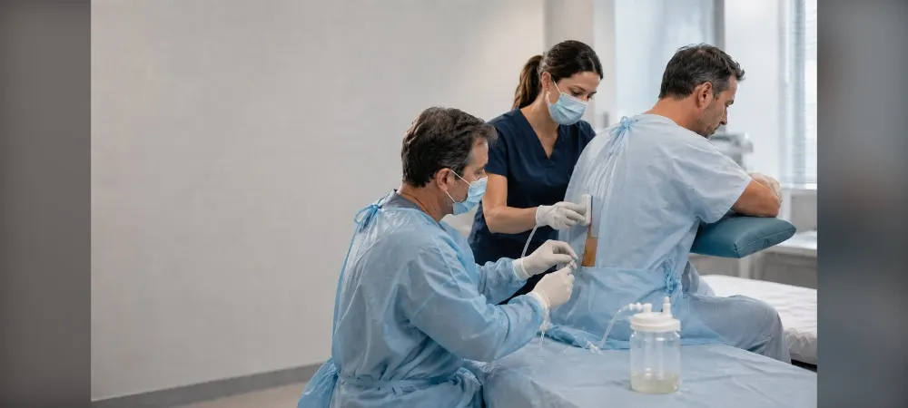
When you are taken to the procedure room, you will be given oxygen via a face mask or nasal tube. You will be in a sitting position on the hospital bed. You will place your arms on the support areas on the side. This position widens the spaces between the ribs and facilitates the insertion of the needle.
With the ultrasound device, the safe area where the fluid is most dense is determined with millimeter precision. The risk of the needle touching the lung tissue, diaphragm, or adjacent organs drops below 1%. Imaging guidance almost completely eliminates complications during the procedure.
The procedure area is sterilized with an antiseptic solution. Then, a local anesthetic drug is applied to the area. It is injected first into the skin, then to the rib periosteum, and finally to the vicinity of the parietal pleura. This ensures that the procedure is painless.
After the area is numbed, your doctor inserts the needle between the ribs. The needle advances from the upper border of the rib. Thus, the vessel and nerve bundle under the rib are not damaged. You may feel slight pressure as the needle advances.
The fluid is slowly drawn into the needle. You may be asked to remain still or take deep breaths during the procedure. If there is a large amount of fluid, a tube is attached to the needle. Vacuum systems or three-way stopcock mechanisms are activated. The fluid is collected into a closed bag in a controlled manner. Instead of rapid evacuation, the lung is allowed to expand gradually.
When enough fluid is collected, the needle is removed. A bandage and dressing are applied to the area. The collected fluid samples are sent to the laboratory. After the procedure, your doctor may request a chest X-ray to check the condition of the lungs.
Procedure Duration
Thoracentesis usually takes 15 to 30 minutes. However, if it is performed for therapeutic purposes and a large amount of fluid is drained, the procedure may take longer. Including the preparation phase, the total time varies between thirty minutes and one hour depending on the amount of fluid.
Thoracentesis is mostly an outpatient procedure. Patients who do not have problems on the chest X-ray after the procedure are discharged after 1-2 hours of observation. In some cases, a catheter may need to be inserted, and you may be asked to stay in the hospital for one or two days.
Post-Thoracentesis Process
When the procedure is completed, a short observation process begins. Your doctor may request a chest X-ray to ensure that the lungs are in good condition. Your blood pressure, pulse, and respiratory values are checked at regular intervals. It is checked whether there is any blood or fluid leakage in the dressing area. This monitoring process can take several hours.
Recovery Process
Recovery after thoracentesis occurs in a short time. In cases that do not require hospitalization, you can go home as soon as your doctor approves. Most patients are discharged on the same day. However, the patient's age and general health status are among the factors that affect the recovery process.
You can return to your normal diet and activities at home. On the other hand, you should not perform heavy physical activities for two days after the procedure. Avoid strenuous movements such as lifting weights. The patient's recovery process is known to be 1 to 2 weeks on average. It is essential that you do not push yourself, especially during the first two days.
Respiratory exercises are recommended after the pleural fluid is drained to allow the lung that has been under fluid pressure to expand. Breathing exercises relax the lung. These exercises should be applied regularly. It is normal to have slight tenderness in the procedure area. When the effect of the anesthesia wears off, slight pain may be felt in the intervention area. In this case, simple painkillers are sufficient.
Things to Consider
Keep the bandage at the entry site dry and clean for at least 24 hours. After the first 24 hours, you can take a shower without irritating the area. The presence of blood and other fluids in the dressing should be checked. If there are stitches in the procedure area, it is important to take local protective measures.
Call your hospital when you observe any of the following situations:
- Fever of 38°C and above
- Redness or swelling in the area where the needle was inserted
- Leakage of blood or other fluids from the needle site
- Shortness of breath or difficulty breathing
- Chest pain
- Coughing up blood
These symptoms may be a harbinger of complications such as infection, bleeding, or pneumothorax. Therefore, apply to a healthcare institution without delay.
Possible Side Effects and Risks
All medical interventions have risks. The most common situations of the thoracentesis procedure include mild pain in the procedure area, small bruises under the skin, or temporary coughing fits during the procedure. Mild chest pain or coughing, which can be seen rarely, is closely monitored.
Pneumothorax is the most common complication of the procedure and is seen in 12-30% of cases. However, this rate drops to 3% in thoracentesis performed under ultrasound guidance. Thanks to imaging technologies, this risk has decreased to negligible levels such as 1%. Pneumothorax is a condition where air enters between the lung membranes as a result of the needle touching the lung tissue. Small air leaks usually heal on their own.
Other risks include bleeding, infection, liver or spleen damage. Empyema and other infections can occur if the needle passes through infected tissue. If fluid is removed quickly or in large amounts, the risk of pulmonary edema increases. Spleen or liver punctures may be encountered, but if a small-diameter needle is used, bleeding usually limits itself.
The possible risks of the procedure may vary depending on your general health and other factors. These risks are minimized in procedures performed under ultrasound guidance by specialist physicians. Consult your doctor about which risks apply to your situation.
Frequently Asked Questions About Thoracentesis
Many patients have similar concerns before the procedure. Questions such as will there be pain, how many times can it be repeated, and exactly where is it performed are frequently asked. Providing clear answers to these questions helps you reduce your pre-procedure anxiety.
Is Thoracentesis Painful?
Before the procedure, local anesthesia is applied to the skin and lung membrane area where the application will be performed. Therefore, you will not feel severe pain during the procedure. Since anesthetic medication is applied, it is not expected that the person will feel pain during the procedure.
Only slight pressure during needle insertion or a temporary feeling of coughing as the lung expands may occur. However, this is a natural part of the procedure. Removing the fluid makes the person feel comfortable and does not cause pain.
After the procedure is finished, when the effect of the anesthesia wears off, slight pain may be felt in the intervention area. In this case, you can use simple painkillers. In fact, slight pain or tenderness is normal and passes within a few days.
How Many Times Can Thoracentesis Be Performed?
The thoracentesis procedure can be performed at intervals of a few days when pleural fluid recurs. It can be repeated when necessary to relieve symptoms and support the treatment process. Since it is performed to ensure the reduction of the person's symptoms and to control their health status, it can be applied every few days.
However, it should be considered that every intervention creates trauma in the pleural membrane. It should not be forgotten that it carries the risk of developing adhesions in the pleural membranes. Therefore, your doctor decides carefully whether the procedure will be repeated.
Alternative treatment methods can be evaluated in patients with constant fluid accumulation. For example, options such as inserting a permanent catheter or chemical pleurodesis can be considered.
Where Is Thoracentesis Performed?
Thoracentesis is performed from the rib spaces. In loculated fluids, the needle entry site can be made from a specific intercostal space. For free fluid drainage, the entry site to be chosen should be preferred 1-2 intercostal spaces below the upper level of the fluid.
Care is taken that the needle entry hole is not too close to the diaphragm. Under ultrasound guidance, the safe area where the fluid is most dense is determined. The needle advances from the upper border of the rib, and the vessel and nerve bundle under the rib are not damaged.
Usually, the procedure is performed from the side of the back, at the level of the shoulder blade. The patient being in a sitting position widens the spaces between the ribs and facilitates the insertion of the needle.
When to Return to Normal After the Procedure?
Patients can usually return to their light daily activities within 24 hours after the procedure. However, it is recommended to avoid heavy physical effort on the first day. Protecting the tapes in the procedure area for 24-48 hours is critical to minimize the risk of infection.
It is important not to perform strenuous movements for about 2 days after the procedure takes place. You should stay away from challenging activities such as lifting weights. The recovery that occurs in a short time may vary depending on the person's health status and age.
Most patients cannot return to their workplace or normal routine on the same day. You will need rest. The full recovery process takes an average of 1 to 2 weeks, and performing regular breathing exercises during this process will help your lung recover quickly.
In Summary, Thoracentesis
Although thoracentesis may seem complicated at first, it is actually a safe and effective procedure. Undoubtedly, thanks to this procedure, both shortness of breath is relieved and underlying diseases can be diagnosed. Therefore, it is important to have this procedure done in situations recommended by your doctor.
The risk of complications is quite low in thoracentesis performed under ultrasound guidance. It is sufficient to rest for a few days after the procedure and follow the symptoms you need to pay attention to.
As a result, thoracentesis can quickly improve your quality of life. If you have concerns, talk to your doctor openly and make the right decision together.