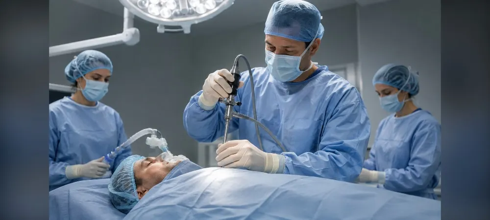
Choosing the right method for diagnosing masses and lymph node enlargements in the chest region is of critical importance. Mediastinoscopy offers a minimally invasive solution, particularly for lung cancer staging. This method is a reliable diagnostic procedure performed under general anesthesia.
In this article, you will find answers to what mediastinoscopy is, how it is performed, and why it is done. You will also learn in detail about who the procedure is performed on, the preparation process, and what to pay attention to after the procedure.
What is Mediastinoscopy?
Mediastinoscopy is a surgical procedure performed to examine the mediastinum region located behind the breastbone and to obtain tissue samples. It is performed with a special instrument called a mediastinoscope, which has a small camera at its tip. This diagnostic method is considered the gold standard in chest diseases.
Mediastinum Region and Anatomy
The mediastinum is an anatomical space located in the middle of the rib cage, between the two lungs. It is bounded by the breastbone (sternum) in the front, the spine in the back, the lungs on the sides, the entrance to the neck at the top, and the diaphragm at the bottom. This region is quite important because it houses the body's vital organs.
The mediastinum contains the heart, major blood vessels (aorta and vena cava), the windpipe (trachea), the esophagus, the thymus gland, and numerous lymph nodes. Because these organs are in very close proximity to each other, even a small mass developing in the mediastinum can lead to serious symptoms.
Surgeons divide the mediastinum into three main sections to better analyze this region. The anterior mediastinum is located behind the breastbone and contains the thymus gland. The middle mediastinum houses the heart, major blood vessels, and the windpipe. The posterior mediastinum is the part containing the esophagus and the descending aorta.
Different diseases are seen in these anatomical regions. For example, tumors originating from the thymus usually develop in the anterior mediastinum, while tumors originating from nerve cells develop in the posterior mediastinum. Additionally, the lymphatic drainage of the lungs and esophagus occurs into the mediastinal lymph nodes.
Key Features of the Mediastinoscopy Procedure
The mediastinoscopy procedure is performed under hospital conditions under general anesthesia. It is ensured that the patient is asleep and does not feel any pain. An endotracheal tube is placed in your mouth to help you breathe.
The mediastinoscope used in the procedure consists of a long, thin, and flexible tube. It has a small camera and a lighting system at its tip. Thanks to this camera, the doctor monitors and evaluates the structures inside the chest on a screen.
A small incision is made in the neck region, 2-3 cm above the breastbone. The mediastinoscope is inserted through here and gently advanced toward the mediastinum region. The device reaches the mediastinum by passing in front of the windpipe.
During the procedure, biopsy samples are taken from the lymph nodes around the airways and from suspicious tissues. The images obtained are recorded on a computer. After the procedure is completed, the mediastinoscope is removed, and the surgical incision is closed with stitches. The patient is awakened and disconnected from the ventilator.
Why is Mediastinoscopy Performed and Who Is It Applied To?
When enlarged lymph nodes or masses are detected in the mediastinum region via imaging methods, it is necessary to obtain a tissue sample to determine the cause. The answer to the question of who undergoes mediastinoscopy is primarily patients who have a suspicion of lung cancer or have a confirmed diagnosis. This method is also used in the evaluation of lymph node enlargements of unknown cause and mediastinal masses.
Lung Cancer Diagnosis and Staging
The most critical factor determining the treatment plan in lung cancer is the stage of the disease. Understanding whether the cancer has spread to the mediastinal lymph nodes directly affects the decision for surgical intervention. Mediastinoscopy is a gold-standard method in the preoperative staging of lung cancer, with a sensitivity of over 75%, a specificity of 100%, and a diagnostic success rate of over 90%.
If no cancer cells are detected in the lymph nodes, the patient is generally considered suitable for surgery. However, if metastasis is detected in the mediastinal lymph nodes, treatments such as chemotherapy or radiotherapy are preferred instead of surgery. Therefore, the most important answer to the question of why mediastinoscopy is performed is to prevent unnecessary surgeries and to choose the correct treatment method.
Mediastinoscopy is resorted to in cases where a diagnosis cannot be made with methods such as bronchoscopy, endobronchial ultrasound (EBUS), or endoesophageal ultrasound (EUS). In some cases, if it is predicted that these procedures will not yield results, a decision for direct mediastinoscopy is made to avoid wasting time.
Examination of Mediastinal Masses
Different types of masses or tumors can develop in the mediastinum. A definitive diagnosis is essential to understand whether these structures are benign or malignant. Although imaging methods show the presence of the mass, they may be insufficient in determining its character.
Mediastinoscopy is an effective method for taking biopsies from mediastinal tumors such as thymoma, teratoma, and lymphoma. This surgical procedure, which provides large tissue samples, is preferred especially in cases where radiological images are insufficient to distinguish diseases from one another.
Diagnosis of Lymphadenopathy (Lymph Node Enlargement)
If enlarged lymph nodes are seen in the mediastinum on a chest X-ray, computed tomography, or PET scan, it is necessary to understand why they are enlarged. Mediastinal lymphadenopathy is not specific to cancer alone and can be a sign of many different diseases.
Mediastinoscopy is necessary to find the underlying cause of lymph node enlargements of unknown origin. Tissue samples taken from the lymph nodes during the procedure are examined under a microscope to reach a definitive diagnosis. In this way, different conditions such as infections, autoimmune diseases, or cancer are distinguished from one another.
Evaluation of Other Chest Diseases
In the diagnosis of sarcoidosis, it is necessary to see special cellular structures called non-caseating granulomas within the tissue. Small samples taken with a fine needle may be insufficient to show this structure; therefore, larger tissue pieces are taken via mediastinoscopy.
Similarly, in diseases such as tuberculosis and lymphoma, taking a piece from the mediastinal lymph nodes is mandatory for diagnosis. These are important areas where mediastinoscopy comes into play when a definitive diagnosis cannot be made with other methods. The procedure's low morbidity rate makes it a reliable and effective diagnostic method.
Preparation for Mediastinoscopy and Pre-Procedure Process
A comprehensive preparation process is required for the success of the procedure. Detailed evaluation of the patient and following the correct steps are fundamental requirements for a safe operation. Tests performed and precautions taken during the preparation stage minimize potential risks.
Tests to be Performed Before the Procedure
Before mediastinoscopy, the patient's general health status and suitability for the procedure are evaluated. Some standard tests are applied for this purpose. Blood tests are performed to check bleeding and clotting functions. Since work will be done in close proximity to major vessels, it is of great importance that clotting values are within normal limits.
Chest X-ray and thoracic computed tomography (CT) are among the imaging tests. Heart rhythm is checked with electrocardiography (ECG). If deemed necessary, pulmonary function tests are also requested. In addition, basic vital signs such as fever measurement, blood pressure measurement, heart and respiratory rate measurement, and weight measurement are recorded.
All of these tests help minimize the risk of operation. Be sure to attend your appointments for other procedures requested by your doctor.
Medications Used and Points to Consider
It is mandatory to inform your doctor about all medications you use (including vitamins and herbal supplements). It is of critical importance to consult your doctor, especially regarding blood-thinning medications. These medications may need to be discontinued or changed under physician supervision for a certain period before the procedure.
The timing for discontinuing blood-thinning medications varies depending on the type of medication. Medications such as Aspirin, Coumadin (warfarin), Plavix (clopidogrel), Eliquis (apixaban), Pradaxa (dabigatran), Lovenox (enoxaparin), and Xarelto (rivaroxaban) are generally discontinued 5-7 days before the procedure. Talk to your surgeon and the doctor who prescribed the medication to find out when to stop taking the medication.
It is appropriate to discontinue vitamin E and non-steroidal anti-inflammatory drugs as recommended by the doctor. Patients using medication for diabetes should consult their doctors about what to do on the morning of the procedure.
Eating and drinking should be stopped at least 6-8 hours before the procedure. This rule includes water, chewing gum, and candy. Having an empty stomach is of critical importance for anesthesia safety.
On the day of the procedure, products such as deodorant, cream, and powder should not be used. Accessories such as metal objects, piercings, earrings, and jewelry should not be worn. If you use contact lenses, you must also remove them.
Anesthesia Evaluation
Mediastinoscopy is performed under general anesthesia. Before the procedure, an anesthesiologist evaluates the patient and questions their medical history in detail. This evaluation ensures the planning of the safest anesthesia method.
The intervention is strictly performed under general anesthesia because the patient being awake, coughing, or making the slightest startle movement can lead to vascular injuries that are difficult to compensate for. The patient is completely put to sleep and connected to a ventilator.
How is Mediastinoscopy Performed?
After general anesthesia is administered, the procedure begins. First, medication is given through an intravenous line, and the patient is completely put to sleep. An endotracheal tube is inserted into the throat and connected to a ventilator. This step is necessary to ensure safe breathing throughout the procedure.
Surgical Stages of the Procedure
The neck and chest area are washed with an antiseptic solution. The surgical area is prepared by covering it with sterile drapes. The surgeon makes a horizontal incision approximately 2-3 cm above the breastbone. This incision is positioned just above the breastbone (sternum).
A surgical path is created in front of the windpipe by entering through the incision. The point to be careful about here is not to cause any damage to the windpipe. The mediastinoscope reaches the mediastinum space by advancing from the outside and in front of the trachea.
Thanks to the camera at the tip of the device, the region is viewed in detail. The images are transferred to a computer screen, and the surgeon works accompanied by these images. The heart, lungs, and surrounding structures are examined. Suspicious areas and enlarged lymph nodes are identified.
Video mediastinoscopy is used in some centers. In this system, the image is magnified on the screen and provides a wider field of view. However, the fact that the device is larger than a standard mediastinoscope can create difficulties in some narrowed areas.
Biopsy Technique
Samples are taken from suspicious tissues and lymph nodes under image guidance. The surgeon collects a sufficient amount of tissue using special biopsy instruments. Pieces are removed from the lymph nodes around the airways with a cutting instrument.
The samples taken are sent to the pathology laboratory. In some cases, a frozen section analysis is performed during the procedure. The surgical time may be prolonged when this analysis is awaited.
Procedure Duration and Anesthesia
Mediastinoscopy takes an average of 45 to 60 minutes. However, this duration changes depending on the number of biopsies to be taken and the scope of the procedure. In patients where a frozen result is awaited, the surgical time extends up to an average of 65 minutes.
After the procedure is completed, the mediastinoscope is gently removed. The incision site is closed with absorbable stitches under the skin. These stitches dissolve on their own over time, and only a faint scar of 2-3 cm remains. The patient is slowly awakened and disconnected from the ventilator.
The mortality rate is between 0.08% and 0.15%. These figures show that the procedure is quite safe. It does not carry major risks when performed by experienced thoracic surgeons.
Post-Mediastinoscopy Process and Points to Consider
After the procedure is completed, you will be taken to the recovery room. As the effect of the anesthesia wears off, you may feel strange or your mind may be foggy for a few hours. Your mouth and throat area will be numb for a few hours. You will be closely monitored to ensure that you do not have any problems.
Length of Hospital Stay and Discharge
Patients are usually admitted to the hospital on the morning of the surgery. Due to medical protocol, it is preferred that the patient be kept under observation in the hospital for one night. However, clinical experience shows that almost all complications occur during the surgery or in the first six-hour period after the surgery.
For this reason, patients who pass the first six hours without problems, whose blood pressure and pulse remain stable, and who can eat and walk on their own can be discharged from the hospital on the evening of the same day. Patients whose clinical condition is stable are discharged the next day. You can go home a few hours after the procedure, but you must have a companion with you due to the medications you received or the anesthesia.
Things to Do During the Recovery Period
The ban on eating and drinking continues until the numbness wears off. If there is a sore throat after the numbness wears off, it is recommended that you consume softer foods. You may experience a sore throat, hoarseness, or coughing one day after the procedure.
A sterile dressing is applied to the incision area, and this dressing must remain in place for 24 hours. At the end of this period, the bandage can be removed, and you can take a shower. During the shower, nothing should be applied to the incision area; only water and soap should be allowed to run over it.
Keep the incision site clean and dry. Use the painkillers recommended by your physician regularly. Avoid strenuous physical activities and heavy lifting for 1-2 weeks. Activities such as swimming should be avoided for 10 days after the procedure. Patients can usually return to their daily lives within 2-3 days.
Potential Risks and Complications
The probability of developing complications related to the procedure is around 0.5-1%. It is a procedure that does not carry major risks when performed by experienced surgeons. The life-threatening risk rate is quite low (0.08%).
The most common complications include hoarseness, bleeding, opening of the lung lining, and injury to the windpipe. There is a risk of bleeding, but this rate is below 1%. Hoarseness can occur due to the nerve going to the vocal cords being affected, and the risk is around 1-2%.
If you notice any of the following symptoms, you should consult your doctor immediately: bleeding from your stitches, high fever, severe chest pain, swelling in the neck, shortness of breath, difficulty swallowing, or hoarseness that lasts for more than a few days or continues to worsen.
Evaluation of Results
Biopsy results are usually available in 5-7 business days. The results will generally be ready within 1 or 2 weeks. The doctor performing the procedure can provide you with information about the pieces sent to the laboratory. The pathology report indicates the definitive diagnosis of the disease. The result waiting process can be worrying; it is correct to definitely contact your doctor regarding this matter.
Frequently Asked Questions About Mediastinoscopy
Is mediastinoscopy a painful procedure? Since you are under general anesthesia during the procedure, you will not feel any pain. After waking up, you may experience mild pain at the incision site, but these are easily controlled with painkiller medications.
How long does mediastinoscopy take? The procedure takes an average of 45 to 60 minutes. However, this duration may extend depending on the number of biopsies to be taken. For example, if a frozen section analysis is awaited, the procedure may take longer.
When are the mediastinoscopy results available? The pathology examination and reporting process usually take 5 to 7 business days. In some centers, the results are available within 1 or 2 weeks. Your doctor performing the procedure will provide information about the samples sent to the laboratory.
Is mediastinoscopy a safe procedure? It is a low-risk and safe method when applied by experienced teams. The probability of developing complications is around 0.5-1%. The risk of death is between 0.08-0.15%, which is a very low rate.
Which complications can be seen? The most common complications are hoarseness, bleeding, opening of the lung lining, and injury to the windpipe. Besides this, conditions such as pneumonia, incision site infection, and lung collapse can rarely develop. In experienced centers, these risks are at a minimum level.
When can one return to daily life after mediastinoscopy? Patients can usually return to their normal daily activities within 1-2 weeks. It is recommended that you avoid strenuous physical activities.
Are there alternative methods to mediastinoscopy? Alternatives such as EBUS and CT-guided needle biopsy are available. However, mediastinoscopy is used in cases where a diagnosis cannot be made with these methods. Your physician will decide on the appropriate method.
Can mediastinoscopy be performed on everyone? It may not be suitable in some cases. The patient's general health status, medications used, and heart problems are evaluated. The physician decides after a detailed evaluation.
How many types of mediastinoscopy are there? Four different techniques are applied. Standard cervical mediastinoscopy is performed most frequently. Others are anterior, extended cervical, and subxiphoid mediastinoscopy. Each has different entry points.
What happens if metastasis is not detected? If no cancer cells are detected in the lymph nodes, the patient's chance of surgery is high. However, if there is spread in the mediastinal lymph nodes, chemotherapy or radiotherapy is preferred.
In Summary, Mediastinoscopy
Mediastinoscopy is a reliable method for reaching a correct diagnosis in chest diseases. Especially in lung cancer staging, it helps your doctor make the most accurate treatment decision. The procedure is low-risk and is safely applied in experienced hands.
In reality, most patients return to their normal lives within a few days. It is important that you carefully follow the pre-procedure preparation instructions and follow your doctor's recommendations afterward. If you have any concerns, talk to your doctor openly. Thanks to mediastinoscopy, uncertainties are eliminated, and the treatment process becomes clear.