Femoroacetabular impingement is a hip joint condition frequently seen in active individuals, particularly those aged 20-40. This condition causes severe hip pain, limited range of motion, and a decrease in your daily quality of life. If left untreated, hip impingement syndrome can lead to osteoarthritis over time. In this article, we will explain everything you need to know about the symptoms, causes, diagnostic methods, and treatment of hip impingement syndrome.
What is Hip Impingement Syndrome (Femoroacetabular Impingement)?
Structure and Function of the Hip Joint
To understand your hip joint, you must first look at its structure. This joint is formed between the upper part of your thigh bone (femur) and the socket area (acetabulum) of your pelvic bone. As a ball-and-socket joint, the hip is one of the largest and strongest joints in your body.
Your hip joint moves in 6 different directions and has a total angular movement of approximately 340 degrees. It carries 4 times your body weight while walking. Especially when sitting, standing up, and kneeling, this load creates shear force on the joint surface.
The triangular-shaped circular structure surrounding the edge of the acetabulum is called the labrum. The labrum ensures that the parts of your joint work in harmony during movement. The cartilage tissue covering the joint surfaces ensures smooth movement and reduces friction.
Definition of Femoroacetabular Impingement Syndrome
Femoroacetabular impingement syndrome is a condition characterized by excessive friction in the hip joint. Basically, your femoral head (ball) and acetabulum (socket) rub against each other in a way that causes abnormal damage to your hip. The damage occurs in the joint cartilage or the cartilage-like structure around the acetabulum called the labrum.
Hip impingement syndrome is divided into three types. CAM-type impingement refers to a femoral head that is not spherical or perfectly round. As a result of the loss of roundness in the head, abnormal contacts occur between the head and the socket.
In PINCER-type impingement, the acetabulum, or joint socket, covers the femoral head more than normal. As a result of this coverage, damage occurs in the anterior labrum due to impingement between the femoral neck and the acetabulum. In many cases, these two forms are seen together, which is called mixed-type impingement.
Mechanism of Disease Onset
Hip impingement syndrome begins when bony prominences strike the other bone during movement, preventing the joint from moving further. The protruding bones rub against each other, causing pain. This friction damages the surrounding tissues.
Femoroacetabular impingement syndrome is associated with cartilage damage, labral tears, early-onset hip osteoarthritis, and lower back pain. Over time, persistent impingement damages the joint and cartilage.
It is not known for certain whether the disease is congenital or develops during growth. It is thought that, in addition to the person's genetic predisposition, environmental factors also play an important role. Various strenuous sports activities performed before skeletal development is complete may increase the risk.
Causes and Risk Factors for Hip Impingement Syndrome
Anatomical and Genetic Factors
The primary cause of hip impingement syndrome is that your hip bones do not develop in the ideal form during growth in adolescence. A combination of genetic and mechanical factors leads to this condition. Hip impingement syndrome is seen as a prominence on the femoral head, excessive coverage of the bone surrounding the hip joint, or a combination of both.
Deformity of your femoral head, abnormal shape of the acetabulum, additional bone growths, or cartilage damage are among the anatomical risk factors. Your family history affects your risk of developing hip impingement syndrome. Those who have had similar problems in family members are more prone to this issue.
Congenitally low femoral anteversion angle can cause limited internal rotation of the hip, leading to the formation of femoroacetabular impingement syndrome. The risk increases in people with a family history of hip impingement syndrome.
The Effect of Sports and Physical Activities
Activities requiring repetitive and intense hip movement during childhood and adolescence can trigger bone remodeling. Sports such as basketball, soccer, ice hockey, and dance are among the activities that strain the hip joint.
Especially sports performed during the growth phase that frequently strain the hip affect bone shaping, laying the groundwork for the development of femoroacetabular impingement. Constantly repeated movements strain the joint, leading to abnormal friction.
Sports where the hip is exposed to constant and excessive bending and rotation movements are among the most important triggers. In athletes involved in disciplines such as ballet, martial arts, and weightlifting, repetitive movements increase impingement, causing symptoms to appear at an early age.
While femoroacetabular impingement syndrome is seen at a rate of 55% in the athlete population, this rate is 23% in the general population. The incidence increases in occupations associated with repetitive hip flexion, internal rotation, and adduction movements.
Age and Gender Factors
Hip impingement syndrome is usually seen between the ages of 20-40. Symptoms generally appear in young adulthood or middle age. The prevalence varies between 10-15%.
Gender differences are significant. Hip bone prominence is more common in young men. CAM-type is more common in men, and PINCER-type is more common in women. In the general population, 4% of healthy women and 24% of healthy men have femoroacetabular impingement morphology.
Excessive coverage of the hip joint in a pincer shape is more common in women. Mixed-type is seen in both genders.
Previous Injuries and Developmental Conditions
A history of childhood hip disease that may have changed the shape of the hip bone head poses a risk. Those who have had hip dislocation, slipped capital femoral epiphysis, or Perthes disease are in the high-risk group.
Conditions such as Legg-Calve-Perthes disease or slipped capital femoral epiphysis distort the shape of the femoral head, laying the groundwork for CAM-type impingement. A history of previous hip fracture increases the risk.
Conditions such as femoral neck fracture and avascular necrosis of the femoral head are among the risk factors. Individuals with developmental hip dysplasia carry the risk of femoroacetabular impingement in later years.
What Are the Symptoms of Hip Impingement Syndrome?
Groin and Hip Pain
Symptoms of hip impingement syndrome can be confused with many different conditions. Especially in young people, complaints are difficult to interpret because they resemble osteoarthritis or injuries seen in older ages. It may be overlooked by doctors.
The most common symptom is groin pain. Patients usually describe the pain by making a "C" shape with their hands, grasping the side and front of their hip. This is called the "C-sign" and is a very typical finding for hip impingement syndrome.
The pain starts as a deep and dull ache. However, it can become sharp and stabbing over time. You feel intense pain in the groin and on the outside of the leg after sports or intense exertion. It increases especially while playing sports and decreases with rest.
Limited Range of Motion and Joint Stiffness
Your hip joint's range of motion decreases. You have difficulty especially when putting on shoes or socks, bending over, or getting up from a low chair. You experience pain and a stabbing sensation when getting in and out of a car.
It is typical to feel pain in the hip when standing up after sitting for a long time. Stiffness occurs after a long car ride or an office meeting. You have difficulty getting up from a deep chair. Symptoms such as inability to sit cross-legged or cross your legs are seen.
Over time, your body begins to restrict movements to avoid pain. A significant stiffness and limitation develop in the hip joint, especially in inward rotation movements. In some patients, joint stiffness and loss of range of motion occur without significant pain.
Clicking, Locking, and Catching Sensation
Clicking, locking, or a "pop" sound caused by a mechanical problem inside the joint is frequently associated. A clicking or locking-like sound is heard with hip movement. These mechanical symptoms are usually caused by the damaged labrum or a loose piece of cartilage getting caught in the joint space.
You describe a catching or impingement sensation in the front of the hip joint or groin area. This sensation becomes prominent especially during hip bending or rotation movements. You experience a feeling of the hip locking during certain movements.
Radiation of Pain and Triggering Activities
Pain may not always be in the groin. There is often a complaint of lower back pain. This pain is usually localized in the sacroiliac region or the greater trochanter. It can radiate from the hip to the middle of the thigh. The pain usually does not go beyond the knee level.
The intensity of the pain increases after long walks, intense exercise, or sitting for a long time. Movements such as squatting or turning trigger the pain. You may feel a sharp, stabbing pain with a sudden turning or squatting movement. A decrease in performance is seen in athletes.
Types and Diagnosis of Hip Impingement Syndrome
CAM-Type Hip Impingement
The problem is located at the head of your thigh bone. There is an abnormal bony prominence at the junction of the femoral head and neck. Normally, this area should be slightly concave, but in the CAM type, it is in the shape of a non-spherical bump.
When your hip is bent, this prominence hits the edge of the socket, first tearing the labrum and then stripping the cartilage. It is more common in young and active men. Cartilage damage is usually at the 1 o'clock position, full-thickness, and severe.
PINCER-Type Hip Impingement
The problem is on the acetabulum side. The edge of the socket covers your femoral head more than necessary, or the angle of the socket is abnormal. When the hip reaches its limits of movement, the femoral neck hits this excessive bone edge, compressing the labrum.
It is encountered more frequently in middle-aged women and adolescent girls. In the PINCER type, cartilage damage is usually between 11 and 1 o'clock, more superficial and mild. In this type of impingement, damage begins primarily in the labrum, and repetitive contact leads to cyst formation within the labrum.
Mixed-Type Hip Impingement
It is the most common form. Structural defects are present in both the femur and the socket. Both mechanisms accelerate joint deterioration. The accumulation of microtraumas increases, and the risk of osteoarthritis rises.
Treatment planning in the mixed type requires a more complex approach. You must correct both problems completely.
Physical Examination and Tests
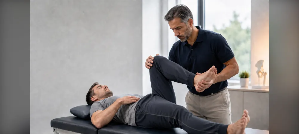
Your doctor measures your hip range of motion. Specifically, the limitation in bending and inward rotation movements is evaluated. The FADIR test is the most important clinical examination method for hip impingement.
While lying on your back, your hip and knee are bent 90 degrees, and then your leg is turned inward. If you feel pain in the groin area, the test is positive. This positivity strengthens the suspicion of femoroacetabular impingement.
In the FABER test, bending, outward opening, and external rotation maneuvers are applied to your hip. The presence of pain indicates an intra-articular problem.
Diagnosis with Imaging Methods
X-ray is the primary diagnostic method. AP pelvis and special hip radiographs are taken to examine the bone structure in detail. The size of the CAM lesion is evaluated by alpha angle measurement, and the presence of the PINCER lesion is evaluated by acetabular coverage and retroversion.
MRI imaging is the most valuable method for showing soft tissue damage. It detects labral tears and cartilage damage. MR arthrography provides more detailed results with contrast material but may show a healthy labrum as torn.
Computed tomography shows the bone structure in three dimensions. It is used for surgical planning. However, even if there are structural problems in imaging, surgery is avoided if impingement tests are negative during examination.
Treatment and Exercises for Hip Impingement Syndrome
Non-Surgical Treatment Methods
In the first stage of treatment, non-surgical methods are tried. Strengthening your hip muscles, activity modifications, and intra-articular injections are applied. Nonsteroidal anti-inflammatory drugs reduce pain and inflammation.
However, conservative treatment does not correct the underlying bony problem. Symptoms often persist or recur. If significant improvement in your pain cannot be achieved despite the use of non-surgical methods, surgery is recommended. Similarly, surgery may be necessary if the pain lasts longer than six months and seriously affects your daily activities.
Intra-articular injections contain cortisone or hyaluronic acid. These injections provide temporary relief. It is recommended that you avoid movements that cause impingement.
Physical Therapy and Hip Impingement Exercises
The current international guideline recommends that the first-line treatment be exercise-based and last at least 3 months. Physical therapy focuses on strengthening the core and hip muscles. Specifically, the gluteus medius, gluteus minimus, and core region muscles are strengthened.
Exercises are aimed at strengthening the muscles around your hip joint. It helps to alleviate symptoms and increase your ability to move. Hip extension, hip adduction, hip abduction, hip flexion, and squatting exercises are applied.
However, you should avoid some movements. Especially flexion above 90 degrees, and hip internal and external rotation should not be strained. You should avoid movements such as deep squat, Bulgarian split squat, and sumo deadlift.
Surgical Treatment Options
In patients who do not respond to conservative treatment, arthroscopic FAI surgery is the gold standard treatment method. Hip arthroscopy is performed with a minimally invasive closed method. Usually, 3-4 incisions of approximately 1.5 cm are made.
The most critical part of the surgery is the removal of the bony impingement. The abnormal bony prominence at the femoral head-neck junction is carefully shaved with an arthroscopic shaver. The torn labrum is sutured back to the bone edge using suture anchors. If there is cartilage damage, additional procedures such as microfracture can be applied.
Recovery Process and Post-Operative Care
After surgery, protected weight-bearing with crutches is usually required for 4-6 weeks. The average length of stay is 1 night. Physical therapy is absolutely necessary and lasts for months.
After surgery, your pain subsides and your hip range of motion increases. Most daily life activities can be performed within 6 weeks at the latest. Return to sports usually takes between 6 to 12 months.
The physical therapy and rehabilitation program in the post-operative period is very important. This program aims to strengthen muscles, increase flexibility, and regain normal range of motion.
Prevention Methods and Things to Consider
Regular exercise and correct movement techniques protect your joint health. You can do low-impact exercises to strengthen the muscles that support the hip joint. Activities such as walking and swimming are suitable.
You should avoid excessive strain, especially in high-impact sports that require rotation movements. You should pay attention to your body mechanics when performing movements such as weightlifting or squatting. Avoiding overloading can slow its progression.
Frequently Asked Questions About Hip Impingement Syndrome
How does hip impingement go away?
Hip impingement does not improve without surgery, but you may benefit from non-surgical treatments for a while. For example, pain may decrease with weight loss. Avoiding movements that cause pain ensures less load on the hip joint.
In mild cases, painkillers and anti-inflammatory drugs are recommended. Cortisone or PRP injections can be made into the joint. However, the mechanical bony prominence does not improve. Surgery is usually required for a permanent solution.
Is hip impingement dangerous?
When left untreated, it causes cartilage destruction and labral loss. The osteoarthritis that develops at the end of this process can condemn the patient to total hip replacement surgery at an early age.
Cartilage damage in the hip joint increases, and osteoarthritis develops at an early age. Limited movement and chronic pain emerge. Processes leading to prosthetic surgery can be experienced. For this reason, people with suspected hip impingement should consult an orthopedist without delay.
In whom is hip impingement syndrome more common?
It is frequently seen in young individuals who play sports. Congenital bone structure defects cause complaints to start at a young age. It is usually seen between the ages of 20-40.
It is more common in athletes, individuals who do intense physical activity, and people with congenital abnormalities in the hip joint structure.
Which doctor should be visited for hip impingement?
A person with hip impingement syndrome should make an appointment with the orthopedics and traumatology department. Surgical procedures performed by orthopedics and traumatology specialists who deal with hip arthroscopy are successful.
How long does it take for hip impingement to heal?
If there is no labral tear and only the bony prominence around the head has been shaved closed, recovery can be achieved within 1 month. If the labral tear has been repaired with the help of an anchor, recovery can take up to 3 months.
Usually, patients begin to return to their daily activities within 4-6 weeks after hip arthroscopy. However, the recovery time for sports and intense physical activities can vary between 3 to 6 months. Return to sports activities is between 4-6 months.
In Summary: Hip Impingement Syndrome
Hip impingement syndrome is a treatable condition when diagnosed early. In summary, if you are experiencing groin pain and limited movement, you should consult an orthopedist without delay. Although non-surgical methods provide relief in mild cases, a permanent solution is usually possible with surgery.
When left untreated, this condition can lead to osteoarthritis at an early age. However, you can return to your normal life with the right treatment and regular exercises. To protect your hip health, you need to have regular check-ups and avoid movements that cause pain. Early intervention brings better results in the long run.
Fill Out the Form for Detailed Information and Contact!
Start with the right assessment for What is Hip Impingement Syndrome (Femoroacetabular Impingement)? What Are the Symptoms?
Share your symptoms with our specialist physicians; let us determine the right diagnosis and treatment plan together.
Warning
If your symptoms started suddenly, are progressing rapidly, or significantly restrict your daily life, consult a physician without delay.
Plan your What is Hip Impingement Syndrome (Femoroacetabular Impingement)? What Are the Symptoms? journey with our specialists.
Fill in the form or book an appointment to learn about treatment options, the process and preparation.