Horner's Syndrome (Claude Bernard)
Horner's syndrome is a rare but notable condition originating from the nervous system, first described through the work of French physiologist Claude Bernard. Causing asymmetry around the face and eyes, this syndrome occurs when a portion of the sympathetic nerve pathways is damaged. Because it typically manifests as a symptom of another underlying disease, diagnosis and treatment are of great importance.
What is Horner's Syndrome (Claude Bernard)?
Horner's syndrome occurs due to damage in a part of the autonomic nervous system known as the sympathetic nervous system. It is a neurological syndrome characterized by specific signs, particularly in the eye and facial regions. Described by Claude Bernard in the 19th century, this condition is sometimes referred to as “Claude Bernard-Horner Syndrome”.
Horner's syndrome is generally unilateral, meaning it is seen on only one side of the face. At the root of this syndrome lies damage or compression occurring at any point along the sympathetic nerves, which originate in the brainstem, pass through the spinal cord, and reach the facial region. As a result of this damage, the sympathetic nervous system fails to function properly on one side of the face, leading to the symptoms outlined below. This syndrome is not a disease in itself, but rather appears as a sign of another disease or trauma. Therefore, investigating the underlying cause following diagnosis is of vital importance.
What Are the Symptoms of Horner's Syndrome?
The most striking aspect of Horner's syndrome is that it causes distinct physical changes in and around the eye. These symptoms usually start suddenly and appear on only one side of the face. The symptoms of Horner's syndrome include:
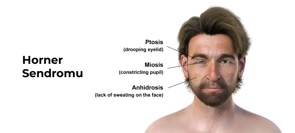
Ptosis (Drooping Eyelid): Among the ocular findings of Horner's syndrome, a slight drooping of the upper eyelid is observed on the affected side. This drooping is usually about 1 to 2 millimeters and creates notable asymmetry. It occurs due to the loss of sympathetic innervation of Müller's muscle, and patients may describe this condition as a "feeling of heaviness in my eye".
Miosis (Constriction of the Pupil): The pupil in the affected eye appears smaller than normal. This constriction becomes even more prominent, especially in a dark environment, because while the pupil in a normal eye dilates in the dark, this dilation is insufficient in the affected eye. Reaction to light continues normally, but the pupil size is asymmetric. The eye symptoms of Horner's syndrome are generally unilateral and may be the first change the patient notices in the mirror.
Anhidrosis (Lack of Sweating): Sweating decreases or disappears completely on the affected side of the face. This condition is particularly noticeable during hot weather or physical activity. The lack of sweating can be seen across the entire face or only in specific regions, depending on the location of the lesion.
Additional symptoms may be observed in some patients. These include flushing on the face due to vasodilation, conjunctival injection (redness in the white of the eye) on the affected side, and in rare cases, a change in iris color. Symptoms of Horner's syndrome in infants can be different, and iris hypopigmentation (lightening of color) may develop on the affected side. The severity of the symptoms varies depending on the level of the damage along the sympathetic nerve pathway.
How Does Horner's Syndrome Occur?
The development of Horner's syndrome is related to an interruption in the three-stage neural pathway of the sympathetic nervous system. This pathway starts from the brain, extends to the spinal cord, then to the chest region, and finally around the eye. Any injury occurring along this pathway can prevent the entire system from functioning properly. The causes of Horner's syndrome can be listed as follows:
First-Order (Central) Lesions: These types of lesions occur at the level of the hypothalamus, brainstem, or cervical spinal cord. The most common causes include stroke, tumors, multiple sclerosis, syringomyelia, and traumatic spinal cord injuries. Brainstem infarctions, such as Wallenberg syndrome, can also cause central Horner's syndrome.
Second-Order (Preganglionic) Lesions: These are conditions where the nerve fibers between the spinal cord and the superior cervical ganglion are affected. Causes of lesions at this level include lung tumors (Pancoast tumor), thyroid cancer, lymphoma, aortic aneurysm, trauma, and surgical complications.
The relationship between Pancoast tumors and Horner's syndrome is quite significant, as these tumors located in the upper part of the lung compress the sympathetic nerve pathways and cause the syndrome. Tumors associated with Horner's syndrome include neuroblastoma, thyroid cancer, lymphoma, and various neck tumors. Surgical interventions in the neck region, particularly thyroidectomy and carotid endarterectomy, can cause damage at this level.
Third-Order (Postganglionic) Lesions: These are conditions where the nerve pathway between the superior cervical ganglion and the eye is affected. Internal carotid artery dissection, cavernous sinus thrombosis, migraines, cluster headaches, and orbital tumors are the primary causes in this category. Inner ear infections and middle ear surgery can also rarely cause damage at this level.
The age factor is also important. Horner's syndrome in infants is generally associated with birth trauma, neuroblastoma, or congenital vascular anomalies. Early diagnosis and treatment are particularly important in infant cases of Horner's syndrome. In adults, malignancies, vascular diseases, and trauma are more frequently seen causes. The spectrum of diseases associated with Horner's syndrome is quite wide and can be a harbinger of serious systemic illnesses.
How is Horner's Syndrome Diagnosed?
The diagnosis of Horner's syndrome is primarily established through clinical observation and physical examination, followed by pharmacological tests and imaging methods. The diagnostic process is shaped around confirming the presence of the syndrome and identifying the underlying cause. Horner's syndrome is diagnosed using the following methods:
Clinical Evaluation: The first step is taking the patient's medical history and performing a detailed physical examination. The physician evaluates the eyelid position, pupil size and light reflex, and the facial sweating pattern. Documentation with photographs is helpful for tracking changes throughout the process.
Cocaine Test: The most reliable test to confirm the diagnosis of Horner's syndrome is the cocaine drop test. A 10% cocaine drop is instilled into both eyes, and pupil sizes are measured after 45 minutes. While cocaine dilates the pupil in a normal eye, there is no dilation or only minimal dilation in the eye with Horner's syndrome. If there is a difference of more than 0.8 mm between the pupils, the test is considered positive.
Apraclonidine Test: In cases where cocaine is unavailable, a 0.5-1% apraclonidine drop can be used as an alternative. This test works on a similar principle and is effective in diagnosing Horner's syndrome.
Localization Tests: Once Horner's syndrome is confirmed, a hydroxyamphetamine test is performed to determine the location of the lesion. When a 1% hydroxyamphetamine drop is instilled, the pupil dilates in preganglionic lesions, whereas no dilation occurs in postganglionic lesions.
Imaging Methods: Appropriate imaging methods are chosen depending on the location of the lesion. The brainstem, spinal cord, and nerve pathways are examined using magnetic resonance imaging (MRI), while the upper region of the lung and neck tumors are evaluated with computed tomography (CT). Ultrasound or angiography can be used to detect carotid artery dissection.
Laboratory Tests: A complete blood count, erythrocyte sedimentation rate, tumor markers, and an autoimmune disease panel may be requested to target the underlying cause.
How is Horner's Syndrome Treated?
The primary goal in the treatment of Horner's syndrome is to find and eliminate the underlying cause, because Horner's syndrome often appears as a symptom of another disease. Therefore, treatment varies from person to person and requires a multi-faceted approach. The following methods are applied for Horner's syndrome treatment:
Treatment of the Underlying Disease: In the treatment of Horner's syndrome, different methods are applied depending on the underlying cause. In the presence of a tumor, oncological treatments such as surgery, chemotherapy, or radiotherapy are preferred. In cases of carotid artery dissection, the vascular tear is brought under control with anticoagulant medications. If infection or inflammation is present, antibiotic or anti-inflammatory treatments are administered. For multiple sclerosis or other neurological diseases, immunosuppressive medications or supportive treatment methods are utilized.
Management of Eye and Facial Symptoms: If eyelid drooping or other visual symptoms cause discomfort, aesthetic or functional interventions can be performed by a plastic surgeon or ophthalmologist; however, in most cases, these symptoms do not cause permanent damage.
Rehabilitation and Follow-up: Because Horner's syndrome can often be an indicator of a serious systemic disease, regular physician check-ups and imaging follow-ups are important. Symptoms may improve over time or remain stable. Complete recovery depends on the course of the underlying disease.
Horner's syndrome is particularly important in the field of pediatrics. Congenital Horner's syndrome (including special cases such as harlequin-type Horner's syndrome) must be followed by pediatric neurologists. The treatment process can be long-term, but in most cases, symptoms may lessen or become controlled over time.
Frequently Asked Questions
Horner's syndrome is not directly a cranial nerve disorder; it arises from damage to the sympathetic nerve chain. This chain originates from the brainstem and reaches around the eye. The affected structure is the autonomic (sympathetic) nervous system rather than the cranial nerves. The lesion leading to Horner's syndrome can be at one of three levels of the sympathetic nerve pathway: central (brainstem, hypothalamus), preganglionic (spinal cord and chest region), or postganglionic (around the carotid artery and eye area). The location of the lesion varies according to the type of underlying disease. Horner's syndrome can be temporary in some cases. Especially in mild nerve injuries developing after trauma, symptoms may decrease over time with treatment; however, if there are serious causes such as tumors, vascular tears, or central nervous system diseases, the symptoms may be permanent. Treatment depends on the underlying cause. Congenital Horner's syndrome occurs as a result of injury to the nerves in the neck or shoulder region during birth in infants. In this case, differences in eye color (heterochromia), ptosis, and a small pupil are generally observed. Early diagnosis can reduce the long-term effects of nerve damage. Anisochoria (a difference in size between the two pupils) is typically observed in Horner's syndrome. Because the pupil is smaller in the affected eye, a more prominent difference is created compared to the other eye. This difference is more noticeable in dark environments because dilation is reduced in the affected eye. In some cases, Horner's syndrome can appear as a paraneoplastic syndrome, meaning as an indirect effect of a tumor. Particularly, certain cancers located in the upper part of the lung, such as Pancoast tumors, can cause Horner's syndrome by compressing the sympathetic nerves. This condition must be investigated. Horner's syndrome itself generally does not pose a life-threatening danger. The underlying cause can be serious, such as a stroke, tumor, vascular dissection, or spinal cord disease. Therefore, Horner's syndrome must always be taken seriously, and its cause must be determined through detailed examinations.
If you are experiencing symptoms of Horner's syndrome, early diagnosis is of great importance.
Hisar Hospital Intercontinental Ophthalmology specialists offer comprehensive evaluations and advanced imaging methods to determine the underlying cause.
Contact us immediately to schedule an appointment or obtain detailed information.
Start with the right assessment for What is Horner's Syndrome (Claude Bernard)? 7 Key Symptoms and Causes
Share your symptoms with our specialist physicians; let us determine the right diagnosis and treatment plan together.
Warning
If your symptoms started suddenly, are progressing rapidly, or significantly restrict your daily life, consult a physician without delay.
Plan your What is Horner's Syndrome (Claude Bernard)? 7 Key Symptoms and Causes journey with our specialists.
Fill in the form or book an appointment to learn about treatment options, the process and preparation.