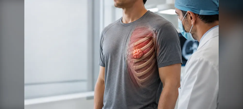
Chest Wall Tumor
What are they, what are the symptoms, and how are they diagnosed and treated?
It is natural to feel concerned when you notice a lump in your chest. At this point, it is important to know that the majority of lumps felt in the chest area are harmless formations such as lipomas (fatty lumps), cysts, or benign bone growths. The chest wall is a structure composed of ribs, the sternum (breastbone), the clavicle (collarbone), vertebrae, and the muscles and connective tissue between them. It acts as an armor protecting vital organs like the heart and lungs and enables us to breathe. A chest wall tumor is an abnormal tissue growth that develops in the bones, cartilage, or soft tissues that make up this structure.
In contrast, true tumors originating from the chest wall's own tissue are relatively rare, and the proportion of malignant ones among them is significant. Therefore, it is important for every new lump to be evaluated by a specialist.
In this article, we explain the types of chest wall tumors, their symptoms, how they are diagnosed, and the treatment process in simple terms.
By origin: primary and secondary tumors
Primary tumors originate directly from the chest wall's own tissue—bone, cartilage, muscle, or connective tissue. They are a rare group among all tumors.
Secondary (metastatic) tumors are formed when cancer from another region reaches the chest wall. This occurs either through the spread (metastasis) of cancer from a distant organ via the bloodstream or by the progression of a tumor from adjacent organs such as the lungs, pleura, or breast toward the chest wall. This group constitutes a significant portion of tumors detected in the chest wall; therefore, individuals with a history of cancer should not neglect new lumps in the chest area.
Benign tumors
Benign tumors grow slowly, do not spread to surrounding tissues, and do not metastasize to distant organs. They are usually painless. However, when they increase in size, they may put pressure on surrounding tissues or the lungs.
The most common ones are:
• Lipoma: Soft, movable masses originating from fatty tissue that slide when pushed with the finger. It is the most common benign tumor in the body.
• Osteochondroma and chondroma: Benign formations developing from bone and cartilage tissue. They are usually seen in the area where the ribs join the cartilage.
• Fibroma: Benign tumors originating from connective tissue.
• Fibrous dysplasia: Occurs when normal bone tissue within the rib is replaced by connective tissue; it is mostly discovered incidentally.
Malignant tumors
Malignant tumors grow faster, can erode bone tissue, and carry the risk of spreading to distant organs. Most malignant tumors originating from the chest wall's own tissue belong to the group of sarcomas, which are cancers of soft tissue and bone origin:
• Chondrosarcoma: Originates from cartilage tissue and is the most common malignant primary tumor of the chest wall in adults. It usually develops in the anterior part of the ribs or the sternum.
• Ewing sarcoma: Mostly seen in children and young adults.
• Osteosarcoma: A tumor originating from bone tissue that can progress rapidly.
• Soft tissue sarcomas: Tumors originating from muscle, fat, and connective tissue (such as rhabdomyosarcoma, fibrosarcoma).
Where are they seen?
The vast majority of chest wall tumors develop in the ribs. Less frequently, they may be located in the sternum, scapula (shoulder blade), or clavicle. As a general tendency, while tumors originating from soft tissue are mostly painless, pain is prominent in tumors originating from bone or those that have progressed to the bone.
What Are the Symptoms?
Symptoms vary depending on the tumor's location, size, and growth rate. While some tumors cause no complaints for years, others can show significant signs in a short time.
Palpable lump or mass
The most frequently noticed finding is a lump in the chest wall that can be felt by hand or seen from the outside. These lumps often start as painless. Masses that grow over time, have a hard consistency, feel as if they are attached to the rib, or have redness or prominent vascularization on the overlying skin must be evaluated.
Pain and tenderness
The tumor stretching the periosteum (bone membrane) or putting pressure on surrounding nerves can cause pain. This pain can be constant or increase with movement. Pain that is dull in character, does not go away with rest, and especially wakes you up from sleep at night is a finding that should be evaluated more carefully.
Shortness of breath
If the tumor grows into the chest cavity and prevents the lung from expanding comfortably, or if it leads to fluid accumulation between the lung membranes (pleural effusion), shortness of breath may develop. This condition first appears with exertion and, as it progresses, becomes noticeable even with less movement.
Deformity in the rib cage
Large tumors can disrupt the symmetry of the rib cage and create a protrusion or asymmetry that is noticeable from the outside.
Other symptoms
In cases where the tumor puts pressure on the airways or progresses into the lung tissue, cough and rarely blood in the sputum may be seen. In advanced stages, general symptoms such as unexplained weight loss, loss of appetite, and constant fatigue may accompany.
When should you consult a doctor? If you have a newly noticed lump in your chest wall that is gradually growing or hardening; if you are experiencing chest pain that lasts longer than a few weeks and wakes you up at night; or if you have previously received any cancer treatment, consult a Thoracic Surgery specialist. Remember that the evaluation is often completed with a physical examination and simple imaging.
Causes and Risk Factors
In most chest wall tumors, a definitive cause cannot be determined. However, some factors may increase the risk:
• Previous radiotherapy: In individuals who have previously received radiation therapy to the chest area, the risk of developing a tumor in this region years later increases slightly. Therefore, long-term follow-up of patients who have received radiotherapy is important.
• Genetic predisposition: A family history of cancer and certain hereditary syndromes may play a role, especially in sarcoma-type tumors.
• Occupational and environmental exposures: Long-term exposure to certain chemical substances may increase the risk.
• History of another cancer: Since a significant portion of tumors detected in the chest wall are formed by the spread of cancer from another organ, new findings in this area in individuals who have received cancer treatment are evaluated carefully.
A frequently asked topic is impact and trauma. It has not been shown that a blow to the chest causes a tumor. However, a previously existing but unnoticed mass may become apparent in the area examined after trauma. For this reason, lumps noticed after an impact should also be evaluated.
How Is It Diagnosed?
The diagnostic process proceeds in steps. The goal is to determine whether the mass is benign or malignant and exactly which tissue it originates from. An accurate diagnosis is the foundation of the correct treatment plan.
Examination and history
The process begins with listening to your complaints. How long the lump has been present, whether it has grown, the nature of the pain, your past illnesses, your history of radiotherapy, and your family history of cancer are questioned. During the examination, the size, hardness, whether it is attached to underlying tissues, and the condition of the skin over it are evaluated by hand.
Imaging
The first step is usually a chest X-ray. Then, necessary advanced examinations are performed:
• Computed Tomography (CT): It is the best method to show bone structures and the damage the tumor has caused to the bone. It reveals the size of the tumor and its spread to surrounding tissues.
• Magnetic Resonance Imaging (MRI): Shows soft tissues in detail. It is of great importance in evaluating the tumor's relationship with large vessels and nerves, and therefore in surgical planning.
• PET-CT: Scans the whole body to show whether the disease has spread to another region. It is used in staging, evaluating the response to treatment, and follow-up after treatment.
Which methods will be used is decided according to the characteristics of the mass and your general condition.
Biopsy
A definitive diagnosis can only be made by examining the tissue under a microscope. The most commonly applied method is taking a small tissue sample with a thick needle under local anesthesia and under the guidance of ultrasound or tomography. In some cases, it may be preferred to remove a small part of the mass with a minor surgical procedure for examination.
Since where and how the biopsy is performed can affect the future surgery, it is important that this decision is made together with the thoracic surgery team that will undertake the treatment.
Staging
When a malignant tumor is detected, staging is performed to determine the extent of the disease. The size of the tumor, whether it has progressed to surrounding tissues, the status of lymph nodes, and whether there is spread to distant organs are evaluated. Staging determines whether surgery is appropriate and whether additional treatment is required.
How Is Treatment Performed?
The treatment plan is determined individually according to the tumor's type, size, location, extent, and your general health status. This decision is made in a council where thoracic surgery, medical oncology, radiation oncology, radiology, and pathology specialists evaluate together.
Surgery
Surgery is the most effective method in the treatment of chest wall tumors. The goal is to completely remove the tumor along with a certain amount of healthy tissue around it. Ensuring that no tumor cells remain at the edges of the removed tissue is decisive for preventing the disease from recurring. For this reason, in malignant tumors, a wider area is removed, and if necessary, one or more ribs or a part of the sternum is removed.
Reconstruction of the chest wall
When a large area is removed, the gap created in the rib cage must be closed and the integrity of the chest wall must be restored. Otherwise, respiratory mechanics will be impaired. This procedure is often performed in the same surgery.
For this, special synthetic patches (mesh) or titanium plates are used to provide both flexibility and durability to the chest wall. When the created area needs to be covered with soft tissue, tissue pieces prepared from your own muscles in the back or chest area are used. In comprehensive cases, work is done together with plastic surgery and orthopedics teams. Which method will be used is decided before the surgery and explained to you in detail.
Radiotherapy
Radiation therapy targets tumor cells and stops them from multiplying. It can be used to shrink the tumor before surgery and to control microscopic cells that may have remained after surgery. In cases where surgery is not appropriate, it can be applied as the primary treatment method. In some tumor types, radiotherapy alone may be sufficient.
Chemotherapy and other drug treatments
In some rapidly progressing tumor types (e.g., Ewing sarcoma), drug treatment is an integral part of the therapy and is often started before surgery. Drug treatment is also used to reduce the risk of spread or to keep the disease under control in cases where surgery is not appropriate.
Follow-up
Regular check-ups are of great importance after treatment is completed. In malignant tumors, examinations, blood tests, and imaging are performed more frequently in the first years and at longer intervals in subsequent years. The purpose of this follow-up is to catch a possible recurrence at the earliest and most treatable stage.
Frequently Asked Questions
Is every lump in the rib cage cancer?
No. The majority of lumps felt in the chest wall are harmless formations such as lipomas, cysts, or benign bone growths. However, a definitive distinction cannot be made by physical examination; therefore, every new lump must undergo a specialist evaluation.
Can a tumor form in the rib?
Yes. Since ribs are composed of bone tissue, a tumor can develop here just as it can in other bones. In fact, the most common site for chest wall tumors is the ribs.
Does a chest wall tumor threaten life?
This depends entirely on the type, location, and extent of the tumor. Benign tumors do not threaten life; they may only need to be removed when they grow and put pressure on surrounding structures. In malignant tumors, very successful results can be achieved with early diagnosis and appropriate treatment. Treatment success is high in tumors that are caught at an early stage and can be completely removed. For this reason, evaluating the lump or pain early instead of delaying it makes a big difference.
How long does the surgery take?
The duration varies greatly depending on the scope of the procedure to be performed. While the removal of a superficial and small soft tissue mass can be completed in half an hour, surgeries requiring wide excision and reconstruction of the chest wall can take several hours.
How does the post-operative process proceed?
In extensive surgeries, thin tubes called drains are placed in the surgical area and the chest cavity to allow the drainage of fluid and air; these are removed within a few days. Depending on the scope of the surgery and your general condition, you may be monitored in the intensive care unit for the first few hours. Afterward, you are followed up in the ward, and your recovery is monitored with X-rays and blood tests. In extensive surgeries, the hospital stay is on average 5–6 days; it is much shorter in minor interventions.
How long does recovery take, when can I return to work?
This varies according to the size of the surgery. You can return to your daily life within 1–2 weeks after the removal of a small mass. In patients who have undergone wide excision and chest wall repair, recovery may take a few weeks, sometimes longer. During this period, pain control and performing the recommended breathing exercises regularly will accelerate recovery. The time to return to work is decided together with your doctor, taking into account the physical load of your job.
Is surgery risky?
As with any surgical intervention, there are risks such as bleeding, infection, and respiratory problems. With an experienced team and correct planning, these risks are minimized. Your doctor will explain the specific risks and expected benefits to you in detail before the surgery.
Does the tumor recur?
Recurrence is rare in benign tumors after they are completely removed. In malignant tumors, there is always a possibility of recurrence; therefore, regular check-ups are important. Early detection of a possible recurrence significantly increases the chance of treatment.
Which department should I apply to?
The first application should be made to the Thoracic Surgery department. The diagnosis and treatment process is carried out together with the participation of medical oncology, radiation oncology, radiology, pathology, and plastic surgery specialists when necessary.
Chest wall tumors are rare diseases but can be successfully treated when evaluated correctly. Most lumps felt in the chest area are harmless; however, distinguishing which lump requires attention is only possible with a specialist examination and imaging.
The most decisive factor is time. Treatment success is high in tumors that are noticed early and can be completely removed. If you notice a new lump in your chest or persistent pain, consult a Thoracic Surgery specialist without delay.
Şule Karadayı
Fill Out the Form for Detailed Information and Contact!
Start with the right assessment for Types of Chest Wall Tumors
Share your symptoms with our specialist physicians; let us determine the right diagnosis and treatment plan together.
Warning
If your symptoms started suddenly, are progressing rapidly, or significantly restrict your daily life, consult a physician without delay.
Plan your Types of Chest Wall Tumors journey with our specialists.
Fill in the form or book an appointment to learn about treatment options, the process and preparation.