At least moderate foraminal stenosis is present in 40% of 60-year-olds. This rate rises to 75% in individuals aged 80 and over. However, the surprising fact is this: only 17.5% of those experiencing severe foraminal stenosis show symptoms. So, what is foraminal stenosis and what does it mean? What is neural foraminal stenosis? In this article, we will explain everything you need to know about foraminal stenosis symptoms, causes, and treatment in plain language.
What is Foraminal Stenosis?
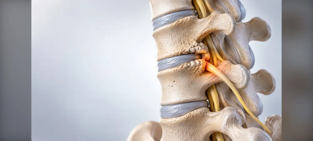
There are small openings on the sides of your spine through which nerves pass. These openings are called the "foramen." Foraminal stenosis is a condition that occurs as a result of the narrowing of these openings. When narrowing occurs, the nerve roots exiting the spinal cord are compressed, and this compression leads to various symptoms.
As the space between the vertebrae through which nerves pass becomes smaller, pressure is exerted on the nerves. This pressure manifests itself particularly in the back, lower back, or neck region. Consequently, you experience discomforts such as pain, numbness, and muscle weakness.
What is Neural Foraminal Stenosis?
The neural foramen is the opening where the spinal nerve exits the spine and branches out to other parts of the body. The size of this opening varies depending on where it is located in the spine. For example, the exit hole in the lumbar spine section is approximately 18-22 millimeters high and 5-8 millimeters wide. In the cervical spine section, it has a height of 10-14 millimeters and a width of 5-7 millimeters.
Neural foraminal stenosis is a type of spinal stenosis that affects these openings. To understand the condition, we can use the following analogy: Imagine an electrical cord trapped between a door and its frame when the door is closed. Over time, the pressure on the cord can damage it and affect how it transmits electricity. Similarly, foraminal stenosis exerts pressure on the affected nerves.
When narrowing occurs in the openings, the spinal nerves are irritated or damaged. Neuroforaminal stenosis is often seen together with spinal canal narrowing. Therefore, symptoms can become more complex.
Types of Foraminal Stenosis
The location of foraminal stenosis also determines its type. The types according to different sections of the spine are listed as follows:
The cervical spine is located in the neck region. This is the second most common area where foraminal stenosis develops. The thoracic spine is found in the upper and middle back. The lumbar spine is in the lower back region, and this is the most common area where foraminal stenosis is seen. The sacral spine is located in the lower back and pelvis region, while the coccygeal spine is at the very bottom.
The region where the narrowing occurs directly affects the symptoms you will experience. While stenosis in the lower back region causes leg pain, stenosis in the neck region can lead to problems in the arms and hands.
Causes of Foraminal Stenosis
The causes of foraminal stenosis can emerge through the combination of multiple factors. While a single cause is sufficient for some people, a combination of several factors is required for others. Any condition that alters the structure of your spine can cause the space through which the nerve roots pass to narrow.
Aging and Degenerative Changes
As you age, a series of structural changes occur in your spine. Radiological findings of canal narrowing are detected in approximately 20% of the population over 60 worldwide. Symptomatic canal narrowing is present in approximately 10-15% of individuals over 50.
The discs between the vertebrae lose water content with age. This leads to a decrease in disc height and a narrowing of the canal. As the discs lose their elasticity, they wear out more easily and become unable to act as shock absorbers.
The small joints at the back of the spine grow and thicken over time. This growth narrows the spinal canal. As the joint cartilage wears down, calcification occurs, and the resulting calcification puts pressure on tissues inside the canal.
The thickening of the ligamentum flavum surrounding the spinal canal reduces the canal diameter. Ligaments are bands of fibers that hold the spine together. Arthritis causes the ligaments to thicken over time and bulge into the spinal canal space.
Bone spurs formed on the vertebral bodies can grow inward into the canal. Bone spurs or excessive bone growth compress the nerves in the spine. Your body responds by producing new bone as the cartilage wears away.
Disc Herniation
Between the spinal bones are cushions called discs. These discs provide flexibility to the movements of the spine. Over time, some of the inner material of the discs leaks outward and exerts pressure on the spinal cord or nerves.
The jelly-like center of the discs passes through a weak or torn outer layer. The bulging disc then presses on the nerves near the disc. Herniated discs are one of the common causes of foraminal stenosis.
Back injuries can cause herniated or bulging discs. Disc herniation is frequently seen in the 30s and 40s when the nucleus pulposus has a gelatinous structure.
Arthritis and Joint Diseases
Osteoarthritis is a wear-and-tear condition that breaks down the cartilage in your joints. Cartilage is the protective coating of the joints. As the cartilage wears down, the bones begin to rub against each other.
Rheumatoid arthritis leads to joint inflammation and contributes to the development of foraminal stenosis. Diseases affecting the muscles, bones, or connective tissues that hold the spine together can cause foraminal stenosis.
Conditions such as ankylosing spondylitis and Paget's disease accelerate this process. Paget's disease is a bone disorder that causes excessive bone destruction. Spondylolisthesis is the slippage of one vertebra over another and contributes to canal narrowing.
Trauma and Injuries
Spinal injuries resulting from various traumatic events such as car accidents can cause foraminal stenosis. Canal narrowing in the lower back may occur as a result of traumas.
Back injuries lead to scar tissue or structural changes. Spinal fractures are some of these changes. Swelling of nearby tissue after back surgery may also contribute to the development of foraminal stenosis.
Cysts and tumors can obstruct the spinal cord and spinal nerves. This causes foraminal stenosis to occur in a place where it normally would not develop.
Symptoms of Foraminal Stenosis
Foraminal stenosis symptoms usually begin slowly and worsen over time. You experience discomforts that increase particularly with movement in the area where the nerves extend in the body. The location of the symptoms is an important clue in the diagnosis and treatment of foraminal stenosis.
Pain Symptoms
Pain radiating to the neck, back, lower back, or arms and legs is the most prominent finding of foraminal stenosis. It radiates from the region where the narrowing is located toward the hip, leg, shoulder, or arm. Narrowing in the lumbar region usually manifests itself with pain radiating toward the hips, legs, and feet.
The character of the pain is in a striking manner toward the legs. It increases with standing or walking. In some patients, a burning or tingling sensation accompanies the clinical picture. You feel relief when you sit down.
Numbness and Tingling
Depending on the affected nerve root, numbness occurs in the arms or legs. Numbness in the legs increases especially when walking or standing. You experience a tingling or "pins and needles" sensation.
Due to nerve compression, burning and stinging can be seen in the legs and feet. Similar numbness problems occur in the hands, arms, and fingers. These symptoms worsen with walking or standing.
Muscle Weakness
Weakness develops in the arms and legs due to nerve pressure. A feeling of "giving way" or weakness may occur in the legs. You feel a sudden giving way in your legs while walking.
Loss of muscle strength may be observed. You experience difficulty when climbing stairs. In advanced stages, loss of muscle control comes into question. In addition, cramps may occur in the legs.
Movement Limitation
Pain and difficulty in movement increase after prolonged sitting or standing. You experience loss of strength in the leg after walking a certain distance. Patients describe it as: "Last year my leg would start hurting after walking for fifteen minutes, this year it starts hurting after walking for five minutes."
Walking distance gradually shortens. A need to stop and rest develops for a while. You experience relief when you rest or lean forward. Some patients start walking by leaning forward to reduce the pressure on the nerves.
Symptom Differences by Region
If there are symptoms in the thumb, index finger, and middle finger, there may be a problem in the cervical spine. Discomfort felt in the upper abdomen and just below the breastbone points to the thoracic spine. Symptoms affecting the outer edge of the foot, the fourth and little toes, are due to stenosis in the lower part of the back.
In the neck region, tingling radiating to the arms, weakness in the hands and fingers, and problems with balance and walking occur. In advanced cases, problems with bladder and bowel control may develop.
Diagnosis of Foraminal Stenosis
Your doctor relies on your symptoms when diagnosing foraminal stenosis. However, they utilize various examinations and tests for a definitive diagnosis. The diagnostic process consists of three main stages.
Physical Examination
Your doctor first listens to your medical history. You explain when your symptoms started, under which conditions they increase, and how they affect your daily life. This information provides a clear picture of the underlying disease.
During the physical examination, your doctor observes your movements. They check your gait, posture, and flexibility. They determine in which positions you feel pain. For example, if your complaints decrease when you walk by leaning forward, this may point to foraminal stenosis.
They test your muscle strength. They check your reflexes. They determine in which areas numbness or tingling is present. This examination provides important clues to find the causes of the symptoms.
Imaging Methods
Magnetic resonance imaging shows the spinal canal and the nerves passing through it. It provides extremely detailed images that allow you to separate bones, nerves, and muscles from each other. The degree of foraminal narrowing is graded with MRI.
X-rays, computed tomography, and MRI scans help determine the narrowing in the spinal canal, the pressure exerted on the nerve roots, and the degree of compression. Although many people have foraminal stenosis visible on MRI, symptoms are not observed.
Computed tomography is used for people with implanted devices such as pacemakers. The metallic components in the pacemaker must not be exposed to the strong magnetic field of the MRI. The CT scan can be combined with a myelogram.
Electrophysiological Tests
Electromyography is used to evaluate the function of nerves. It examines the electrical activity of the nerve and muscular system. EMG clearly shows which nerve root is under what degree of compression.
Nerve conduction studies measure the conduction velocity of the current in nerves. Conclusions about nerve activity are drawn from this. In individual cases, diagnosis is supported by neurological examination and electrophysiological evaluations.
EMG results are frequently pathological. This condition is characterized by findings pointing to bilateral multiradicular neurogenic involvement. Your doctor may recommend other tests depending on your special medical history or needs.
Treatment of Foraminal Stenosis
Foraminal stenosis treatment varies according to the severity of your symptoms and the location of the stenosis. If symptoms are mild, your doctor monitors the condition with regular follow-up appointments. Conservative treatments are almost always the first approach.
Conservative Treatment Methods
Non-surgical treatments are applied in the initial stage. Reducing movement can be helpful, especially when it occurs along with inflammation and swelling caused by injury. However, complete bed rest is generally not beneficial. Changes in activities and non-invasive approaches are prioritized during this process.
Physical Therapy and Exercises
Physical therapy helps increase physical strength and flexibility by reducing the impact of foraminal stenosis symptoms. It is applied to strengthen muscles, reduce pressure on the spine, and increase mobility. Although there is no standardized regimen, many therapists focus on stretching and strengthening core muscles.
Exercises focused on forward bending provide relief by opening the openings through which nerves pass. Strong abdominal and back muscles take the load off the spine and act as a natural corset. Exercise increases the amount of blood coming to the nerves, reducing the symptoms of narrowing.
Medication Treatment
Nonsteroidal anti-inflammatory drugs alleviate swelling and pain. They can reduce inflammation around the affected spinal nerve. Pain relievers alleviate mild and moderate pain. Muscle relaxants are used to reduce muscle spasms. Antidepressants and anticonvulsants can be applied to alleviate nerve pain.
Oral steroid medications reduce inflammation and swelling around the affected area. Oral opioid medications help treat acute pain but are usually intended for short-term use only.
Injection Treatments
Your doctor may recommend cortisone injections. The anti-inflammatory medication is injected directly into the foraminal stenosis area. Cortisone significantly alleviates inflammation and pain. Its effects may be temporary.
Transforaminal injection reduces radicular pain and inflammation by delivering medication directly to the nerve root at the foraminal level. Pain-relieving effects usually last from a few weeks to a few months. It can be repeated at certain intervals when necessary. Injections include X-ray guidance.
Surgical Treatment
When conservative treatment is not sufficient or symptoms are severe, surgical intervention may be required. Foraminotomy is performed to widen the neural foramina. Laminectomy is the procedure of removing a part of the bony structure called the lamina to widen the spinal canal. This procedure alleviates pain by reducing pressure on the nerves.
Spinal fusion is the procedure of joining two or more vertebrae together. Laminectomy with fusion is typically reserved for patients with concurrent spondylolisthesis. Most modern spinal decompression surgeries involve a minimally invasive approach.
Post-Treatment Recovery Process
The recovery period depends on the patient's general health status and the treatment method applied. Recovery after conservative treatment supported by physical therapy can be seen within a few weeks. Full recovery after surgical intervention can take several months.
One or two days after the surgery, the patient is started to be walked. You can go outside after two to three weeks. You can return to work after a month, provided you adhere to certain movement restrictions. Full recovery occurs after an average of six months. Regular physical therapy performed under the supervision of a specialist physician helps the spine rebalance by increasing muscle strength.
Frequently Asked Questions
Is foraminal stenosis dangerous?
Foraminal stenosis is a condition that significantly reduces the quality of life and progresses if left untreated. The spinal cord, having lost its natural structure, does not return to its former state over time. However, pain remains a manageable problem.
The degree of danger depends on the severity of symptoms. In mild cases, it can be controlled with conservative treatments. In advanced stages, the risk of permanent nerve damage increases. Therefore, early diagnosis and an appropriate treatment approach are of vital importance.
Does foraminal stenosis go away on its own?
It does not heal on its own. Degenerative changes in the spine are irreversible processes. Canal narrowing is anatomically permanent.
Nevertheless, pain can be managed. Conservative methods such as medication, physical therapy, and weight loss alleviate complaints. The disease can be treated by eliminating nerve compression with surgical methods. In addition, lifestyle changes help relieve pain.
Which exercises should be done in foraminal stenosis?
Exercise does not destroy ossified tissues or physically widen the canal. However, it increases the functional capacity of the spine, strengthens supporting muscles, and corrects posture, allowing nerves to be more comfortable in a narrow space.
Giving a slight incline to the treadmill works. When walking uphill, the torso naturally leans forward slightly, which opens the spinal canal and allows you to walk more comfortably. Avoid walking downhill.
Bicycling can be preferred to strengthen the body. It is important to keep the spine in an upright and natural position. Swimming is also frequently used since it works all muscle groups and strengthens the lower back, back, and abdominal muscles.
If you feel sharp pain, numbness, or a sensation of electricity radiating from the hip to the leg during exercise, this indicates that nerve compression has increased. You must stop that movement immediately and consult your physiotherapist.
Is foraminal stenosis surgery risky?
Since the professional approaches of modern medicine aim to anticipate and minimize possible risks and complications, these risks are encountered quite rarely. Post-operative infection may occur. Damage to surrounding tissues or organs may occur during the surgical procedure.
Bleeding may occur during surgery. Nerve damage can lead to loss of sensation. Tears may form in the spinal dura mater. This risk is slightly higher, especially in patients undergoing surgery for the second or third time.
In elderly patients, conditions such as hypertension, diabetes, and heart problems can increase risks both during anesthesia and the post-operative period. Complaints such as numbness, lack of sensation, and tingling may be experienced after surgery. However, these complaints are not expected to be permanent.
Foraminal Stenosis in Summary
Foraminal stenosis can lead to serious symptoms, but it is a manageable condition with the right approach. Early diagnosis carries great importance, especially in this problem that emerges with age. If you are experiencing pain radiating to the legs, numbness, or muscle weakness, you should definitely consult a specialist.
Treatment options are in a wide range. Conservative methods such as physical therapy and medication give effective results in most cases. In advanced stages, surgical intervention can completely eliminate the pressure on the nerves. Therefore, a treatment plan to be applied under the control of a specialist physician significantly increases your quality of life.
Fill Out the Form for Detailed Information and Contact!
Start with the right assessment for What is Foraminal Stenosis? What Are Its Symptoms? How Is It Diagnosed and Treated?
Share your symptoms with our specialist physicians; let us determine the right diagnosis and treatment plan together.
Warning
If your symptoms started suddenly, are progressing rapidly, or significantly restrict your daily life, consult a physician without delay.
Plan your What is Foraminal Stenosis? What Are Its Symptoms? How Is It Diagnosed and Treated? journey with our specialists.
Fill in the form or book an appointment to learn about treatment options, the process and preparation.