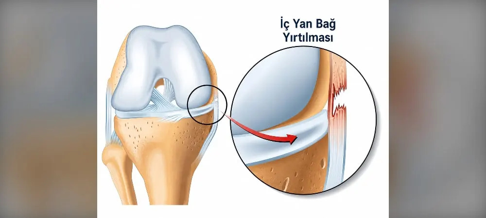
The most common ligament injury seen in the knee is a medial collateral ligament injury. When the medial collateral ligament of the knee is damaged, your knee may overextend or begin to bend in directions different from normal. Medial collateral ligament injuries of the knee usually occur due to sports traumas. In this article, you will find everything you need to know about medial collateral ligament injury symptoms, diagnosis, and treatment of the knee.
What is the Medial Collateral Ligament of the Knee and Where Is It Located?
The medial collateral ligament of the knee is known in medical terminology as the medial collateral ligament (MCL). It is the most frequently injured ligament of the knee joint in both adults and children. This ligament is a strong and flexible tissue band extending between the thighbone (femur) and the shinbone (tibia).
Located on the inner part of the knee joint, this structure plays an important role in ensuring knee stability. The primary function of the ligament is to prevent excessive inward movement of the knee and to protect the joint. It ensures that your knee remains stable and functions properly while moving.
Anatomical Structure of the Medial Collateral Ligament
The medial collateral ligament of the knee consists of three main structures. These are the superficial medial collateral ligament, the deep medial collateral ligament, and the posterior oblique component. This complex structure ensures that the knee is protected at different movement angles.
The ligament can be examined in three to four layers. The outermost layer contains the protective sheath located just beneath the skin. Beneath it lies the superficial medial collateral ligament. In the innermost layer, the joint capsule and the deep medial collateral ligament are positioned.
Between these two ligaments lies a small fluid-filled sac (bursa) that allows them to glide smoothly over each other during movement. Structures such as the tendons of the three important muscles known as the "pes anserinus" and the saphenous nerve, which carries sensation to the anteromedial part of the leg, also pass through this region.
Difference Between the Superficial and Deep Medial Collateral Ligaments
The superficial part is the largest and strongest part of the system. It originates from the medial femoral epicondyle and extends to four centimeters below the inner surface of the tibia. It starts right above the bony prominence on the inner part of the thighbone and attaches to the shinbone at two different points.
The deep part originates from the medial joint capsule and attaches to the medial meniscus. There is no direct connection between these two ligaments. However, the deep medial collateral ligament has meniscofemoral and meniscotibial divisions, through which it connects with the medial meniscus.
Location of the Medial Collateral Ligament in the Knee
The medial collateral ligament is located on the inner part of the knee and has a wide, flat membranous band structure. It is positioned slightly behind the inner side of the knee joint. Its primary role is to resist valgus forces (inward bending) applied to the knee.
The ligament limits abduction and rotation of the knee joint. It is considered the primary static stabilizer of the knee and helps passively stabilize the joint. In this way, it prevents the knee from excessively bending in unexpected directions.
How Does a Medial Collateral Ligament Injury of the Knee Occur?
Medial collateral ligament injuries occur in two main ways. The first of these is a direct blow to the outside of the knee, and the second is non-contact strains. The majority of injuries develop as a result of a sudden valgus force.
Injuries Caused by Direct Impact
An acute blow to the outer side of the knee creates tensile forces on the inner side. This situation is the most common cause of injury. This mechanism is frequently seen, especially in contact sports such as soccer, American football, or rugby.
When a player's foot is planted on the ground and an opponent interferes from the outside, sharply forcing the knee inward, it causes the ligament to suddenly stretch. Direct blows to the lateral part of the knee create significant valgus force in the joint.
There are classic examples in elite athletes. A direct blow to the lateral side of the athlete's knee while the foot is in contact with the ground leads to injury. Injuries occurring through contact generally have a higher potential to cause more severe damage to the ligament.
Injuries Caused by Non-Contact Strains
Injuries also occur without a direct blow to the knee. This situation is usually observed during sudden direction changes, stopping, or pivoting movements.
Sudden rotation of the torso while the foot is planted on the ground is one of the main injury mechanisms. When skiing, getting the ski caught while the body continues to rotate creates severe tension on the ligament. Scenarios such as the foot remaining fixed on the ground while the body rotates over the knee can lead to injury.
This type of injury is frequently seen during sudden maneuvers in sports like soccer and basketball. Likewise, when the foot is in contact with the ground and the knee is flexed, extreme internal rotation of the knee causes injury.
It has been reported that the most common form of injury in athletes results from falling to the ground while the foot is in plantarflexion and the knee is in flexion. These forces, which stretch or tear the structures on the inner side of the knee, mostly occur as a result of indirect trauma.
Risk Factors and Injury Mechanisms
Medial collateral ligament injuries of the knee usually occur due to sports traumas. A sudden valgus force that stretches the structure of the ligament to the point of micro- or macroscopic damage constitutes the vast majority of injuries.
Contact sports stand out as the most important risk factor. Falls, impacts, and sudden movements during sports can lead to injuries. While injuries can develop over the long term due to reasons such as improper or missing equipment, they can also occur suddenly as a result of accidents.
Damage to the ligament can occur as a result of the knee receiving a blow from the outside or being forced inward. The injury can occur alone or together with other ligament injuries. The structures most frequently injured along with the medial collateral ligament are the anterior cruciate ligament and the meniscus.
Medial Collateral Ligament Injury Symptoms and Grading of the Knee
Different symptoms appear depending on the degree of injury. Pain is felt along the line where the ligament runs on the inner part of the knee and is the most fundamental finding. Localized pain on the inner side of the knee is the most common finding. Swelling, tenderness, bruising, and a feeling of instability in the knee are frequently encountered conditions in injuries.
Grade 1 Injury Symptoms
This is the mildest degree of injury. A mild stretch or tear occurs in the ligament, but the ligament is still intact and its functions are not impaired. Mild pain, very little swelling, and mild tenderness are observed. A valgus stress test is accompanied by joint opening of less than 5 mm and a firm endpoint. In such cases, a few fibers are torn, but the ligament is structurally intact.
Knee stability is preserved. Findings are generally mild, and the patient complains of pain when pressing on the collateral ligament. Stretching has occurred and there is no tear. Return to sports is possible with treatment within 1-2 weeks.
Grade 2 Injury Symptoms
This is a moderate injury. A partial tear occurs in the ligament, and the stability of the ligament decreases. There is moderate pain on the inner part of the knee, noticeable swelling, and tenderness. Joint opening between 5 and 9 mm is detected during the valgus stress test. There is usually an intact or perceptible end-point.
There is a limitation of movement and a noticeable loss of knee stability. The patient may complain of the knee giving way during jumping or pivoting movements. A feeling of the knee "giving way" may be experienced from time to time. Pain and swelling are more pronounced, and a 3-4 week treatment period is required to return to daily activities.
Grade 3 Injury Symptoms
This is a complete rupture of the ligament and is the most severe degree of injury. Joint opening of 10 mm or more is seen on the valgus stress test without a perceptible end-point. This degree of laxity suggests that other knee ligaments may also be injured.
Severe pain may be felt at the moment of injury, but sometimes the pain can subside. There is intense swelling, bruising, and a significant loss of stability in the knee. There is marked tenderness and swelling over the ligament. The patient has difficulty bending the knee, and the knee giving way is observed even during normal walking. A serious feeling of the knee "giving way" is experienced when the knee is moved.
Initial Findings After Injury
At the moment of injury, especially in severe tears, patients may express feeling a sound coming from inside their knee or feeling something snap. The patient may hear or feel a pop sound at the moment of injury.
The timing of swelling is an important clue. If noticeable and tense swelling develops in the knee within the first few hours after the injury, this situation usually points to bleeding inside the joint. Swelling that develops more slowly, within 12-24 hours, is generally seen only in cases where the medial collateral ligament is damaged. Tenderness along the course of the ligament can be detected during the examination.
How is a Medial Collateral Ligament Injury Diagnosed?
The diagnostic process begins with a detailed injury history. In most cases, a medial collateral ligament injury can be diagnosed solely through a detailed history and physical examination. How the injury occurred, in which direction the knee was forced, and the onset time of symptoms are evaluated.
Performing the examination as soon as possible after the injury, before swelling and muscle spasm increase, yields the most accurate results. Therefore, early evaluation is of great importance in terms of diagnosis.
Physical Examination and Valgus Stress Test
The physician first observes whether there is any bruising, swelling, or deformity on the inner part of the knee. Then, using their fingers, they press along the line where the ligament extends from its attachment on the thighbone to the shinbone to find the point where tenderness is most intense.
The most critical part of the examination is a special maneuver called the valgus stress test. In this test, the physician applies a controlled force to the inner side of the knee while holding the patient's leg at specific angles. It is checked how much the joint space opens and how loose the ligament is.
The test performed with the knee bent at 30 degrees relaxes other supportive structures in the knee, allowing the superficial medial collateral ligament to be evaluated in isolation. The physician evaluates whether there is an opening on the inner side of the knee and whether an endpoint is obtained during this opening. When used as a single test with pain as the outcome measure, it has 78% sensitivity and 67% specificity. When used with laxity as the outcome measure, it has 91% sensitivity and 49% specificity.
The test performed when the knee is in a fully straight position is particularly important. If laxity is detected in this position, it indicates that not only the medial collateral ligament but also other important structures in the posteromedial corner of the knee and possibly the cruciate ligaments are torn.
MRI Imaging and Other Radiological Methods
X-ray is the first method applied in every severe knee injury. Although it does not directly show the ligaments, it is essential to rule out possible bone fractures, small bone fragments pulled off by the ligament from its attachment site, or growth plate injuries in children. Avulsion fractures can be seen on anteroposterior and lateral radiographs.
Magnetic resonance imaging is the most effective method for imaging soft tissue injuries. With MRI, the location, degree, and completeness of the tear in the medial collateral ligament are clearly seen. It is valuable to show whether there are accompanying injuries in other intra-articular tissues.
Detection of Accompanying Other Injuries
A medial collateral ligament injury of the knee can occur alone or together with other injuries. It is important to distinguish an MCL tear from other knee injuries. During the examination, the status of the anterior and posterior cruciate ligaments and menisci must be checked carefully.
Among the most common accompanying injuries are anterior cruciate ligament tears, meniscus tears, and bone marrow edema. MRI is invaluable in diagnosing these secondary accompanying injuries.
Treatment Options for Medial Collateral Ligament Injury of the Knee
Most medial collateral ligament injuries heal successfully with non-surgical methods. The main reason for this is that the ligament is located outside the joint and has a very good blood vessel network. Thanks to this biological advantage, the self-healing potential of the ligament is quite high.
Non-Surgical Treatment Methods
Treatment options vary depending on the severity of the injury. While rest, cold application, and bandages are sufficient in mild cases, different approaches may be required in advanced cases. Excellent results can be achieved with non-surgical treatment in isolated grade 1, grade 2, and even most grade 3 injuries where the knee is stable in the straight position.
Applying ice at 20-minute intervals for the first 48 hours reduces swelling and pain. Compression with an elastic bandage or knee brace is recommended. Keeping your knee elevated above heart level reduces swelling. Bandage application, movement restriction, and ice application in the early period are important for relief.
Physical Therapy and Rehabilitation
The basic principle of non-surgical treatment is early and controlled movement. In the past, keeping the knee immobilized for weeks was practiced, but this approach caused the healing ligament tissue to be weak and disorganized. In the modern approach, bending and straightening the knee are permitted in a controlled manner.
The main goals of the rehabilitation program include controlling pain and swelling, protecting the healing ligament, preventing muscle wasting, and regaining full range of motion in the knee. Strengthening the quadriceps and hamstring muscles plays an important role in increasing knee stability. Proprioceptive exercises and balance work can also be part of the program.
Use of Knee Braces and PRP Injection
Using a hinged knee brace prevents the knee from opening sideways while allowing it to be bent and straightened in a controlled manner. Generally, there is no need for a knee brace in grade 1 injuries. However, using a knee brace is necessary in grade 2 and non-surgical grade 3 injuries.
PRP injections under ultrasound can accelerate healing[271]. Most grade 1 and grade 2 medial collateral ligament injuries heal without the need for surgery with a well-implemented physiotherapy program, 2 doses of PRP injections under ultrasound, and the use of a knee brace[271].
Conditions Requiring Surgery
Although most cases heal without surgery, surgical treatment is inevitable in some cases. The decision for surgery is made based on the severity and location of the injury, other accompanying problems, and the patient's activity level.
The presence of valgus laxity when the knee is fully straight requires surgery. In multi-ligament injuries, especially when accompanied by an anterior cruciate ligament injury, both ligaments must be operated on at the same time[271]. Laxity in the medial collateral ligament will cause anterior cruciate ligament surgery to fail[271]. Bone fractures due to avulsion and tears completely separated from the thighbone or shinbone also require surgical intervention.
Recovery Times and Return to Sports
The recovery time varies depending on the degree of injury. While mild injuries heal within 1-2 weeks, a return to sports is possible within 5-10 days in grade 1 injuries[271]. In moderate injuries, this period can extend to 3-4 weeks. A 2-4 week treatment period is required in grade 2 injuries[271].
Healing can take 6-8 weeks in severe tears. A period of 4-8 weeks is anticipated in grade 3 injuries[271]. These durations are averages and vary from patient to patient and depending on the sport performed[271].
Frequently Asked Questions
Can a person with a medial collateral ligament injury of the knee perform daily activities?
Depending on the degree of injury, daily activities can be affected. Careful walking is possible in mild injuries. However, in severe injuries, the use of a knee brace and support is required.
How can a medial collateral ligament injury be prevented?
Risk can be reduced by doing regular muscle-strengthening exercises. Do warm-up exercises before sports. Use appropriate sports equipment and move with correct techniques.
When should you consult a doctor?
If you experience severe pain, noticeable swelling, or difficulty walking in your knee, consult a doctor immediately. Evaluation is required if you feel your knee giving way or cannot bend your knee fully.
Does the injury leave permanent damage?
With proper treatment, most medial collateral ligament injuries of the knee heal completely. Early intervention and an appropriate rehabilitation program improve outcomes. If left untreated, chronic knee instability can develop.
How is a medial collateral ligament injury symptom distinguished from other knee injuries?
Pain in a medial collateral ligament injury is localized to the inner part of the knee. In other ligament injuries, pain is felt in different regions. Examination and imaging are required for a definitive medial collateral ligament injury diagnosis of the knee.
In Summary, Medial Collateral Ligament Injury of the Knee
A medial collateral ligament injury of the knee heals successfully with correct treatment and rehabilitation. Most cases do not require surgery, and complete recovery is achieved with a physical therapy program.
If you feel pain or swelling in your knee, make sure to consult a doctor for early diagnosis. Timely evaluation shortens the healing process and prevents permanent problems.
Regardless of the degree of injury, when you apply regular exercises and the correct rehabilitation program, you can safely return to sports and your daily activities. Be patient and compliant with the treatment process.
Fill Out the Form for Detailed Information and Communication!
Start with the right assessment for What is a Medial Collateral Ligament (MCL) Injury of the Knee? What are the Symptoms? How is it Diagnosed and Treated?
Share your symptoms with our specialist physicians; let us determine the right diagnosis and treatment plan together.
Warning
If your symptoms started suddenly, are progressing rapidly, or significantly restrict your daily life, consult a physician without delay.
Plan your What is a Medial Collateral Ligament (MCL) Injury of the Knee? What are the Symptoms? How is it Diagnosed and Treated? journey with our specialists.
Fill in the form or book an appointment to learn about treatment options, the process and preparation.