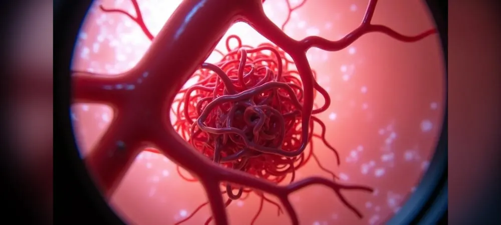
The answer to the question of what AVM disease is points to a vital vascular problem. This disease consists of lesions formed by the direct connection of arteries and veins when a capillary network fails to develop between them for an unknown reason. This condition, which carries a risk of brain hemorrhage if left untreated, can lead to serious health problems.
Research shows that bleeding occurs in 2-4% of AVMs every year. Headache, in particular, emerges as the most common symptom of this disease. Also known as a vascular tangle, this condition occurs with the formation of a blood vessel tangle in the vessels, although the exact cause is not fully known. In this article, we will share with you what AVM disease is, its symptoms, diagnostic methods, and treatment options in clear language.
What is AVM disease and how does it form?
Arteriovenous malformation (AVM) is a complex vascular structural disorder consisting of abnormal connections between arteries and veins. As a significant anomaly in the circulatory system, this condition is most commonly seen in the brain and spinal cord, although it can also appear in different parts of the body. While AVM disease is mostly congenital, it can in some cases develop later in life.
What is a vascular tangle?
AVM, known as a vascular tangle, forms when arteries and veins, which normally should function separately, connect directly with each other without capillaries. In a healthy circulatory system, arteries transport oxygen-rich blood to organs, capillaries allow this blood to be used by tissues, and veins send oxygen-depleted blood back to the heart. However, in the presence of an AVM, this order is disrupted.
In this abnormal structure, the vessels form a tangle resembling a bird's nest, and the blood flow passing through it is much greater and faster than in a normal vessel. Vessel walls are structurally weak and fragile. Because high-pressure blood flow causes thinning in these weak vessel walls, the vascular tangle may not withstand the pressure created by the blood passing through it and can rupture at its weakest point.
What is AVM disease in the brain?
AVM disease in the brain is a congenital vascular anomaly thought to result from the faulty development of cerebral vessels in the womb. Causing a significant disruption in the brain's feeding system, this condition means the absence or lack of capillaries that should normally exist between arteries and veins.
Normally, oxygen- and nutrient-rich blood arriving at the brain passes from arteries to capillaries and then to veins to complete its circulation. However, in AVM disease, this transition is disrupted, and blood bypasses the capillaries, passing directly from arteries to veins. Brain AVMs are a rare condition occurring in about 1 out of every 5000 people (1/5000).
AVMs in the brain usually consist of a vascular tangle called a nidus and one or more feeder arteries supplying blood to this tangle. This structure can "steal" the blood supplying the surrounding brain tissue, leading to inadequate nourishment of that region.
Effects of AVM on the body
AVM disease primarily causes significant changes in blood flow. Because high-pressure blood coming from the arteries passes directly to the veins without capillaries, excessive pressure builds up on the veins. This situation leads to weakening and dilation in the vessel walls.
One of the most important effects of AVM is the risk of hemorrhage. Because the vessels forming the vascular tangle are much weaker than a normal artery, they can rupture at any time and cause severe bleeding. Especially in brain AVMs, this condition can lead to life-threatening brain hemorrhages. Brain AVMs carry a risk of bleeding of approximately 2% each year.
Additionally, AVM prevents surrounding tissues from receiving adequate oxygen. Because blood passes directly to the veins, tissues cannot be nourished and may lose function over time. When oxygen is insufficient, brain tissues weaken or may die completely. This condition can cause various neurological problems depending on the region where the AVM is located.
Nevertheless, although AVM disease is congenital, in the vast majority of patients, the first symptoms appear between the ages of 20 and 40. In some patients, it may show no symptoms at all and can be discovered incidentally during brain imaging examinations performed for other reasons.
Causes of AVM disease
AVM disease is generally considered one of the mysterious subjects of the medical world because a clear cause for the formation of this disease has not yet been fully explained scientifically. However, scientific research offers some clues.
Congenital causes
Medical experts generally accept that AVM disease is a congenital condition. It is thought to arise as a result of errors occurring during the formation process of brain vessels in the womb during the development of the baby, particularly in the embryonic period.
Under normal conditions, blood vessels form within a specific program during fetal development. The failure of the capillary network that should exist between arteries and veins to develop healthily leads to the disruption or absence of this structure. This situation causes blood flow to become unbalanced through direct connections.
Researchers believe that AVM occurs as a result of the incorrect formation of vessels in the brain tissue during fetal development in the womb. Normally, your heart sends oxygen-rich blood to the brain via arteries, and this blood slows its flow by passing through increasingly narrowing vessel networks. However, in AVM, this process is disrupted, and blood passes directly from arteries to veins.
Acquired conditions
Although arteriovenous malformations are mostly congenital, in some cases they can also develop in adulthood. Traumatic AVM disease can rarely be seen as a result of post-traumatic brain injuries.
Also, though rare, acquired AVM-like structures may form in the following situations:
Head trauma
Infections
Vascular inflammation
However, structures formed later in this manner exhibit different characteristics from classic congenital AVMs. Maternal exposure to radiation during pregnancy, use of medications without a physician's recommendation, various hormones taken through foods, and certain viruses can be counted among risk factors.
Is it hereditary?
AVM disease is generally not a hereditary disease. That is, the risk of this disease occurring in the children of a brain AVM patient is not higher than in other people. In other words, having someone with AVM in the family does not increase the likelihood of other family members having it.
However, a predisposition to AVM formation may occur in some rare genetic syndromes. For example, a genetic disorder called Hereditary Hemorrhagic Telangiectasia (Osler-Weber-Rendu disease) can increase the risk of vascular malformations in the brain or other organs.
Scientific studies show that genetic factors may play a role in the formation of AVM at a rate of approximately 10-15%. However, AVM can develop in many people who have no history of any similar disease in their family. Research shows that a set of genes and potential environmental factors are effective in the formation of the malformation in question.
The frequency of AVM in males is higher than in females. Furthermore, although the presence of a family history is considered a risk factor in some cases, it has not been fully determined whether this is coincidental or a true genetic connection.
In conclusion, AVM is a vascular formation disorder that generally develops outside the individual's control and during the prenatal period. Although the exact cause is not known, problems related to development in the womb are thought to be the most likely cause.
What are the symptoms of AVM disease?
AVM disease manifests different symptoms depending on the affected area. Interestingly, in about half of the patients, the first symptom appears as a brain hemorrhage and requires urgent intervention. In some patients, however, AVM can remain silently without showing any symptoms for years and can be discovered incidentally during imaging examinations performed for other reasons.
Brain AVM symptoms
The symptoms of AVM disease formed in the brain are quite varied and differ depending on the location. Even when there is no bleeding, the following symptoms may occur:
Headache: It is the most commonly observed symptom. Particularly recurrent and severe headaches are one of the important signs of AVM.
Seizures: Epileptic seizures are one of the frequently seen symptoms of AVM. Sometimes they may also appear in the form of contractions in only a single arm or leg (focal seizures).
Neurological problems: Symptoms such as weakness on one side of the body, numbness, and imbalance can be observed.
Visual disturbances: Visual field losses, blurred vision, or double vision may be experienced.
Speech disorders: Speech difficulty and comprehension difficulties may arise.
In the case of hemorrhage, symptoms suddenly intensify. Sudden and severe headache (described as the most intense pain the person has ever felt in their life), altered state of consciousness, nausea, vomiting, and even conditions progressing to coma may occur. This clinical picture requires emergency intervention.
Brain AVM symptoms can begin at any age, but they mostly appear between the ages of 10 and 40. Its effect accumulates over time and causes symptoms in early adulthood. Upon reaching middle age, brain AVMs generally remain more stable and the likelihood of showing symptoms decreases.
Spinal cord AVM symptoms
The symptoms of AVMs in the spinal cord differ from those in the brain. Primarily, spinal cord AVM symptoms include the following:
Back and lower back pain: Among the most frequent symptoms of AVM located in the spinal cord.
Numbness and weakness in the legs: Spinal cord AVMs can cause loss of sensation and decreased strength in the legs.
Urinary incontinence: It can appear as the loss of bladder control.
Walking difficulty: Balance disorders and instability in gait may occur.
In spinal cord AVMs, there is a risk of paralysis development in the event of sudden hemorrhage. Therefore, spinal cord AVM symptoms must be taken seriously and a specialist should be consulted without losing time.
General body symptoms
AVM can be seen not only in the nervous system but also in other regions of the body and displays symptoms specific to its location:
Tinnitus: Pulsatile tinnitus synchronized with the pulse can be observed, especially in dural AVMs.
Shortness of breath: If the AVM is located in the lungs, shortness of breath may be experienced during exertion.
Abdominal pain: AVMs in the abdominal region may cause pain.
Coughing up blood: A symptom that can be seen in AVMs in the lungs.
Thickening in the arm and leg: Swelling and thickening can occur in the affected limb in body AVMs.
Skin discoloration: Changes in skin color may be observed at the site where the AVM is located.
An important point is this: In some pregnant women, AVM symptoms may worsen during pregnancy due to changes in blood volume and blood pressure.
The absence of any symptoms does not mean that the AVM carries no risk. Quietly progressing AVMs can also bleed at an unexpected moment. Therefore, diagnosed AVM patients should undergo regular check-ups even if they show no symptoms.
How is AVM disease diagnosed?
Doctors utilize various imaging methods when evaluating patients with suspected AVM. These methods enable a detailed examination of the vascular structure, confirming the presence of the disease and guiding the treatment plan. There are four main methods used in the diagnosis process, and each offers different advantages.
Magnetic Resonance (MR)
Magnetic Resonance imaging is the primary method resorted to in AVM diagnosis. MR clearly images the internal structures of the body using radio waves and a magnetic field. Especially in contrast-enhanced MR examinations, the vascular tangle (nidus) structure can be easily distinguished.
In the diagnosis process, MR angiography is usually added to the MR examination. Thanks to this additional test, the vascular malformation can be identified with high accuracy. The MR angiography test is often performed without injecting any medication into the patient and takes about 15-20 minutes.
MR provides detailed information about brain tissue, vascular structures, and traces of hemorrhage. Additionally, by showing the exact location of the AVM in the brain, it makes a major contribution to treatment planning.
Computed Tomography (CT)
Computed Tomography is preferred especially in patients with suspected hemorrhage due to providing rapid results. CT uses a series of X-rays to create detailed cross-sectional images of the brain.
In some cases, CT Angiography can also be performed to increase diagnostic accuracy. In this procedure, a contrast dye is used to make the vessel visible, allowing the arteries supplying the arteriovenous malformation to be seen in more detail. CT angiography can accelerate treatment decisions, especially in acute hemorrhage cases.
Because a CT scan clearly shows acute hemorrhage, it is a preferred diagnostic method in emergency situations. However, it does not provide as detailed soft tissue differentiation as MR.
Catheter Angiography
Catheter angiography or Digital Subtraction Angiography (DSA) is accepted as the "gold standard" method in AVM diagnosis. This method both gives certainty to the diagnosis and provides the most appropriate information for treatment planning.
During the procedure, the doctor places a long, thin tube (catheter) into an artery in the groin and advances the catheter to the brain vessels under X-ray imaging guidance. Then, dye is injected into the vessels to make them visible under X-rays.
With catheter angiography, the entry points of arteries, direction of flow, map of the vascular tangle, and structure of the draining veins are imaged in the most detailed manner. Thus, the most accurate and appropriate treatment method for the patient can be determined. Today, 3-dimensional images of vessels can be obtained thanks to angiography devices equipped with advanced digital technology.
Transcranial Doppler Ultrasound
Transcranial Doppler (TCD) is a non-invasive ultrasound technique used to measure blood flow velocity in major brain arteries. Using sound waves, it provides real-time data regarding the movement of blood in brain vessels.
This method is used to detect abnormal blood vessel connections in AVMs. TCD is a diagnostic method that can be easily applied at the bedside, is repeatable, and poses no risk to the patient.
In the evaluation report published by the American Academy of Neurology, it is stated that the sensitivity of TCD in diagnosing circulatory arrest and brain death is 91-100%, and its specificity is 97-100%. This shows that the method is quite reliable.
TCD provides great benefits in evaluating cerebral blood flow, especially in critically ill patients. Additionally, because it is an inexpensive, non-invasive, and easy-to-use method, it is also widely used for AVM screening.
All these imaging methods performed for AVM diagnosis are of great importance in determining the exact location, structure, and characteristics of the disease. Accurate diagnosis is vital for the formulation of an effective treatment plan.
How is AVM disease treated?
AVM treatment is planned individually and may require different approaches for each patient. The primary goal in treatment is the complete elimination of the vascular tangle or minimization of the risk of hemorrhage. The treatment decision is made by a multidisciplinary team, considering the size and location of the AVM, bleeding history, and the patient's general condition.
Surgical intervention
Microsurgical treatment is accepted as the primary option in AVM disease. In this method, definitive and rapid treatment is provided by completely removing the vascular tangle. It is ideal especially for vascular tangles of appropriate size located in superficial and silent regions of the brain. During surgery, microscopic techniques are used to reach the vascular tangle through an opening made in the brain and it is removed from the body.
During the procedure, the vessels feeding the vascular tangle are first controlled, and then the AVM is completely removed. Neuromonitoring is used to avoid damaging healthy brain tissues during surgery. When performed by an experienced neurosurgeon, it has high success rates. Full recovery at a rate of 98.2% has been achieved with surgical treatment in 38.5% of Spetzler–Martin Grade I AVMs and 27.2% of Grade II AVMs.
Endovascular Embolization
Endovascular embolization is the procedure of closing AVM vessels by entering through a groin vessel. In this method, a catheter is placed into the patient's groin artery and the lesion area is reached under angiography guidance. Then, depending on the type of lesion, occlusive materials (coils, embolizing particles, vascular adhesives) are placed inside the vessel.
This treatment is usually not sufficient on its own and is used to make surgery safer or to reduce AVM size before Mr Linac treatment. Following embolization, patients can usually be discharged on the same day, the recovery period is short, and they experience less pain. It is a method that can be preferred especially in patients with coexisting health problems.
Medication treatment
Today, it is not possible to completely cure AVM disease with drug treatment. Medications are used only to alleviate symptoms such as headache and epileptic seizures. In patients experiencing epileptic seizures, seizure-preventing medications are used to try and keep the patient under control.
Applied in addition to active treatment methods such as surgery, Mr Linac, or embolization, drug treatment helps in managing symptoms. However, it cannot correct the structural disorder caused by the vascular tangle and does not eliminate the risk of hemorrhage.
Conclusion
In conclusion, AVM disease is a serious health problem consisting of abnormal connections between arteries and veins. This congenital condition is most commonly seen in the brain and spinal cord, although it can also appear in different parts of the body. Its main symptoms include headache, seizures, neurological problems, and visual disturbances, but some patients may show no symptoms at all.
Imaging methods such as MR, CT, catheter angiography, and transcranial doppler ultrasound are used in AVM diagnosis. Thus, the location, size, and structure of the vascular tangle can be determined. Different treatment approaches may be required for each patient. Among options such as surgical intervention, the Mr Linac treatment method, or endovascular embolization, the one most appropriate for the patient's condition is chosen.
AVM disease, which can lead to serious health problems like brain hemorrhage when left untreated, can be brought under control when diagnosed early and treated appropriately. Therefore, it is of vital importance for those experiencing symptoms such as headache or seizures to consult a specialist without wasting time. It should not be forgotten that every AVM case is unique, and the treatment plan must be prepared specifically for the individual.
Today, thanks to developments in the medical field, the treatment options offered to AVM patients are increasing steadily. Especially advancements in surgical techniques and the widespread adoption of minimally invasive methods have raised treatment success and accelerated the recovery process. Therefore, it is of great importance for patients diagnosed with AVM to remain hopeful and under regular follow-up with an expert team.
AVM disease stems from abnormal connections between arteries and veins. Learn about its symptoms, diagnostic methods, and treatment options.
Start with the right assessment for What is AVM Disease? What Are Its Symptoms? How Is It Diagnosed and Treated?
Share your symptoms with our specialist physicians; let us determine the right diagnosis and treatment plan together.
Warning
If your symptoms started suddenly, are progressing rapidly, or significantly restrict your daily life, consult a physician without delay.
Plan your What is AVM Disease? What Are Its Symptoms? How Is It Diagnosed and Treated? journey with our specialists.
Fill in the form or book an appointment to learn about treatment options, the process and preparation.