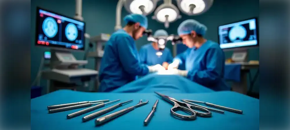
Microsurgical techniques play a very important role in the treatment of brain tumors and allow tumors to be removed with the greatest precision. Microsurgery not only makes surgery safer, but also significantly shortens the length of hospital stay and the time needed to return to normal life. This technique aims to remove the tumor completely while causing minimal damage to brain tissue.
In brain tumors, microsurgery increases your chances of survival and significantly improves your quality of life. The ability to work with high precision is one of the greatest advantages of microsurgery. By reducing the risk of damage to surrounding tissues, it allows the maximum possible amount of tumor to be removed. In this article, we will explain in clear language what microsurgery is, how it is applied in brain tumors, and the advantages it provides.
What Is Microsurgery in Brain Tumors?
The use of microsurgery in neurosurgery has significantly increased the success rate of delicate and complex operations. In this section, we will examine in detail what microsurgery is, its role in brain tumors, and the special instruments used.
What is microsurgery?
Microsurgery is the name given to delicate surgical procedures performed using high-magnification microscopes. This technique allows surgeons to work on very small structures that are difficult to see with the naked eye. Microsurgery is particularly successful in operations on extremely complex and delicate organs such as the brain.
Microsurgery makes it possible for the surgeon to work under a microscope and operate in extremely delicate and small areas. In operations performed with this method, the surgeon aims to remove the tumor completely while causing minimal damage to brain tissue. As a result, the surgical success rate increases and the patient's recovery process is faster.
The role of microsurgery in brain tumors
Brain tumors are a highly diverse group of diseases and usually present with various symptoms, such as signs related to increased intracranial pressure (headache, nausea, vomiting), partial or complete paralysis that varies according to the location of the tumor, and seizures (epileptic seizures, epilepsy). Microsurgery is an indispensable component in the treatment of brain tumors.
The greatest advantage of microsurgery in the treatment of brain tumors is that it makes it possible to work with high precision. Because brain tissue is extremely complex and delicate, the surgeon's margin for error is very small. Thanks to microsurgery:
- Targeted intervention focuses only on the tumor area, and healthy brain tissue is preserved
- Because there is a lower risk of bleeding, the patient recovers faster
- Patients can usually return to their normal lives in a shorter time
In addition, this method makes it possible to work without damaging the vital nerve and vessel structures surrounding the tumor. Reaching the brain tumor tissue, separating the tumor from the surrounding neural and vascular structures, and removing as much tumor tissue as possible with as little damage as possible form the basis of this treatment.
Use of the microscope and special instruments
The vast majority of operations in neurosurgery are performed under a surgical microscope. The microscope delivers the image of the area to be operated on to the surgeon under focused light, magnified 6, 12 or 25 times. In this way, the surgeon can see details that normally cannot be seen with the naked eye.
The surgical microscope greatly magnifies the area where the surgeon will work, allowing details to be seen clearly. Equipped with high-resolution optical lenses, these microscopes can magnify from 6x up to 40x. This visual support is indispensable for distinguishing and removing only the tumor area without disturbing the natural structure of the brain tissue.
The special instruments used in microsurgical procedures include brain spatulas, dura dissectors, nerve root retractors and micro brain knives. These instruments are extremely precise and fine, allowing safe work in delicate tissues such as the brain.
Today, for some special tumor types (especially tumors of the pituitary region), treatment can be performed through the nose, without opening the skull, with the help of a microscope and an endoscope. This is another important advantage offered by microsurgery.
Advantages of Microsurgery
Microsurgery, one of the most important advances in modern medicine, provides many benefits to the patient in brain tumor treatment. Performed with an enlarged field of view and delicate instruments, this surgical procedure offers far more advantages than conventional methods.
Precision and targeted intervention
The greatest advantage of the microsurgical method is that it makes it possible to work with high precision. Because brain tissue is extremely complex and delicate, the surgeon's margin for error is very small. Thanks to this technique, the risk of damage to surrounding tissues is reduced and the maximum possible removal of the tumor is achieved.
With microsurgery, the focus is only on the tumor area and healthy brain tissue is preserved. This targeted intervention is preferred especially for tumors located in delicate regions. It also makes it possible to work without damaging the vital nerve and vessel structures surrounding the tumor.
Neuronavigation systems used during the operation allow the surgeon to see the tumor in real time. As a result, the risk of surgical error is significantly reduced.
Less bleeding and tissue damage
Microsurgical procedures are minimally invasive. This allows surgery to be performed through a smaller incision. Thanks to small incisions:
- Less scarring occurs
- The risk of infection decreases
- Pain after surgery is reduced
- Less damage occurs in brain tissue
The risk of bleeding is lower in operations performed with the microsurgical method. Thanks to the enlarged field of view, the surgeon can see and protect the blood vessels more clearly. As a result, the patient recovers faster.
Short recovery time
Compared with conventional neurosurgery, patients can return to daily life sooner after microsurgery. The procedure being less invasive and damage to healthy tissue being kept to a minimum are factors that speed up the recovery process.
After surgery, discharge is planned following an average of 3 days of follow-up on the ward. In patients without wound problems, the stitches are removed after 10–14 days. Particularly in cases where minimally invasive methods are used, because they involve a smaller incision and less disruption of brain tissue, there is less risk, less discomfort and a shorter recovery period compared with classical surgery.
Reducing the risk of recurrence
One of the most important advantages of microsurgery is that it makes it possible to remove the tumor completely. Removing the entire tumor significantly reduces the risk of the disease recurring.
In meningiomas in particular, the main goal of surgical treatment is complete removal of the tumor. When complete resection is achieved in Grade 1 cases, the risk of recurrence is quite low, and the patient can continue his or her life without problems with long-term follow-up.
Advanced imaging techniques such as intraoperative MRI, ultrasound (USG) and CT help doctors gain better control over the delicate structures surrounding the tumor by creating a real-time map of the brain anatomy during surgery. In addition, special filters used in the microscope make it possible to check during surgery whether any tumor tissue remains. Thus, the tumor is completely removed before the patient wakes up from the operation.
Advanced Technologies Used in Microsurgery
Technological advances have been made available to surgeons to increase the success of microsurgical operations. Thanks to these technologies, safer and more effective operations have become possible for both patients and surgeons.
Preoperative 3D modeling
Preoperative 3D modeling creates a three-dimensional model of the patient's brain structures, allowing the surgeon to plan the operation in advance. This technology clearly visualizes the vessels, nerves and healthy brain tissue around the tumor. Thus, before the operation, the surgeon can simulate how to remove the tumor in the safest and most effective way.
Neuronavigation system
Neuronavigation is a technology that enables the surgeon to find his or her way within the internal structures of the brain with millimetric accuracy during surgery. This system generally works in a way similar to a GPS system; it shows the surgeon the location of the tumor and the position of the surgical instruments in real time. By determining the exact location of the tumor within the brain with 1 mm precision, it allows the surgeon to proceed during the operation without damaging surrounding tissues.
Neuromonitoring
Neuromonitoring makes it possible to continuously monitor the patient's neurological functions during brain surgery. This technology helps the surgeon minimize the risk of damaging nerve pathways or important brain regions. Even the slightest change in the nervous system during the operation is noticed immediately, and the surgeon can take the necessary measures.
Intraoperative angiography
Intraoperative angiography is an imaging technique used to evaluate the condition of blood vessels during brain tumor surgery. Intraoperative angiography performed using ICG (indocyanine green) dye visualizes the blood flow of the brain's vessels during surgery. In this way, the surgeon can clearly see the vessels around the tumor during the operation and adjust the surgical plan accordingly.
Intraoperative fluorescence
Intraoperative fluorescence is a technology used to make it easier to distinguish tumor tissue from healthy brain tissue. Fluorescent agents such as sodium fluorescein (NaFl) produce a green fluorescent color in tumor tissues under a special filter (FL 560 nm). In studies, NaFl staining has been observed to be effective in all patients with glioblastoma multiforme and meningioma and in 90.4% of metastatic tumors.
Intraoperative ultrasonography
Intraoperative ultrasonography (IOUS) is a technique that provides real-time imaging of brain tissue during surgery. This technology allows the surgeon to see the location and size of the tumor instantly. The use of intraoperative ultrasonography helps determine the surgical resection margins and shows the relationship with surrounding structures; it reduces operative time, blood loss and length of hospital stay while increasing gross total resection rates.
The Brain Tumor Surgery Process with Microsurgery
Microsurgery in brain tumor treatment is a comprehensive process that extends from the planning stage to long-term follow-up. This process requires an individualized approach for the patient and includes steps that must be carried out meticulously at every stage.
Preoperative planning
The microsurgical treatment process begins with detailed planning. In the first step, the patient's general health status is evaluated, and the location, size and type of the tumor are determined. Using imaging techniques (such as MRI and CT), the location of the tumor and the surrounding structures are clearly identified. At this stage, strategies to be followed to preserve brain functions are also planned.
The neurosurgeon requests the necessary imaging tests and examines the location, structure and behavioral characteristics of the tumor in detail. Before the operation, your doctor will inform you about which medications you should or should not take and will also tell you the required fasting period before anesthesia.
Stages of the surgical procedure
A microsurgical operation generally consists of the following stages:
- Patient preparation and anesthesia: The procedure is performed under general anesthesia; since the patient is asleep, there is no pain or awareness
- Opening of the skull: The scalp is incised in a way appropriate to the tumor location, and then a section of the skull is opened to gain access to a specific area
- Removal of the tumor: The surgeon carefully removes the tumor using a microscope and delicate surgical instruments
- Inspection and control of bleeding: After the tumor is removed, the area is checked and any bleeding is stopped
- Replacement of the skull bone and closure
Risks and complications
As with every surgical procedure, microsurgery carries some risks. These include bleeding, infection, brain edema, and temporary or permanent impairment of neurological functions. However, when the advantages of microsurgery are taken into consideration, these risks are minimized.
Postoperative recovery process
After surgery, patients are usually closely monitored in the intensive care unit for the first 24 hours. They are then transferred to the ward and begin to move first with assistance and then without assistance. After an average of 48–72 hours of ward follow-up, the patient becomes ready for discharge. In patients who do not develop complications, discharge can be achieved within a total of 4 days.
The stitches are usually removed on the 10th day. Headache or discomfort experienced during this period is normal and can be controlled with the medications recommended by your doctor.
Long-term follow-up and outcomes
Long-term results after microsurgery depend on the type and stage of the tumor and the success of the surgical intervention. Regular check-ups after surgery are critically important for detecting possible recurrences or newly developing complications at an early stage.
The patient's condition is monitored with regular MRI and CT check-ups. After brain tumor surgery, most people show functional improvement compared with before the operation. However, for patients who experience functional loss, comprehensive and individualized physical therapy programs are recommended in the early period.
Success Rates and the Future of Microsurgery
Microsurgical techniques offer high success rates in the treatment of brain tumors. So what lies behind this success? The outcomes of microsurgical procedures performed in brain tumors vary depending on various factors. In this section, we will examine the factors that affect the success of microsurgery and future developments.
Factors affecting success
The success rate of the microsurgical method is directly related not only to the surgical procedure itself but also to many critical factors that influence the process. Factors such as the location, size, type and aggressiveness of the tumor are important elements affecting success. In addition, the patient's lifestyle and general health status also determine the results.
The success of microsurgery requires a multidisciplinary approach, and every detail can directly affect the outcome of the operation. Whether the tumor can be removed completely, whether the patient has few complications in the postoperative period, and the patient's response to treatment determine the success rate.
Microsurgery is preferred especially for small and medium-sized tumors or tumors located in delicate regions. However, it can also be used successfully in large tumors. It should be remembered that some tumor types may be more aggressive and difficult to treat.
Surgeon's experience and quality of equipment
Because microsurgery is an extremely delicate procedure, the surgeon's manual skill and experience are decisive. For surgical success, it is important that the surgeon:
- Has received special training in microsurgery
- Has demonstrated a high success rate in similar cases
- Has long-term case experience
The technological infrastructure of the center where the operation will be performed also plays a major role in the success of microsurgical treatment. Using the right equipment is the foundation of success. High-resolution microscopes, meticulous hand instruments and advanced suture materials are of vital importance.
With the advancement of modern medicine, the success of microsurgery has increased manyfold with technological support. The neuromonitoring system informs the surgeon instantly by preventing damage to brain and nerve tissues during the operation. Thanks to this early warning system, the surgeon proceeds more safely or changes strategy.
Future developments
Robotic technology holds significant promise for the future of microsurgery. Robot-assisted microsurgery eliminates hand tremor, precisely guides millimetric movements and can distinguish complex tissue structures more clearly. In addition, artificial intelligence and genetic advances are determining the course of neurosurgery.
Genetic research is becoming increasingly important in the diagnosis and treatment of brain tumors. Tumors can now be defined not by their appearance under the microscope but by their genetic characteristics. In this way, how long and how a patient will live can be predicted.
Innovations such as nanopore technology analyze the DNA of the tumor within minutes and contribute greatly to tumor typing. In addition, methods such as next-generation sequencing and methylation profiling are replacing standard diagnostic and treatment methods. Thanks to these, microsurgery is becoming both more successful and safer.
In Summary: Microsurgery in Brain Tumors
In conclusion, microsurgical techniques stand out as a groundbreaking development in the treatment of brain tumors. Thanks to microscopes providing high magnification and delicate instruments in particular, surgeons can see and operate on structures that cannot be seen with the naked eye. Thus, it becomes possible to remove tumors without damaging surrounding tissues.
The advantages provided by microsurgery are quite clear. First of all, this method, which offers the ability to work with high precision, aims to remove the entire tumor while protecting healthy brain tissue. It also provides important benefits such as a lower risk of bleeding and a faster recovery process. In addition, reducing the risk of recurrence is among the valuable contributions offered by this technique.
Today, microsurgery is supported by advanced technologies such as 3D modeling, neuronavigation and intraoperative imaging. Thanks to these developments, preoperative planning can be done in greater detail, and during surgery it is possible to proceed with millimetric precision.
The success of your treatment is directly related not only to the characteristics of the tumor but also to the experience of your surgeon and the quality of the equipment used. For this reason, it is important to choose experienced surgeons who specialize in microsurgery and centers with a strong technological infrastructure for brain tumor treatment.
It is anticipated that microsurgical techniques will develop even further in the future with the support of robotic systems and artificial intelligence. These developments mean higher success rates and fewer complications in brain tumor treatment.
Remember that early diagnosis and the right treatment approach are the most critical factors in brain tumor treatment. Microsurgical techniques not only increase your chances of survival but can also significantly improve your quality of life after surgery. An individualized treatment plan for each patient is indispensable for achieving the best result.
Frequently Asked Questions
We answer your questions about microsurgery in brain tumor treatment.
What is microsurgery and how does it work?
Microsurgery means performing the operation under a microscope. With this method, the area being operated on can be seen in great detail and very fine-tipped instruments can be used. In this way, the procedure is performed without damaging the surrounding healthy tissues.
When is microsurgery preferred in brain tumors?
It is preferred especially for small and medium-sized tumors or tumors located in delicate regions. However, it can also be used successfully in large tumors.
How long does a microsurgery operation take?
Brain surgeries take an average of 3–6 hours. Approximately 2–3 hours of this is the active surgical procedure. The remaining time is related to anesthesia, preparation and waking up.
How long does the recovery process take?
Discharge is planned after an average of 3 days of ward follow-up. Generally, 1 month of rest at home is recommended. You can return to jobs that do not require physical exertion, such as desk work, within 2–3 weeks.
What is the risk of recurrence after microsurgery?
When the tumor is completely removed, the risk of recurrence is low. In meningiomas, the 10-year recurrence rate is around 9%, whereas the recurrence rate is higher in malignant tumors such as glioblastoma.
What is the difference between microsurgery and classical surgery?
Microsurgery is performed through a smaller incision, recovery is faster, and the risk of infection is lower. The patient can be helped to stand up on the day of surgery and can be discharged in 2–3 days.
What should be paid attention to after microsurgery?
You should not bathe for 3 days after surgery, you should take care to use your medications regularly, and you should avoid excessive physical activity. In addition, you should not drive for at least 2 months and should abstain from sexual intercourse for 1 month.
Can microsurgery be applied to every brain tumor?
It may not be suitable for every brain tumor. The most appropriate treatment method is determined according to the location, type and size of the tumor.
Microsurgery enables tumors to be removed safely in the treatment of brain tumors by using delicate surgical techniques. This method shortens the recovery time and improves quality of life.