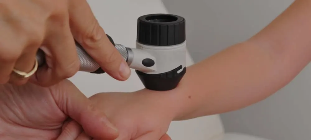
Hemangioma
Hemangiomas, also known as vascular birthmarks, are benign tumors caused by abnormal blood vessel growth. As the most common benign tumors of infancy, hemangiomas usually disappear on their own after growing for a certain period. However, they may persist and continue to grow. Sometimes these benign tumors can bleed or cause open sores. In such cases, it may be necessary to intervene with treatment methods such as various medications, creams, or laser therapy.
What is Hemangioma?
Hemangiomas are benign tumors that occur as a result of the abnormal growth and dilation of blood vessels for any reason. Hemangiomas typically appear red or purple in people with fair skin tones, and brown in those with dark skin tones. They are also known as the most common benign vascular tumors in infants. According to studies, hemangiomas are found in approximately 4-5% of newborns. However, the occurrence of hemangiomas in adults is also a common condition. Some research shows that 3 out of every 4 adults over the age of 75 have a hemangioma.
Hemangiomas, which are also commonly referred to by different names such as vascular birthmarks or vascular tangles among the public, are classified into different types based on characteristics such as size, appearance, and location on the skin. Accordingly, the main types of hemangiomas are:
Capillary hemangioma: Capillary hemangioma is the most common type of hemangioma. It is formed by the clustering of numerous small capillaries. Infantile hemangiomas, which are common in infants and also called strawberry hemangiomas, and cherry hemangiomas seen in adults, fall into this category. Because they are close to the surface of the skin, capillary hemangiomas usually appear bright red.
Cavernous hemangioma: Cavernous vascular birthmarks, on the other hand, consist of larger vascular spaces. There are wide blood-filled spaces between these vessels. They can cause various bleeding and clotting problems. Cavernous hemangiomas usually develop around the eyes and in the deeper layers of the skin. This condition can lead to vision problems such as glaucoma, cataracts, and amblyopia (lazy eye).
Additionally, some hemangiomas may exhibit both capillary and cavernous characteristics. Hemangiomas incorporating features of both types are called combined hemangiomas.
What Are the Symptoms of Hemangioma?
Hemangiomas may not cause any complaints when they appear or thereafter. However, certain symptoms may be observed depending on their location and size. Especially if the hemangioma is large, located in a sensitive area (such as the genital region), or if there are multiple hemangiomas, certain symptoms may arise. Hemangiomas on the skin usually start as small red scratches or blister-like swellings. As they grow, they look like a birthmark turning from dark red to burgundy. Due to this appearance, they are sometimes called "strawberry hemangiomas". When close to the skin surface, they can be bright red, and deeper down, they can be purple or bluish.
Hemangiomas generally range in size from 0.64 cm to 5.1 cm. A hemangioma may occur in a single place on the body, such as a single leg, or they may appear in clusters on the body. Although skin hemangiomas are usually painless, they can sometimes become tender.
Accordingly, the main symptoms of hemangiomas are:
Red, purple, or bluish spots on the skin,
A raised bump on the skin surface,
Narrowing of the visual field or drooping of the eyelid if there is a hemangioma on the eyelid.
Hemangiomas can also develop in internal organs without showing any symptoms. The liver is the most common organ for the formation of liver hemangiomas. The exact cause of liver hemangioma formation is unknown. Hemangiomas generally do not affect liver functions.
However, if the hemangioma in an organ is large or multiple in number, it can adversely affect the function of the organs.
What Causes Hemangioma?
Although the exact cause of hemangiomas is unknown, studies indicate that certain factors may increase the risk of hemangioma. Accordingly, the main risk factors for hemangioma are:
Age: Hemangiomas are generally seen in newborns. Hemangiomas are more common in infants, particularly between the ages of 2 months and 2 years.
Premature birth: Studies show that vascular birthmarks are more frequent in premature infants.
Gender: Although the exact reason is not known, studies show that the risk of developing hemangiomas is higher in females compared to males.
Fair skin: Hemangiomas are more frequently encountered in fair-skinned infants.
Additionally, it is thought that in babies who do not receive enough oxygen while in the womb, excessive blood vessels form in certain areas of the body, leading to hemangiomas.
How is Hemangioma Diagnosed?
Since hemangiomas are spots visible on the skin surface, diagnosis can mostly be made easily with a physical examination. Raised, red, purple, or brown formations seen on the skin provide doctors with important clues about hemangiomas. These lesions, which are particularly common in infants, appear within a few weeks after birth and can start growing rapidly. Doctors can easily make a diagnosis most of the time by looking at characteristics such as the growth rate, color, and shape of these formations.
Hemangiomas sometimes appear in organs such as the liver, kidneys, or brain. In such cases, imaging methods such as ultrasonography, magnetic resonance imaging (MRI), or computed tomography (CT) may need to be used. As a result of the tests performed, if doctors detect a suspicious formation, they may decide to perform a biopsy (taking a small tissue sample). In most patients, organ hemangiomas may also be detected incidentally during examinations performed for another reason.
How is Hemangioma Treated?
Hemangioma treatment may vary depending on factors such as the type, size, location of the hemangioma, or whether it causes any complaints. Under normal conditions, most small hemangiomas disappear on their own over time without causing any problems. However, the baby needs to be followed up regularly and checked for any abnormal growth. Since hemangiomas benefit most from treatment during the first year of life, they should be closely monitored and treated without delay.
Some hemangiomas, however, can grow rapidly and cause various symptoms ranging from vision and hearing problems to digestive system issues such as nausea and loss of appetite, depending on where they form. Additionally, though rare, problems such as ulceration and bleeding can develop on top of hemangiomas. Furthermore, hemangiomas seen in areas like the face can be cosmetically distressing.
The first option in treatment is a medication called propranolol. It is a medication available in syrup form for infants. Before starting treatment, imaging with ultrasound should be performed to determine if there are hemangiomas in the liver and inside the head. Furthermore, a pediatric cardiology examination is recommended before starting the medication.
Most of the time, treatment methods such as medication therapy, topically applied gels, or laser therapy for superficial lesions can help shrink the lesion. In addition, in resistant cases, techniques such as sclerotherapy or embolization can be used to cut off the blood flow going to the hemangioma, ensuring the lesion shrinks. For larger and deeper vascular birthmarks, surgical removal of the hemangioma may be necessary.
Frequently Asked Questions
Although hemangiomas affect people of all age groups, they are generally more common in infants. Studies show that approximately 4-5% of newborns have a hemangioma. They are more frequently encountered, especially in premature babies and those with low birth weight. Furthermore, the risk of developing hemangiomas is higher in fair-skinned babies and females. Whether a hemangioma is evident at birth varies depending on its type. While congenital hemangiomas are evident at birth, infantile hemangiomas, which are the most common type of hemangioma, are usually not seen at birth and appear as a small redness or spot within 1 to 4 weeks after birth. Especially small hemangiomas can resolve on their own without requiring any treatment. However, this is not the case for every patient and every type of hemangioma. Therefore, it is necessary to consult a pediatric hematology specialist to determine whether the hemangioma in your baby requires treatment. Hemangiomas are benign vascular tumors and do not carry a risk of turning into cancer. Although hemangiomas are formed by the excessive growth of blood cells, this growth is neither uncontrolled nor harmful. Small hemangiomas can heal on their own over time without requiring treatment. Hemangiomas around particularly sensitive areas such as the face, ears, nostrils, and perianal region should be treated promptly without wasting time. Moreover, since the greatest benefit from treatment is achieved in the first year of life, infants should be closely monitored by a pediatric hematology specialist from birth. Hemangiomas progress in two phases in infants. In the proliferation phase, which is the first phase, the hemangioma grows rapidly, and then it begins to shrink. This stage, also called involution, usually begins when the baby is 12-18 months old and can take years to complete. Laser therapy can be effective, especially for superficial, small, and skin-close hemangiomas. However, laser may not be sufficient in treating deeper-seated hemangiomas. Hemangiomas are benign tumors, meaning most of them are not dangerous. However, depending on their location, hemangiomas can affect vital functions such as vision, breathing, or feeding. Additionally, hemangiomas with ulcerations on them may carry a risk of infection. Hemangiomas can grow rapidly, particularly in the first year of life. Once hemangiomas appear, they usually regress and disappear over time. In most cases, they do not recur in the same area. For hemangiomas seen in infants, pediatricians refer patients to a pediatric hematology specialist after making an initial evaluation. The decision for treatment, duration, and the type of treatment to be administered are determined by the pediatric hematology specialist. Resistant cases and patients who do not respond to medical treatment should also be evaluated by dermatology and pediatric surgery specialists.
If you or a loved one has symptoms of hemangioma, consult a specialist physician without delay. You can make an appointment with our Hisar Hospital Pediatric Hematology Department specialists for early diagnosis and correct treatment. Contact us immediately for a healthy future.
Start with the right assessment for What is Hemangioma? Symptoms, Diagnosis and Treatment Methods
Share your symptoms with our specialist physicians; let us determine the right diagnosis and treatment plan together.
Warning
If your symptoms started suddenly, are progressing rapidly, or significantly restrict your daily life, consult a physician without delay.
Plan your What is Hemangioma? Symptoms, Diagnosis and Treatment Methods journey with our specialists.
Fill in the form or book an appointment to learn about treatment options, the process and preparation.