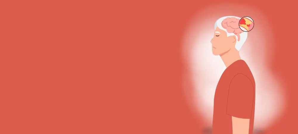
The brain is one of the most vital organs in the body and requires a constant supply of oxygen and nutrients to maintain its functions. This need is met by blood circulation provided through cerebral vessels; however, when an occlusion occurs in one of these vessels, serious health problems can arise. Cerebral major vessel occlusion is a serious condition also known as a "stroke" or "paralysis" among the public, and early diagnosis and treatment can be life-saving.
What is Cerebral Vascular Occlusion?
Cerebral vascular occlusion is a condition where a vessel carrying blood to any part of the brain becomes blocked due to a clot or narrowing. As a result of this occlusion, the brain tissue does not receive sufficient oxygen and glucose, which leads to damage or death of the cells in the affected area.
The occlusion usually occurs in the arteries and is the most common type of stroke (ischemic stroke). In rare cases, a hemorrhagic stroke (brain hemorrhage) caused by a ruptured blood vessel may occur, but this condition is associated with vessel rupture rather than vascular occlusion.
If left untreated, cerebral vascular occlusion can lead to permanent disability, speech and movement disorders, and even death. Therefore, early recognition and treatment of the disease are of vital importance.
What Are the Symptoms of Cerebral Vascular Occlusion?
Symptoms of cerebral vascular occlusion vary depending on the localization, size of the occlusion, and the functions of the affected brain region. Symptoms usually appear suddenly and progress rapidly. Symptoms of cerebral vascular occlusion can be listed as follows:
Motor System Symptoms: Motor system symptoms include weakness or complete paralysis on one side of the body. This condition is usually seen in the face, arm, and leg simultaneously. The patient cannot lift their arm, cannot walk, and one side of their face may droop. Speech disorders are also frequently seen among the symptoms of left cerebral vascular occlusion. Patients may experience difficulty finding words, articulation problems, or a complete inability to speak.
Sensory Symptoms: Sensory symptoms include numbness, tingling, and loss of the sense of touch on one side of the body.
Cognitive Symptoms: Cognitive symptoms include sudden confusion, distractibility, memory problems, and disorientation. Patients may become unable to understand simple commands, recognize familiar people, or know where they are. Balance and coordination disorders, difficulty walking, and a tendency to fall are also characteristic symptoms.
What Causes Cerebral Vascular Occlusion?
The causes of cerebral vascular occlusion are diverse and usually arise from a combination of multiple factors. The causes of cerebral vascular occlusion can be listed as follows:
Atherosclerosis: Atherosclerosis, or hardening of the arteries, is the most common underlying cause. With age, plaque accumulates in the vessels, and these plaques grow over time, narrowing the vessel diameter or breaking off to cause an occlusion.
Heart Diseases: Heart diseases, especially rhythm disorders such as atrial fibrillation, are significant risk factors for embolism formation. Clots formed after heart valve diseases, heart muscle diseases, and previous heart attacks can travel to the cerebral vessels and lead to occlusion. Congenital heart diseases can also be a cause of stroke in young ages.
Vascular Problems: Hypertension, diabetes, high cholesterol, and obesity play an important role among vascular risk factors. These conditions cause damage to the vessel walls and accelerate the development of atherosclerosis. Smoking significantly increases the risk of occlusion by increasing blood viscosity and damaging vessel walls.
Hematological Causes: Hematological causes include blood clotting disorders, polycythemia vera, and blood diseases such as leukemia. Hormone use, especially birth control pills containing estrogen and hormone replacement therapies, can increase the risk of clotting. Pregnancy and the postpartum period are also among the risk factors.
Inflammatory Conditions: Inflammatory conditions, especially vascular inflammation diseases called vasculitis, can cause occlusions in cerebral vessels. Autoimmune diseases such as rheumatoid arthritis and systemic lupus erythematosus are also considered risk factors. Additionally, drug use, especially substances such as cocaine and amphetamines, can lead to vasoconstriction and occlusion.
How Is Cerebral Vascular Occlusion Diagnosed?
Diagnosis of cerebral vascular occlusion begins with clinical evaluation and is supported by emergency imaging methods. The patient's medical history, the onset time, duration, and characteristics of the symptoms are questioned in detail. Activities in the last 24 hours, medication use, and similar past events are particularly important. The following methods are applied for diagnosis:
Neurological examination includes a systematic evaluation of the patients' state of consciousness, motor functions, sensory functions, speech, and visual abilities. Standardized stroke scales such as the NIHSS (National Institutes of Health Stroke Scale) are used to objectively evaluate the severity of the disease.
Brain imaging methods are considered the gold standard in diagnosis. Computed tomography (CT) is the first preferred method because it is fast and widely accessible. With CT, hemorrhage is primarily excluded, and early signs of ischemia are evaluated, although CT may appear normal in the first few hours.
Magnetic resonance imaging (MRI) is more sensitive than CT in showing cerebral vascular occlusion. Diffusion-weighted imaging can show ischemic areas even in the early stages. With MR angiography, vascular anatomy and the localization of the occlusion can be evaluated in detail.
Laboratory tests include blood counts, biochemical analyses, clotting tests, and inflammatory markers. Glucose levels, electrolyte balance, and kidney functions are evaluated as a priority. Electrocardiography and echocardiography are performed for cardiac evaluation.
Cerebral vascular occlusion can also be detected by angiography. Conventional angiography is the most precise method for showing the exact localization and severity of the occlusion. Treatment can also be applied during angiography.
How Is Cerebral Vascular Occlusion Treated?
Treatment of cerebral vascular occlusion is a race against time and requires a multidisciplinary approach. The main goal of treatment is to restore blood flow to the brain tissue and prevent secondary damage. It is handled in two main parts: acute phase treatment and long-term preventive treatment.
Thrombolytic therapy plays an important role in acute phase treatment. Intravenous thrombolysis aims to ensure vessel patency through the use of clot-dissolving drugs. This treatment must be administered within the first 4.5 hours from the onset of symptoms. Patient selection must be careful, and the risk of hemorrhage must be evaluated.
Endovascular treatments are applied as mechanical thrombectomy, especially in large vessel occlusions. The clot is physically removed or aspirated with the help of a catheter. This method can be effective even within 24 hours from the onset of symptoms. Although the success rate of the treatment is high, it should be performed in experienced centers.
Cerebral vascular occlusion can also be treated with stent therapy. A stent is used to keep the narrowed vessel open and is particularly effective in chronic narrowing.
Antiplatelet therapy is used to prevent clot formation. Drugs such as aspirin and clopidogrel are used long-term. Anticoagulant therapy is applied with warfarin or new-generation anticoagulants, especially in cases of cardiac embolism. Treatment for cerebral vascular occlusion is performed by taking into account patient characteristics and the risk of hemorrhage. The recovery time for cerebral vascular occlusion varies depending on the severity of the occlusion, the affected brain region, age, and general health status.
Every second is of vital importance in cerebral vascular occlusion. If you or your relative have even one of these symptoms, you should immediately apply to the emergency department of the nearest hospital.
Frequently Asked Questions
The main risk factors for cerebral vascular occlusion include high blood pressure, diabetes, high cholesterol, smoking, obesity, sedentary lifestyle, excessive alcohol consumption, heart diseases (especially rhythm disorders), and genetic predisposition. Additionally, advanced age, male gender, and stress can increase risk factors. Cerebral vascular occlusion usually manifests itself with symptoms that appear suddenly. The most common signs include facial asymmetry, sudden weakness or numbness in an arm or leg, difficulty speaking, visual disturbances, loss of balance, and severe headache. Cerebral vascular occlusion causes undesirable conditions such as paralysis. When brain cells remain without oxygen due to the occlusion, the functions in the relevant area are lost. This leads to speech, movement, perception, and balance disorders. The risk of permanent damage can be reduced with early intervention. Since such symptoms require emergency intervention, the emergency department should be consulted first. After the diagnosis is made, patients are usually monitored in the neurology department. Neurology specialists should also be consulted for long-term treatment and follow-up. Cerebral vascular occlusion is a condition where time is of the essence. With every passing minute, the risk of permanent damage to the brain tissue increases. If intervention is performed within the first 4.5 hours, significant improvement can be achieved with clot-dissolving treatments. For this reason, 112 emergency services should be called even at the slightest suspicion. The treatment process varies depending on the severity and duration of the occlusion. In the first stage, drugs aimed at dissolving the blood clot are given. In suitable patients, the clot can be removed with mechanical thrombectomy. In the subsequent process, blood thinners, blood pressure and cholesterol-regulating drugs, lifestyle changes, and rehabilitation treatments are applied. In some special cases, especially in large vessel occlusions, cerebral vascular occlusion surgery can be performed. For example, if there is severe narrowing in the neck vessels, the plaque can be cleaned with a surgical procedure called "carotid endarterectomy." Additionally, interventional procedures such as stent placement can also be performed. Not every patient may be suitable for these methods. The risk of vascular occlusion can be significantly reduced by controlling risk factors (blood pressure, sugar, cholesterol), quitting smoking and alcohol, regular exercise, healthy nutrition, maintaining an ideal weight, and regular doctor check-ups. If underlying causes are not treated or lifestyle is not changed, the occlusion may recur. The risk of having a second or third stroke is higher after the first one. Therefore, regular treatment and follow-up are of great importance. Symptoms include facial drooping, numbness or loss of strength in the arm or leg, difficulty speaking and understanding, double vision or vision loss, dizziness, imbalance, and sudden severe headache.
If you or your relative have symptoms of cerebral vascular occlusion (stroke), apply to the emergency department without delay. Early diagnosis and treatment save lives. For more information and appointments, you can contact the Hisar Hospital Neurology Clinic.
Start with the right assessment for Symptoms, Causes, and Treatment of Cerebral Vascular Occlusion (Stroke)
Share your symptoms with our specialist physicians; let us determine the right diagnosis and treatment plan together.
Warning
If your symptoms started suddenly, are progressing rapidly, or significantly restrict your daily life, consult a physician without delay.
Plan your Symptoms, Causes, and Treatment of Cerebral Vascular Occlusion (Stroke) journey with our specialists.
Fill in the form or book an appointment to learn about treatment options, the process and preparation.