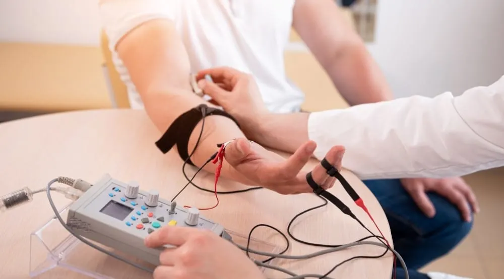
EMG (Electromyography) is a test used to measure the response of a muscle when a nerve stimulates it, as well as the activity (electrical activity) of nerve cells. This diagnostic test is used to detect nerve and muscle diseases. Electromyography is divided into two types: surface EMG and needle EMG. In surface EMG, also known as nerve conduction studies, electrical stimulation pads are attached to the skin over the muscles in order to measure the speed and strength of signals moving between two or more points. Nerve impulses are sent to the muscles and the muscle's response to these stimuli is recorded. In the needle EMG stage, which is performed by inserting needles called electrodes directly into the muscle, the electrical activity within the muscle is measured. The signals are displayed on an oscilloscope (a screen that graphically reflects the electrical wave signal). The data obtained from the electromyography procedure are analyzed by a neurologist to diagnose muscle and nerve disorders.
What Is EMG?
EMG is a medical test that measures the response of a muscle to stimulation by a nerve cell and the electrical potential. If you have persistent complaints such as numbness, tingling, cramps and pain in your muscles, you may need to have an EMG. Electromyography can detect neuromuscular diseases (diseases of the nerves and muscles). It can also be used to determine the loss of function or muscle fatigue in your muscles resulting from excessive exercise. If you have complaints related to your muscles, it is very important to see a doctor and have an EMG without delay, as early diagnosis of nerve and muscle diseases is of great importance.
The first stage of the two-stage EMG procedure is surface EMG. At this stage, the aim is to determine whether the nerve transmits the signal, and if it does, its speed, as well as the strength of the muscle's response to this signal. Surface electrodes are placed on your skin and a nerve stimulus is applied to the muscle. Your doctor evaluates the muscle's response to the stimulus and diagnoses any abnormalities. Then the needle EMG stage is performed, which measures the direct electrical activity of the muscle at rest and during contraction. During needle EMG, very small needles called electrodes are inserted into the muscle. You may feel slight discomfort when the needle is inserted. When the electrodes are first placed, there should be no signal on the electromyography while the muscle is at rest, because under normal conditions a muscle does not produce an electrical signal at rest. You are then asked to contract your muscles by bending or raising your leg. As the contraction strength of the muscle increases, an increase in the fluctuations on the monitor is observed. In this way, your doctor evaluates the size and shape of the waves on the screen and measures the muscle's response to the stimulus. EMG is generally a painless procedure that can be performed while standing. Therefore, it is important to inform your doctor if you feel any pain during the procedure.
How Is EMG Performed?
First, you are asked to remove jewelry, glasses or other metal items that could interfere with the procedure. The procedure can be performed lying down or sitting. Then the muscle to be examined is determined by the specialist physician performing the EMG. In the first stage, special patches are attached to the skin surface. In this procedure, known as surface EMG, the aim is to measure the speed and strength of signals moving between two or more points. In this way, nerve conduction is measured. You are then asked to contract the muscle in the area being examined, first lightly and then strongly. In this way, the activity of the working muscle is measured and the data are displayed on a screen called an oscilloscope. Next, a thin, sterile needle called an electrode is inserted into the muscle. This needle differs from ordinary syringe needles; it is a thin needle specially designed for electromyography. With this method, known as needle EMG, the needle placed into the muscle records the electrical activity in the muscle. Your neurologist can analyze this data and make a diagnosis of a nerve and muscle disease.
Which Diseases Is EMG Used to Diagnose?
Electromyography (EMG) is a very important diagnostic method used to identify problems in the muscles. Neurologists can make a diagnosis of many nerve and muscle diseases by examining electromyography results. Some of these diseases are listed below:
Muscular dystrophy (loss of muscle mass)
Cervical disc herniation
Lumbar disc herniation (herniated disc in the lower back)
Cervical spondylosis (neck arthritis)
Lambert-Eaton syndrome (proximal muscle weakness concentrated in the legs)
Guillain-Barré syndrome (muscle weakness resulting from an attack on the nervous system by the immune system)
Polymyositis (inflammatory muscle rheumatism)
Sciatic nerve dysfunction (compression of the sciatic nerve extending from the lower spine to the hip and leg)
Radiculopathy (nerve compression)
Polio
Peripheral neuropathy (nerve pain resulting from damage to a peripheral nerve)
Myasthenia gravis (muscle weakness resulting from a malfunction of the immune system)
Carpal tunnel syndrome (tingling, numbness and pain in the hand and forearm due to nerve compression)
Amyotrophic lateral sclerosis, also known as ALS (muscle weakness due to damage to the brain and spinal cord)
Focal neuropathies due to damage to a single nerve
Diseases that cause nerve dysfunction (diabetes, kidney disease)
Nerve injuries (stab wounds, gunshot wounds, etc.)
Facial palsy
Early diagnosis of such diseases can be achieved with electromyography. As with all health problems, early diagnosis is of great importance. With this diagnostic method, nerve and muscle diseases are detected, and treatment planning and the treatment process are carried out early, which helps improve quality of life.
What Do EMG Test Results Mean?
Electromyography (EMG) results can help reveal muscle and nerve function disorders and problems with transmission from nerve to muscle. The neurologist evaluates the examination results and prepares a report. Your doctor reviews the data displayed by EMG, and based on this data, may diagnose a muscle disease. You should speak with your doctor to have the results evaluated.
Data arising from medical tests of this kind can be complex and may require professional evaluation. By communicating with your doctor, you can get more information about your health condition, your diagnosed muscle disease and your treatment plan. Therefore, if you have any questions or concerns about your test results, it is important to speak openly with your doctor.
How Is EMG in Adults and Children?
Electromyography (EMG) is widely used across all age groups because of its reliability. Although EMG is generally based on the same principles in adults and children, there are technical differences. The range of diagnoses in pediatric patients is quite wide. Because of diagnostic differences, more consideration is needed in children's age groups than in adults. Performing EMG in children can sometimes be more difficult than in adults, because children may have difficulty understanding the test or may be very active. It is important that children feel comfortable and know they are safe during the EMG test. Extra effort may be needed to communicate with children and make them feel secure. Nerves in infants and children differ anatomically and physiologically from those in adults. Accurate diagnosis requires competent electromyography techniques. Because the skin structure and muscle depths of children differ from those of adults, the choice of electrodes must be made carefully. Since children move their muscles voluntarily, high-rate nerve stimulation may be required. Also, given that children's limbs are smaller than those of adults, using adult measurements would not be appropriate. Test results are evaluated according to the child's growth and development, and the treatment plan is created accordingly.
Frequently Asked Questions About EMG:
The things you need to do before an EMG are as follows:
You should inform your doctor about any medications you are taking and any medical devices, such as a pacemaker.
You should not apply lotion or cream to your skin, because these substances can prevent the electrodes from making proper contact with the skin.
If you have had a recent infection or any health problem, you should inform your doctor before the procedure.
You should avoid smoking before the test.
You should choose comfortable clothing that is easy to put on and take off and allows easy access to the area being tested.
Electromyography (EMG) is generally completed in between half an hour and 1 hour. However, the procedure time may vary because factors such as the areas being examined, the number of muscles and nerves in the area, and the patient's general health status may differ. If your doctor wishes to perform additional examinations, the procedure time may be extended. The EMG result does not take long: it reaches your doctor as a report in about half an hour. It may take several days for your doctor to analyze the report in detail and plan the appropriate treatment. Within a few days, specialist physicians evaluate the results and make a diagnosis by taking into account your symptoms, health status and medical history. They then plan appropriate treatment for you. EMG is a diagnostic method used to read the electrical signals that enable muscle function. It measures the response of muscles to stimuli and their electrical activity, and records the resulting data as a graph or film. This film shows the activity of electrical signals in the muscles. The EMG film plays an important role in the diagnosis and evaluation of diseases related to the muscles and nervous system. Doctors can use this data to diagnose muscle and nerve diseases and determine treatment plans.