


Interventional Radiology
Today, thanks to major technological advances in medical imaging, it is possible to obtain highly detailed images of any part of the human body. These methods are easier to perform than surgical procedures, are generally less painful and less risky, and also shorten hospital stays.
About the Department
The scope, approach and highlighted services of the department.
What Is Interventional Radiology?
Interventional radiology covers a broad range of interventional procedures performed with the help of catheters, guidewires, stents and balloons, alongside radiological imaging techniques such as angiography, computed tomography (CT), magnetic resonance imaging (MRI) and ultrasonography. Interventional radiology is an approach used to recognise, diagnose and treat a patient's disease.
In addition to conventional radiological imaging techniques, interventional radiologists can also perform interventional procedures using catheters or devices for the diagnosis and treatment of patients.
In which areas is interventional radiology used?
Interventional radiology is used to examine patients' internal organs and tissues through minimally invasive intervention. Alongside surgical interventions, interventional radiology makes it possible to monitor treatments, use medical imaging and intervene in the internal organs of patients.
Interventional radiology is used for a variety of conditions affecting the brain and the vascular, oncological, gastrointestinal, musculoskeletal and urogenital systems.
While increasing patient comfort and safety, interventional radiology also enables treatments to be delivered more efficiently and effectively. Interventional radiology prolongs patients' lives while reducing medical costs.
For which diseases is interventional radiology used?
Interventional radiology is used to diagnose any lesion detected in the body and to treat cancer, vascular diseases, heart diseases, kidney diseases, digestive system diseases and many other medical conditions.
These procedures include fine- and core-needle biopsies; targeted treatment of tumours or cancerous cells in superficial and deep organs; relieving obstructions in biliary or renal drainage; opening blocked blood vessels; abscess treatment; closing bleeding vessels; opening vessels blocked by clots; treatment of varicose veins; port catheter placement for oncology patients; varicocele treatment; treatment of prostate enlargement; and treatment of thyroid nodules. In addition, many other procedures can be performed through the skin with a needle under local anaesthesia or sedation.
What are image-guided diagnostic interventional radiology procedures?
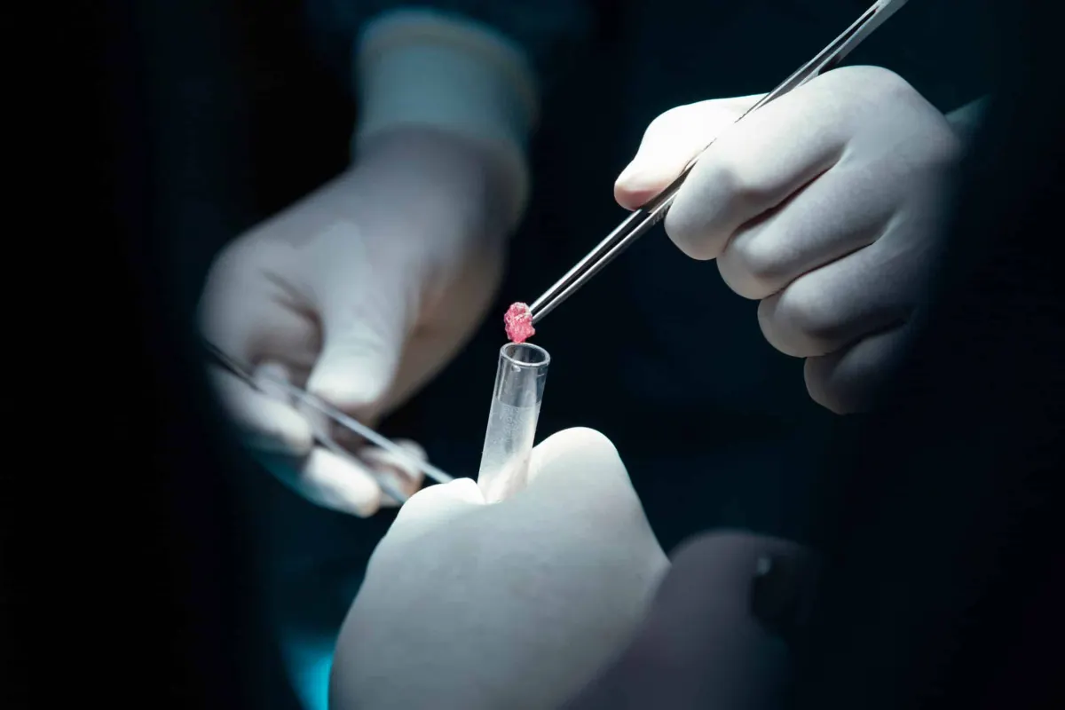
Biopsies are the foremost diagnostic interventional radiology procedures. A biopsy is a procedure performed through the skin under imaging guidance (CT, ultrasonography or angiography) and local anaesthesia, in which a sample is taken from the tissue with special needles and sent to pathology. Aspirating cells from tissue with special needles is called fine-needle biopsy, while removing a piece of tissue with a specialised needle is called core-needle biopsy (tru-cut). The material obtained is then placed in a special solution and sent to the pathology laboratory for examination.
What are therapeutic interventional radiology procedures?
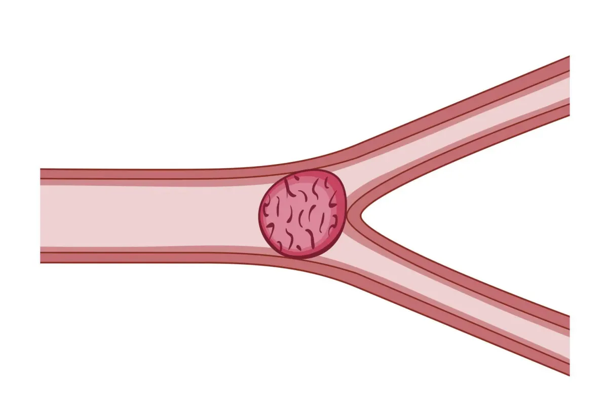
Percutaneous Prostate Artery Embolisation; In benign prostate enlargement, this procedure is performed under local anaesthesia through a blood vessel in the groin to close the vessels supplying the enlarged part of the prostate, thereby shrinking the prostate. It is a day-case procedure. Read more..
Varicocele Treatment; Under local anaesthesia, the groin or neck vein is accessed with a special needle and the draining veins causing the varicocele are closed with coils using special catheters. Read more..
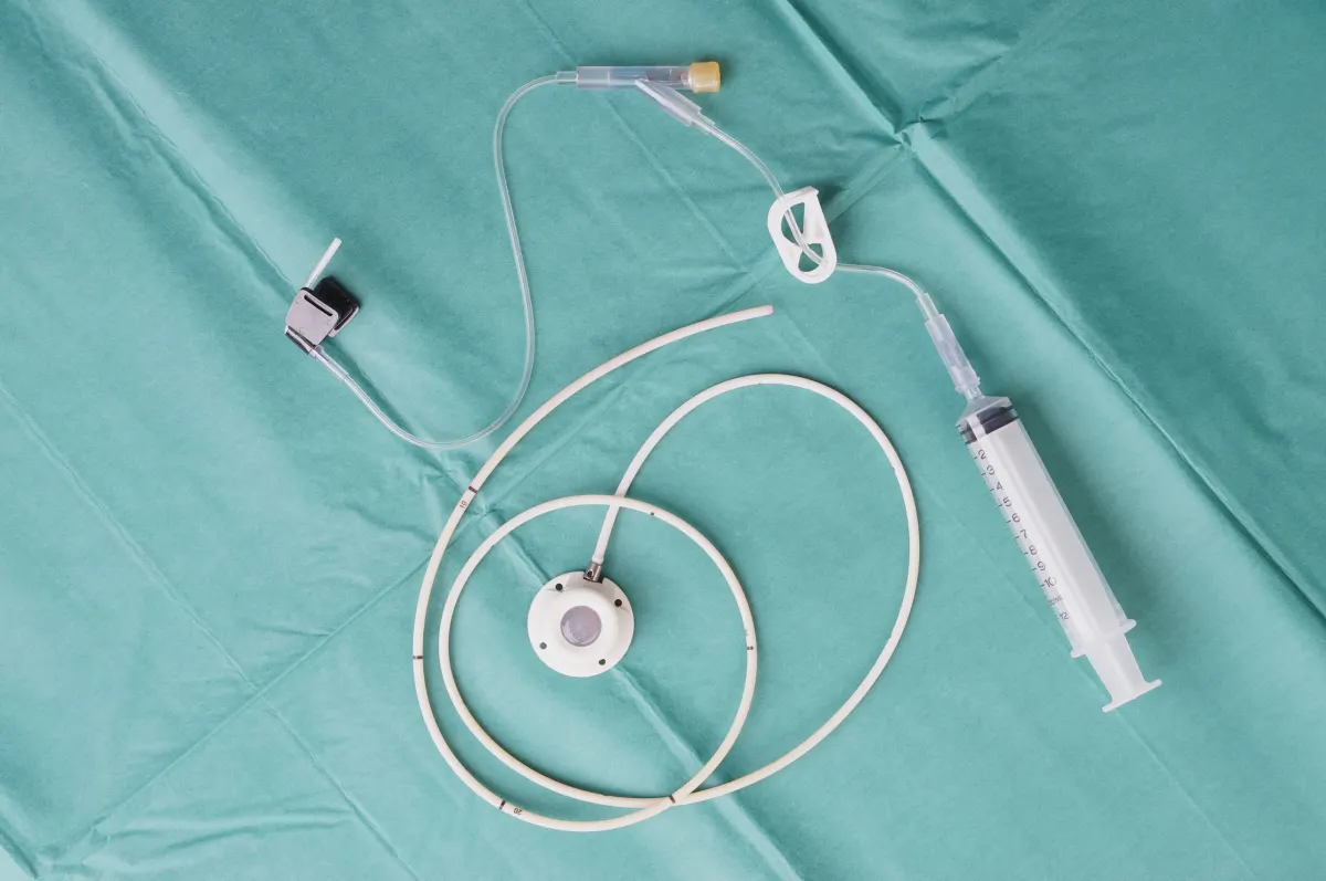
Percutaneous Port Catheter Placement; For oncology or haematology patients who need continuous injections, a special catheter is placed with its reservoir in the upper chest and its tip inside the vein next to the heart; the procedure is performed under local anaesthesia in the angiography laboratory. Read more..
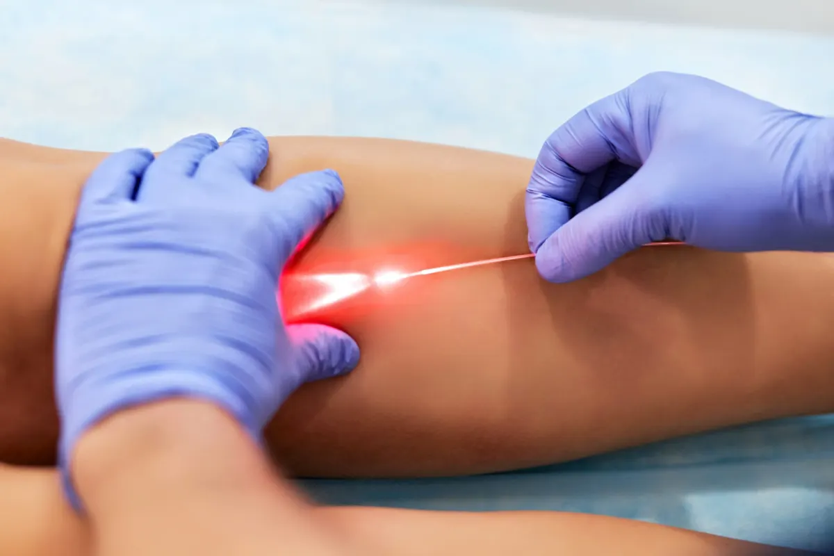
Percutaneous varicose vein treatment; To treat varicose veins of the leg and thigh, the main veins are closed with radiofrequency, laser or medical adhesives when they are insufficient or leaking. Read more..
Percutaneous fibroid treatment; Performed under local anaesthesia through a blood vessel in the groin, this procedure uses special catheters and embolic agents to close the vessels feeding uterine fibroids, causing the fibroids to shrink and soften. Read more..
Biopsies; The most frequently performed procedure in interventional radiology, carried out to determine whether a lesion is a tumour, inflammatory tissue or normal tissue. Read more..
Percutaneous abscess drainage; Draining abscesses in the body under imaging guidance and local anaesthesia using special needles.
Percutaneous pleural effusion drainage; Draining fluid between the membranes of the lungs with special catheters under imaging guidance and local anaesthesia.
Percutaneous pleurodesis; In recurrent pleural effusions, the pleural layers are adhered together using special sclerosing agents (most commonly talc).
Percutaneous catheter drainage of ascites; Draining fluid accumulated in the abdominal cavity with special catheters under local anaesthesia.
Percutaneous nephrostomy; When urine cannot flow normally due to obstruction of the kidneys or urinary tract, a catheter is placed into the urinary collecting system through the skin under local anaesthesia, allowing urine to drain externally.
Percutaneous cyst treatment; Cystic lesions (thin-walled, fluid-filled lesions) in the kidney, liver or anywhere in the abdomen are drained with special catheters under imaging guidance and then, where appropriate, the cells of the cyst wall are treated with special solutions so they can no longer produce fluid.
Percutaneous biliary drainage; In bile duct obstructions, a catheter is placed into the bile ducts through the skin under local anaesthesia to restore bile flow.
Percutaneous cholecystostomy; In patients with gallstones and severe inflammation, special catheters are placed into the gallbladder through the skin under local anaesthesia to drain bile externally.
Percutaneous balloon angioplasty and stenting; Opening blocked blood vessels by accessing them through a vessel in the groin or arm.
Percutaneous aneurysm treatment; When a balloon-like bulge forms in a blood vessel, the vessel is accessed through the skin with special needles and catheters and the aneurysm is closed with a stent or special coils.
Percutaneous tumour embolisation; The vessels supplying the tumour are reached through the skin under local anaesthesia and closed, cutting off the tumour cells' blood supply and stopping bleeding.
Percutaneous radiofrequency/microwave ablation and percutaneous cryoablation treatments; Treatment of thyroid nodules, breast fibroadenomas and tumoural lesions of internal organs (lung, liver, kidney, spleen, etc.) by entering through the skin with a special needle.
Transarterial chemoembolisation (TACE); Used for primary and metastatic liver tumours, this procedure accesses the liver artery through a vessel in the groin under local anaesthesia, delivers special chemotherapy drugs into the vessels feeding the tumour and then closes those vessels.
Transarterial radioembolisation (TARE); Used for primary and metastatic liver tumours (especially neuroendocrine tumours), this procedure accesses the liver artery through a vessel in the groin under local anaesthesia, delivers special radionuclide agents into the vessels feeding the tumour and then closes those vessels.
Percutaneous arthritis treatment; In knee osteoarthritis, to prevent the increase in synovial tissue that causes pain, the vessels supplying the synovial tissue are accessed through the skin under local anaesthesia and closed with special materials.
Percutaneous foam treatment of varicose veins – sclerotherapy; Closing the small capillary veins of the leg and thigh with special solutions injected through fine needles.
Percutaneous thyroid nodule treatment; Treatment of thyroid nodules: solid nodules with radiofrequency or microwave ablation and fluid-filled ones with alcohol. It is performed under local anaesthesia with ultrasound guidance.
Percutaneous treatment of nosebleeds; For nosebleeds that cannot be stopped, particularly those from the back of the nose, the bleeding vessel is reached with special fine catheters through a vessel in the groin under local anaesthesia and closed to stop the bleeding.
Percutaneous treatment of calcific tendinitis; Calcium deposits in and around tendons, particularly in the shoulder, are removed using special needles under ultrasound and fluoroscopy guidance and local anaesthesia, promoting tendon regeneration.
Percutaneous pain treatment; Once the cause of the pain has been identified, the source of the pain or the nerve fibres transmitting it are blocked under imaging guidance using special solutions or devices. It is particularly effective for cancer-related pain.
Percutaneous breast clip marking; In breast cancer, the cancerous tissue is marked with special clips before chemotherapy so that the central point can be located for surgery after chemotherapy.
Percutaneous breast localisation; Marking breast lesions that cannot be felt through the skin because they are deep-seated or small, using special wires under ultrasound or mammography guidance before surgery.
Percutaneous treatment of haemoptysis; Haemoptysis, the coughing up of blood from the lungs, is most commonly seen as a sequela of tuberculosis, in bronchiectasis and in lung cancer. The lung vessels are reached with special catheters through a vessel in the groin, the bleeding vessel is identified and closed with special agents.
Adrenal venous sampling; Performed in patients with hypertension to identify which adrenal gland is secreting aldosterone, this procedure accesses the adrenal veins through a vessel in the groin, takes blood samples and sends them to the laboratory for hormone level measurement.
Percutaneous placement of temporary and permanent dialysis catheters; In patients requiring dialysis, temporary (21-day) or long-term (permanent, usable for months) dialysis catheters are placed into the main veins under ultrasound and fluoroscopy guidance and local anaesthesia.
Quick Appointment Form
Thanks to advanced technological progress in medical imaging, it is now possible to obtain highly detailed images of any part of the human body. In parallel with these developments, methods are being developed that aim to cause the least possible harm to the body and allow diagnosis and treatment while minimising surgical intervention. These methods are easier to perform than surgery, are generally less painful and less risky, and also shorten hospital stays. This discipline, called interventional radiology, involves diagnosis under the guidance of imaging systems and, more often, direct therapeutic procedures performed by radiologists using special instruments. The goal of interventional radiology is to provide an alternative to surgery in many cases, to offer treatment to patients who are not candidates for surgery, or to improve the patient's condition in preparation for surgery and make surgery easier. For interventional radiology procedures to be effective, making the fullest use of all imaging methods is essential. The algorithm determining which imaging method or combination of methods to use for each disease is important.
All imaging methods (except scintigraphy) can be used in interventional radiology. Ultrasonography (USG), computed tomography (CT), fluoroscopy and specially designed MR scanners are used as guidance methods for interventions. While the choice of imaging method lies with the operator, showing the intervention site in the best way, suitability for the patient and cost are also important. These methods are often used together, and such combinations make a significant contribution to diagnosis and treatment. Nevertheless, angiography units form the core of this discipline. Angiography units acquire fully digital images and process them at a high level, allowing images to be subtracted from one another or used as a guide in what can be called a mapping method (roadmapping). Detailed evaluation of the imaging methods used must be accompanied by very careful pre-procedure planning and mastery of the materials to be used. In addition to the experience and manual skill developed over time, the operator must master the use of various materials and drugs and their interactions with tissue. Besides instruments such as needles, catheters and guidewires made of various materials, drugs that aid diagnosis in imaging and therapeutic drugs are also used. A wide variety of materials are used, such as stents that keep vascular and non-vascular channels open, and coils or adhesive agents that, conversely, are used to block them. Interventions are generally performed under regional, limited numbing (local anaesthesia). Although anaesthesia machines integrated into the system are available in the intervention units, low-dose anaesthesia is generally sufficient because the procedures cause little pain. Interventional radiology units are complemented by observation rooms where post-procedure monitoring takes place.
Image-Guided Diagnostic Interventions
The main diagnostic method in interventional radiology is needle biopsy applied directly through the skin to various parts of the body. It has been performed since 1883 and has become ever safer. Thanks to technological developments, procedures today are performed with thinner, more flexible needles, increasing success and minimising side effects. The most common sites are the liver, pancreas, abdominal cavity and its membranes, kidneys and adrenal glands, lower abdomen and genital region, chest, all bones and the neck. It must be performed for a definitive diagnosis in cases where diagnoses such as benign or malignant tumour or abscess cannot be made with clinical findings and imaging methods. Samples can also be taken for diagnosis and culture from formations thought to be of inflammatory origin or from dense fluid collections, and if necessary these can be drained through a catheter (a thin, long tube made of special material).
Therapeutic Interventional Radiology Procedures
In interventional radiology, procedures are applied in two groups: vascular and non-vascular. Vascular procedures are divided into methods aimed at opening or blocking vessels. Blocking procedures are called embolisation and opening procedures are called angioplasty. Blocking is most often performed for aneurysms (balloon- or spindle-shaped widening of a vessel), vascular malformations, stopping bleeding and shrinking tumours. Generally, the feeding artery is blocked. Its most important contribution is in bleeding. Although it is possible to tie off the bleeding vessel surgically, bleeding located very deep or at the far end of an organ or tissue can be blocked as distally as desired thanks to interventional radiology. In addition, depending on the technique and materials used, temporary or permanent blocking is possible. In the common tumour cases of today and their spread, in addition to blocking the vessels feeding the tumour, chemotherapy drugs are also delivered to the tumour to increase the effect of chemotherapy and minimise its side effects. Narrowed vessels are widened with the help of a balloon. The most common cause of vessel narrowing is atherosclerosis (hardening of the arteries). Narrowing or even blockage of vessels can also occur due to certain congenital disorders. First, the narrowed vessel is imaged. If intervention is required, angiography must be performed. After the location and degree of the narrowing are determined, the vessel is widened with a balloon. During this, the innermost layer of the vessel is, in a controlled way, effectively cracked, while its middle layer is stretched. If the blocked segment is very long, if there is complete occlusion, or if sufficient success cannot be achieved with the balloon or the narrowing recurs, stents are placed inside the vessel. Stents are durable, cylindrical, mesh-like devices made of metallic or synthetic materials, used to keep the vessel open over the long term. Today there are stents made of special materials that prevent blood from leaking outside the vessel wall, and stent grafts (vessel patch-covering) are applied. Although they are best known for use in the coronary arteries, they are also used in the renal arteries, the vessels extending to the legs (lower extremities), the main abdominal vessel (abdominal aorta) and the neck vessels supplying the brain. Delivering drugs to the desired area of the body in the required amount (usually in high doses) is needed especially for suddenly blocked vessels or widespread heavy bleeding, and this is called drug infusion. This procedure, performed selectively at the desired site, is one of the procedures of interventional radiology. Placing tubes extending into the main veins required for long-term systemic drug treatments, making treatment easier to tolerate and ensuring its effectiveness, is successfully performed thanks to interventional radiology. Filters are placed inside vessels to prevent clots from moving, breaking off and travelling elsewhere. Their most common use is to prevent clots originating from the deep veins of the legs and lower abdomen from entering the circulation, most often from travelling to the lungs. They can be applied preventively before a clot forms, and permanent or temporary filters can be used. In this way they are placed through the skin and a vein without the need for major surgery, and after a short period of monitoring patients can quickly return to an active life. In addition to preventing clot migration, clots can also be softened, broken up and removed. As in uncontrollable oesophageal bleeding, connections can be created between vessels to regulate circulatory dynamics and pressures in different systems (TIPS = transjugular intrahepatic portosystemic shunt). The most common areas of non-vascular procedures are the kidneys and urinary tract, the bile ducts, abscesses and cysts that may form in various places, and tumours treated directly through the skin. All cysts that form in the abdomen can be drained through the skin and, if necessary, treated to prevent them from forming again. In urinary tract obstruction, if inflammation has developed together with the obstruction or if there is bilateral obstruction, the collecting systems of the kidneys are entered and outflow is provided with tubes (percutaneous nephrostomy). Small kidney stones can be pushed into the bladder, attempts can be made to dissolve them with chemical agents, or in suitable cases they can be removed. Blocked bile ducts can be opened with balloons and stents, inflammation of the obstructed system can be treated, and gallstones can be removed. Abscesses in the body are drained directly through the skin under imaging guidance after their clinical suitability has been established. The abdominal cavity, liver, kidneys, lungs, bile ducts and pancreas are the most common sites. Hydatid cysts, which are common in our country and may form in the liver and throughout the body, are also treated with this intervention. Treating tumours directly through the skin with interventional radiology methods is among the most important procedures of modern medicine. Although these methods vary between different types of cancer, we can give the following examples:
In liver cancers smaller than 3 cm, it is possible to kill tumour cells and completely stop their progression with ethanol injection. Likewise, in many abdominal tumours and tumours that have spread to the liver, especially colorectal tumours, it is possible to destroy the tumour by vaporising it with radiofrequency. In pancreatic tumours, bile duct tumours and malignant liver tumours, the vessel feeding the tumour is selected by advancing to its very tip, and radioactive agents can be delivered directly to the tumour. This protects healthy tissue while causing maximum damage to the tumour (selective internal radiation therapy = SIRT).
For the treatment and control of pain, nerve roots and nerve groups are treated with heat and chemical agents. Image-guided diagnostic and therapeutic procedures are becoming increasingly widespread in modern medicine thanks to advancing technology. By achieving the highest treatment success in the shortest time with minimal tissue damage, they make a significant contribution to returning to an active life.
VIDEOS
Interventional radiology covers a broad range of interventional procedures performed with the help of catheters, guidewires, stents and balloons, alongside radiological imaging techniques such as angiography, computed tomography (CT), magnetic resonance imaging (MRI) and ultrasonography. Interventional radiology is an approach used to recognise, diagnose and treat a patient's disease.
Treatment Methods
The main treatments and procedures performed in our department.
What Is a Port Catheter?
Interventional Radiology
Varicose Vein Treatment in Interventional Radiology
Interventional Radiology
What Is Prostate Embolization?
Interventional Radiology
What Is a Biopsy, How Is It Performed?
Interventional Radiology
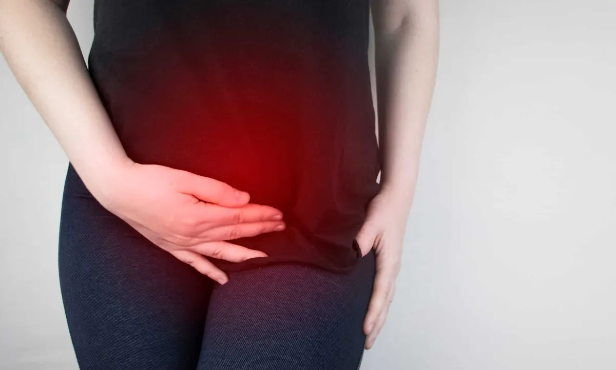
Myoma Embolization
Interventional Radiology

Ablation Therapy in Cancer
Interventional Radiology
What is Axillary Biopsy? Procedure, Risks, and Results
Interventional Radiology
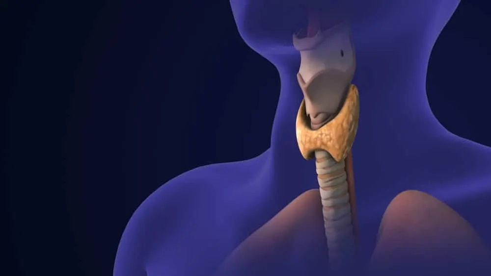
What is Thyroid Biopsy? How is it Performed? Procedure Process, Risks, and Results
Interventional Radiology
Liver Biopsy
Interventional Radiology
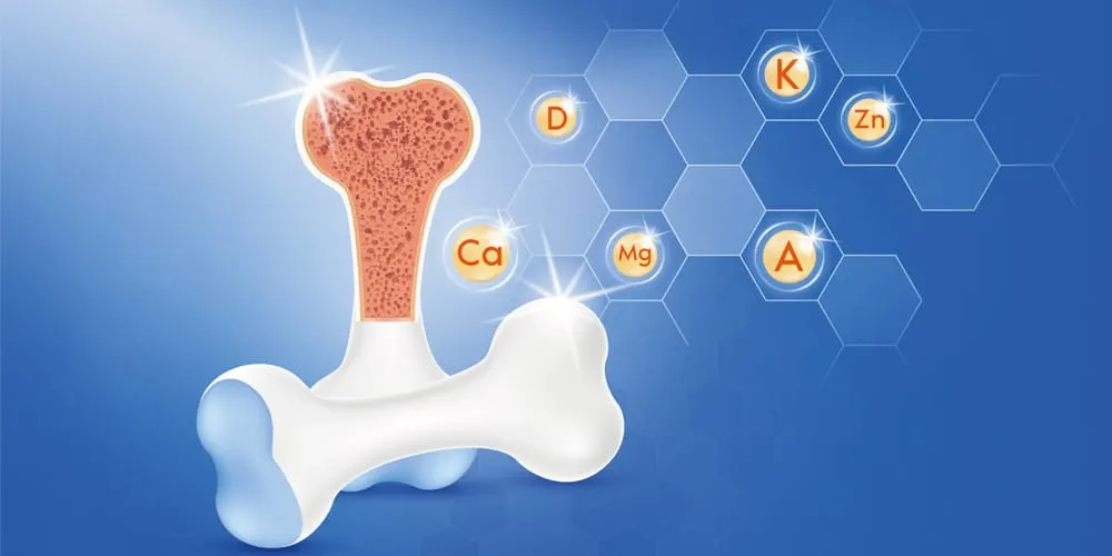
Bone Biopsy
Interventional Radiology
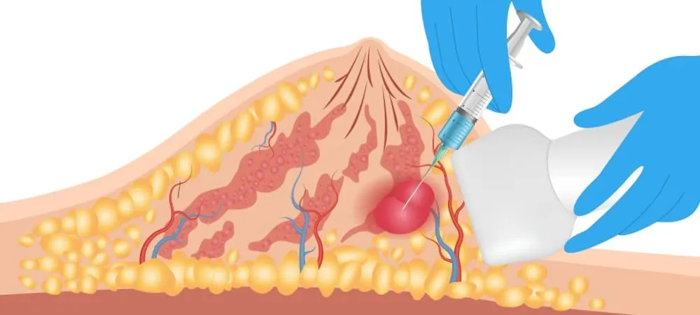
What You Need to Know About Breast Biopsy
Interventional Radiology
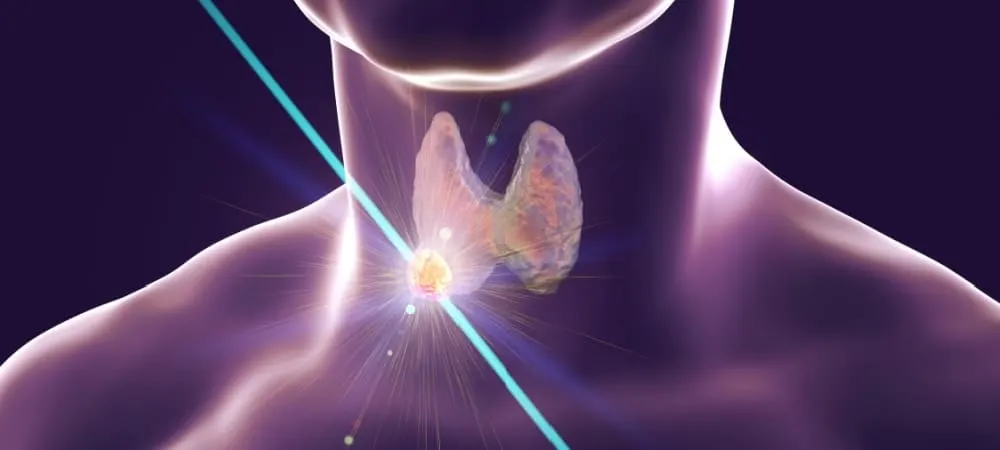
Non-Surgical Thyroid Nodule Ablation: Methods, Advantages, and Risks
Interventional Radiology
Conditions Covered
The main conditions within the department’s diagnosis and treatment scope.
Department Doctors
Our specialist team working in the Interventional Radiology department.
Related Videos
Informative videos from our specialists related to the Interventional Radiology department.


Healthy Living Guide
Featured content related to the department.
Locations
At Which Hospitals?
The Interventional Radiology department is available at the following locations.
Author
Web and Medical Editorial BoardReviewed by a specialist
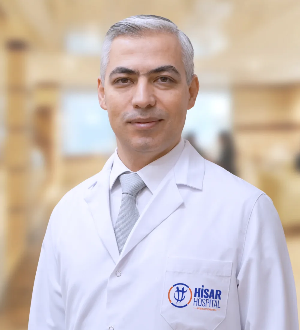 Assoc. Prof. Dr. S. Hilmi AksoyInterventional RadiologyReview date: 31 October 2026
Contact
Interventional Radiology Department — Get in Touch
Fill out the form with your questions; our team will call you back as soon as possible.
The Hisar Hospital Web and Medical Editorial Board contributed to the development of this content. All content on this website is provided for general informational purposes only. Please consult a physician for the evaluation, diagnosis and treatment of your condition.
Last Updated: