

Endoscopy Laboratory
Endoscopy is the examination of the esophagus, stomach, bile ducts and intestines using a flexible instrument with a light and micro-camera system at its tip…
About the Department
The scope, approach and highlighted services of the department.
Endoscopy is the examination of the esophagus, stomach, bile ducts and intestines using a flexible instrument with a light and micro-camera system at its tip.
In the endoscopy unit:
Gastroduodenoscope
Colonoscope
Duodenoscope (ERCP): services are provided with video endoscopic models, and images and CD recordings can be made.
The endoscopy unit is used not only for diagnostic purposes but also for therapeutic purposes. Cleaning, disinfection and sterilization of the endoscope and other instruments used are very important. In our unit, procedures are carried out with automated washing and sterilization devices. Procedures can be reported, photographed, recorded onto a CD when necessary and archived, and a copy is given to the patient. Although complications in endoscopic examination procedures are reduced to a very low level, a CRASH CART (emergency intervention drug and equipment cart) is available in our department to respond promptly to any complications and reactions that may occur. It is equipped with oxygen, suction, monitor, defibrillator and intubation equipment. Perhaps the most important aspect of endoscopic examinations is their superiority in the early diagnosis of cancer. Difficulties in treating late-stage cancers of the digestive system, and in some types even their impossibility, remain a major problem. Being able to diagnose cancers before they grow or spread is also encouraging for treatment. Although gastric cancer in Japan and colon cancer in America are seen much more frequently than in our country, the percentage of deaths from these diseases is also lower than in ours. The reason for this is the performance of periodic endoscopic screening in Japan and America. Treatment of stomach and colon cancers diagnosed at an early stage is generally possible. In the diagnosis of digestive system diseases, imaging methods other than endoscopy and biochemical tests also make a major contribution. Imaging techniques such as ultrasound, tomography and magnetic resonance imaging are frequently used. Our hospital's radiology unit assists our department in diagnosing complex cases, and at the same time enables biopsy to be taken under visualization from organs such as the liver and pancreas (invasive radiology) and provides the opportunity to make a definitive diagnosis. In addition to routine tests, biochemistry tests, including hormone and cancer marker tests, support the work of our gastroenterology unit.
Diagnostic Endoscopy
Esophagogastroduodenoscopy: Used to examine the esophagus, stomach and duodenum.
Colonoscopy: Used to examine the anal canal, rectum and large intestine.
Endoscopic Retrograde Cholangiopancreatography (ERCP): Used to examine the bile ducts.
Therapeutic Endoscopy
Polypectomy: Endoscopic removal of polyps of the large intestine and stomach without surgery.
Sclerotherapy: Stopping variceal bleeding and ulcer bleeding by injection using an endoscopic method.
Band ligation: Treatment of esophageal varices and hemorrhoids by ligating them with elastic bands using an endoscopic method
Dilatation and stent placement: Dilation of strictures of various causes in the digestive system with a bougie or balloon via endoscope, and placement of stents suitable for the strictures.
Percutaneous Endoscopic Gastrostomy (PEG): In patients with esophageal strictures or those who cannot feed orally, an opening into the stomach is made by endoscopic method to establish a feeding route for long-term use, and the patient is fed through it.
Foreign body removal: Removal of accidentally swallowed objects such as needles, coins, etc. via endoscope.
Endoscopic Retrograde Cholangiopancreatography (ERCP): Used for removing gallstones without surgery, relieving bile duct strictures and placing stents.
Endoscopy is the examination of the esophagus, stomach, bile ducts and intestines using a flexible instrument with a light and micro-camera system at its tip.
Author
Web and Medical Editorial BoardReviewed by a specialist
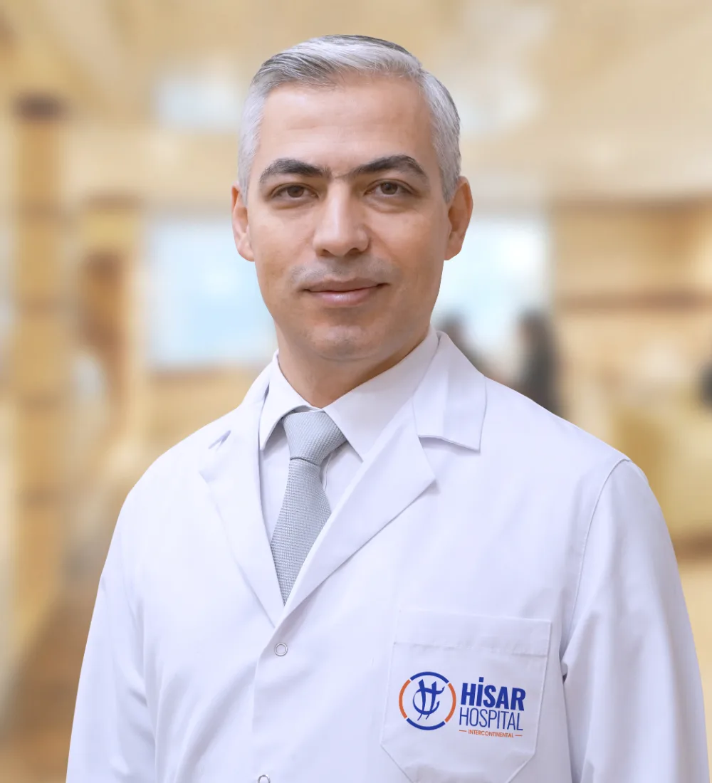 Assoc. Prof. Dr. S. Hilmi AksoyInterventional RadiologyReview date: 02 November 2026
Contact
Endoscopy Laboratory Department — Get in Touch
Fill out the form with your questions; our team will call you back as soon as possible.
The Hisar Hospital Web and Medical Editorial Board contributed to the development of this content. All content on this website is provided for general informational purposes only. Please consult a physician for the evaluation, diagnosis and treatment of your condition.
Last Updated: