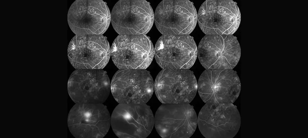
Eye angiography is a safe and painless diagnostic method that allows the visualization of vascular structures in the eye with the help of special dyes. This procedure is based on dyeing and photographing the retinal and choroidal vessels.
Eye angiography is usually a quick procedure and is completed in approximately 10-20 minutes. It is used in the diagnosis of major eye diseases such as diabetic retinopathy, age-related macular degeneration, and retinal vascular occlusions. In addition, in most cases, it is recommended to be performed after a meal.
This article covers how eye angiography is performed, which diseases it is used to diagnose, and important details that should be known before the procedure. In particular, you will find answers to all your questions regarding the risks, duration, and patient preparation for the procedure.
What is Eye Angiography?
Eye angiography is an effective diagnostic method used to visualize the vascular structure in the eye with the help of special dyes. It provides a detailed examination of the fundus, which is called the fundus, and is of critical importance in the diagnosis of retinal diseases.
Definition of Fundus Fluorescein Angiography (FFA)
Fundus Fluorescein Angiography (FFA) is a fundamental diagnostic technique that allows the visualization of retinal vessels using a special substance called fluorescein. In this procedure, fluorescein dye is injected into the vein in the patient's arm and reaches the eye vessels within seconds (approximately 10-15 seconds). Fluorescein is a dye that glows yellow-orange under blue light, and serial photographs are taken with a special camera as it circulates in the eye vessels.
The FFA procedure takes place in four stages:
Pre-arterial phase
Arterial phase
Capillary phase
Venous phase
When needed, late photographs can also be taken after 10 and 20 minutes.
Difference of Indocyanine Green Angiography (ICG)
Although Indocyanine Green Angiography (ICG) is a similar application to FFA, it carries important differences. In this method, a dye called indocyanine green is used instead of fluorescein. ICG is more successful in showing choroidal neovascularizations and leaks that occur especially in cases of age-related macular degeneration.
The main difference between the two techniques is the eye layers they image. While FFA mainly images retinal vessels, ICG is an ideal method to show the choroidal circulation behind the retina . Therefore, when problems in the choroid layer are suspected, ICG is preferred, especially in conditions such as polypoidal choroidal vasculopathy, retinal angiomatous proliferation, and central serous retinopathy.
Which parts of the eye are imaged?
With eye angiography, the vascular structure of the retina and choroid layers located at the back of the eye is examined in detail. Thanks to this imaging:
In the retina: The optic nerve, macula, retinal vessels, and related abnormalities are imaged. Leaks, blockages, and abnormal vessels in the retinal vessels can be clearly detected.
In the choroid layer: Circulation disorders, abnormal vessels, and leaks in this layer under the retina are evaluated.
In the images taken during angiography, hyperfluorescent or hypofluorescent areas are seen depending on the fluorescein density, and each of these has important clinical meanings. Today, the images obtained can be digitally recorded and stored.
Which Diseases Are Diagnosed Using Eye Angiography?
Eye angiography is an important diagnostic method used in the diagnosis of many eye diseases. This technique helps expert physicians detect diseases by examining the vascular structure in the eye.
Diabetic Retinopathy
Diabetic retinopathy is one of the most common eye complications encountered by diabetic patients. Eye angiography detects changes in retinal vessels and helps determine:
Bleeding in the retinal tissue
Vascular occlusions
Leaks and abnormal vessels
Problems such as these. Thus, doctors get the opportunity to determine the treatment plan and locate the areas where laser treatment will be applied.
Macular Degeneration (Yellow Spot Disease)
Age-related macular degeneration (yellow spot disease) is seen in proportion to increasing age, especially after the age of 55. Eye angiography is used in this disease to detect:
Yellow-ish deposits called drusen formed under the macula
Abnormal vessels seen in the wet type
Fluid leaks occurring in the subretinal region
Findings such as these are used to detect. Especially in wet-type macular degeneration, clear visualization of abnormal vessels is of critical importance in treatment planning.
Retinal Vascular Occlusions
Retinal vascular occlusions are among the important causes of sudden vision loss. With eye angiography:
Occlusion points in retinal arteries and veins
Ischemic areas
Areas of bleeding
Are examined in detail. In light of this information, physicians can determine the treatment protocol for retinal vascular occlusions.
Uveitis and Inflammatory Diseases
Uveitis is a type of intraocular inflammation in which the uvea, the middle layer of the eye, becomes inflamed. Eye angiography helps determine the inflammation in the eye by identifying:
The severity of inflammation
Involvement areas
Possible complications
Understanding whether uveitis is of autoimmune or infectious origin can completely change the treatment approach.
Intraocular Tumors and Abnormalities
Eye angiography plays an important role in the diagnosis of intraocular tumors. With this method:
Vascular structures of tumors
Vascularization characteristics
Margins and relationship with neighboring tissues
Can be evaluated clearly. This evaluation helps doctors determine the type of tumor and formulate the treatment strategy.
How is Eye Angiography Performed?
Eye angiography is an effective diagnostic procedure used for a detailed examination of the eye vessels. This procedure is generally a short, painless, and safe procedure. But exactly how is the procedure carried out?
Dilatation of the pupil
In the first stage of the procedure, the pupils are dilated with special drops to see the back of the eye better. Thanks to these drops, the retina and vessels can be imaged more clearly. Generally, it takes 15-20 minutes for the drops to take effect.
Opening a vascular access
After the pupil is sufficiently dilated, a vascular access is opened from the patient's arm or hand. The dye substance will be administered through this vascular access. Before the procedure, the allergy history of some patients is questioned, and allergy medication can be administered as a precaution if necessary.
Dye injection
After the vascular access is opened, a dye called Fluorescein (yellow colored) or indocyanine green is administered intravenously. These dyes glow under light, enabling detailed visualization of the vessels. During the injection, patients may experience a brief sensation of warmth, but no pain is felt.
Photographing
The patient is seated in front of a special FFA camera and their chin is placed on the rest part. After the dye starts to be administered, the progression in the retinal vessels is recorded in the following order:
0-10 seconds: Initial arterial phase photographs
10-30 seconds: Venous filling phase
1-3 minutes: Images showing possible leaks and occlusions
5-10 minutes: Late phase images
The total procedure time usually takes 10-15 minutes. During the procedure, the patient stays still by looking at the target light inside the device.
Evaluation of results
The captured images are examined in detail by an ophthalmology specialist. The findings obtained are digitally recorded and used in preparing the treatment plan. In this evaluation, possible vascular occlusions, leaks, or abnormal vessels are detected.
The eye angiography procedure has no harm to the eyes and can generally be performed on an empty or full stomach. However, some doctors may prefer the patient to have a light meal before the procedure. After the procedure, temporary blurred vision and sensitivity to light may occur because the pupils remain large.
Risks and Application Details of Eye Angiography
Although eye angiography is generally a safe diagnostic method, like every medical procedure, it brings some risks along with it. In this section, we will examine the side effects and application details of the procedure.
Common mild side effects
Most patients may experience mild and temporary side effects after eye angiography. These effects usually pass on their own in a short time:
Nausea and dizziness caused by the dye
Temporary yellowing of the skin or urine (due to the dye)
Mild bruising or swelling at the injection site
Mild stomach discomforts
The dark color of the urine is caused by the contrast medium and may continue decreasing for up to 10 days. This condition is harmless and does not require concern.
Rare allergic reactions
Rarely, allergic reactions to the dye may develop. These reactions are classified according to their severity as mild (1.24%), moderate (0.2%), and severe (0.04%). The total reported side effect rates are generally 0.083%.
Allergic reactions may include:
Itching and skin rashes
Shortness of breath (very rare)
Drop in blood pressure (very rare)
If you have an allergy to iodine or contrast media, be sure to inform your doctor before the procedure.
How long does the procedure take?
The eye angiography procedure takes an average of 10-15 minutes. However, this duration may vary depending on the structure of the vessels and the area to be imaged. In some cases, the procedure can extend up to 20-30 minutes. The recovery of the pupils takes approximately 2-3 hours, and blurred vision is normal during this period.
Should it be performed on an empty stomach?
In most cases, it is recommended to perform eye angiography on a full stomach. When on an empty stomach, the risk of nausea associated with dye injection may increase. However, there may be different recommendations for diabetic patients. Diabetic patients are generally directed to come on an empty stomach and without taking their medications.
Is it a surgery or not?
Eye angiography is definitely not a surgery. It is only an imaging method for diagnosis and evaluation purposes and does not contain surgical intervention. It is very different from cardiac angiography; it does not require hospitalization and vascular opening interventions are not performed. After the procedure, patients can generally return to their daily lives immediately.
In conclusion, eye angiography is a valuable diagnostic method that allows us to examine the retina and choroid vessels. Thanks to this procedure, doctors can detect serious eye diseases such as diabetic retinopathy and macular degeneration at early stages. In addition, thanks to angiography, problems such as vascular occlusions, leaks, and abnormal vessels can be clearly visualized.
The advantages of eye angiography are:
It is a painless and safe diagnostic method
It is completed in a short time (approximately 10-20 minutes)
It does not require hospitalization
It does not involve surgical intervention
However, mild side effects may be seen after eye angiography. These are:
Temporary nausea and dizziness
Temporary yellowing in urine and skin
Mild bruising at the injection site
It should be remembered that eye angiography is not a treatment method, but an imaging technique used for the diagnosis of the disease. Since the recovery of the pupils after the procedure may take a few hours, it is recommended to have an accompanying person with you.
When it comes to eye health, early diagnosis is quite important. Undoubtedly, eye angiography is accepted as the gold standard for making a definitive diagnosis in many eye diseases. Regular eye examinations and advanced tests such as angiography when necessary carry great importance in preventing vision loss.
Frequently Asked Questions About Eye Angiography
Eye angiography is used in the diagnosis of serious eye diseases such as diabetic retinopathy, age-related macular degeneration, retinal vascular occlusions, and intraocular tumors. It is especially necessary to detect abnormalities in the retina and choroid vessels. The eye angiography procedure usually takes 10-15 minutes, but in some cases, it can extend up to 20-30 minutes. The recovery of the pupils may take approximately 2-3 hours. Eye angiography is generally a safe procedure, but mild side effects may be seen. These include temporary nausea, dizziness, temporary yellowing of the skin or urine, and mild bruising at the injection site. Allergic reactions may develop in rare cases.
This test cannot be performed in case of kidney failure. In most cases, it is recommended to perform eye angiography on a full stomach. This can reduce the risk of nausea associated with dye injection. However, there may be different recommendations for diabetic patients, so it is important to consult your doctor before the procedure. Temporary blurred vision and sensitivity to light may occur because the pupils remain large after the procedure. Therefore, it is recommended to have an accompanying person with you. In addition, the dark color of the urine is normal and does not require concern. It is important to consult your doctor in case of any serious side effect.
Do Not Neglect Your Eye Health
Eye angiography is the gold standard in the early diagnosis and correct planning of the treatment of retinal diseases. Especially if you are a diabetic patient or experiencing sudden vision losses, a detailed retina examination is of vital importance to protect your vision.
You can contact us for your questions regarding your eye health, detailed examination, and eye angiography appointment, and immediately schedule an appointment with our expert physicians.