Fibroblast activation protein inhibitor (FAPI) PET is an advanced imaging method used in cancer diagnosis and treatment monitoring. FAPI works by inhibiting an enzyme called fibroblast activation protein (FAP). This procedure uses a radiopharmaceutical called Ga-68 FAPI. Ga-68 FAPI binds to cancer-associated fibroblasts, which are common in the stroma of many cancer types. This makes Ga-68 FAPI PET/CT scanning extremely useful for imaging various cancer types with high sensitivity. Ga-68 emits positively charged electrons (positrons) that can be detected by PET imaging devices. FAPI molecules bind to fibroblasts in the tumor microenvironment where FAP is abundant, imaging cancerous tissues with high sensitivity. Unlike conventional PET scans, FAPI PET can visualize not only cancer cells but also the tumor microenvironment. The most important advantage of FAPI PET is that it can detect small tumors and metastases that cannot be detected by other methods. This method also shows very low binding in non-cancerous tissues, which increases the accuracy of imaging. This technology offers promising results in many cancer types, especially breast, pancreatic and colon cancers.
For Which Patients Is FAPI PET Performed?
PET scanning is used especially in cancer patients to examine the tissues in the area where the tumor has developed in more detail and to evaluate the extent of the disease. It is very useful in the diagnosis and staging of different cancer types and in monitoring the response to treatment. Some of the patient groups for whom FAPI PET may be recommended by doctors are:
Patients with cancer: This method images cancerous tissue by targeting the presence of fibroblast activation protein (FAP) in the tissue surrounding the tumor. It is widely used in many different tumor types, such as breast, pancreatic, colon, lung and liver cancers. FAPI PET can detect the size of tumors and their spread to surrounding tissues and organs more precisely.
Patients suspected of having metastatic cancer: FAPI PET is ideal for detecting the spread of cancer throughout the body in cancer diseases. It can detect small metastatic foci that cannot be noticed with conventional imaging methods. It offers significant advantages for cancers that spread to organs such as bone, liver and lung.
Patients who need follow-up after treatment: FAPI PET may be preferred to evaluate the response to treatment. Whether the tumor tissue has shrunk or been completely destroyed after radiotherapy, chemotherapy or immunotherapy can be clearly visualized with this method.
Patients at risk of recurrence: In patients at risk of cancer recurring, FAPI PET can detect recurrent tumor tissue at an early stage. Recurrences that are small and not yet detectable by other methods can be visualized with this method in particular.
How Is FAPI PET Performed?
PET scanning is a type of imaging used in nuclear medicine. This method uses small amounts of radioactive substances called radioactive tracers. The steps of the FAPI PET procedure may include:
Patient preparation: Before the procedure, the patient is informed not to eat for the period specified by the doctor and not to take certain medications. Drinking water is usually permitted, but the patient should still obtain information from the doctor regarding water consumption. This process is important for the more effective distribution of the radioactive isotopes in the body.
Injection: The patient is injected with a FAPI molecule labeled with gallium-68 (Ga-68). The injection is usually given intravenously, and it distributes in the body so that it binds to tissues containing fibroblast activation protein (FAP). Ga-68 emits positrons that can be detected by PET scanners. After the injection, a wait of usually 30-60 minutes is observed to allow the isotope to reach cancerous tissues in the body and accumulate to a sufficient level.
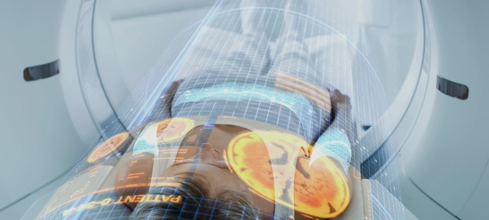
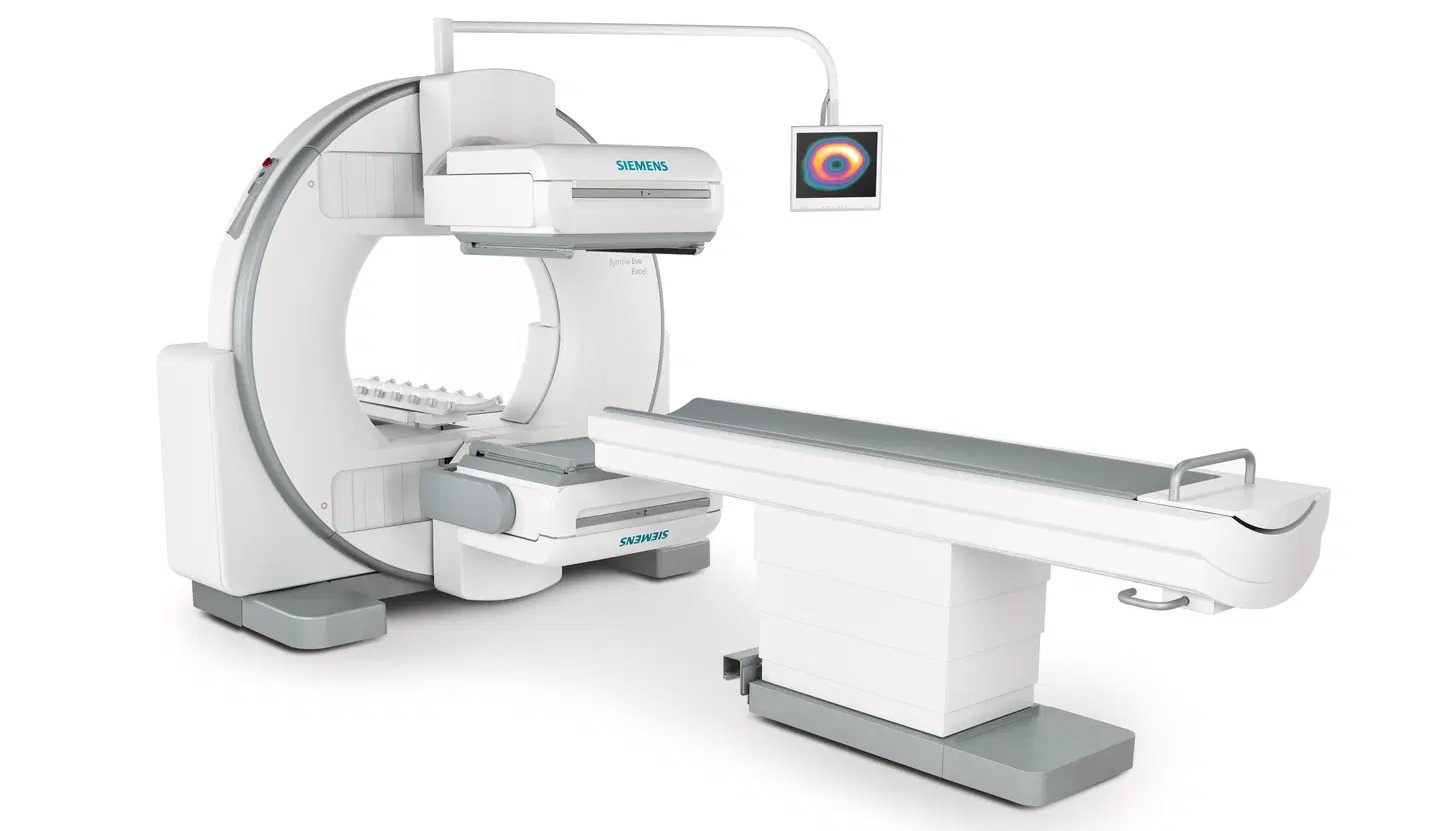
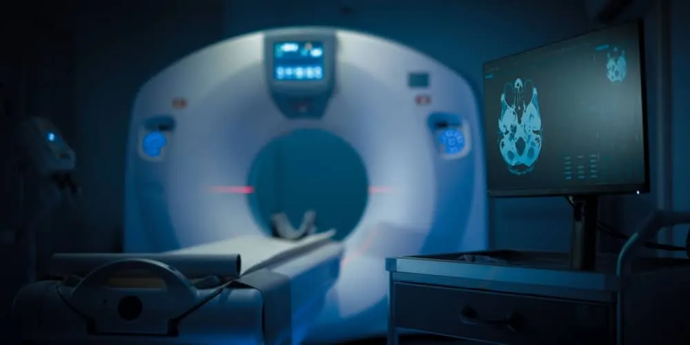
PET scanning procedure: After the injection, the patient is placed in the PET scanner and asked to lie completely still. The PET scanner creates images by identifying the areas where Ga-68 FAPI molecules concentrate. This process usually takes 20-30 minutes, and it is important for the patient to remain as still as possible. In this process, it images in detail the sites where metastases are located as well as tumors.
Image analysis: Scan results are reviewed by radiology specialists. FAP-rich tissues to which FAPI molecules bind clearly show cancerous areas and the tumor microenvironment. Analysis of the images helps determine the stage of the disease and create the treatment plan. The doctor explains the results of the scan to the patient in detail and determines the necessary treatment steps.
Post-procedure follow-up: After the FAPI PET scan, the patient can usually return to daily activities immediately. Since the radioactive substance is eliminated from the body in a short time, it does not pose a long-term risk. However, the doctor determines the post-procedure monitoring plan and recommends further tests or treatment if necessary.
In Which Cancer Types Is FAPI PET Used?
FAPI PET is a next-generation imaging method that gives very successful results, especially in cancer types with high fibroblast activity. This is because FAPI targets the fibroblast cells surrounding cancer cells, and these cells play an important role in cancer growth. Some of the cancer types in which FAPI PET is commonly used are:
Breast cancer
Pancreatic cancer
Colon cancer
Lung cancer
Liver cancer
What Are the Advantages of FAPI PET?
FAPI PET is an imaging method that offers significant advantages in cancer diagnosis and treatment. Some of the advantages of FAPI PET are:
Precise tumor imaging: FAPI PET directly targets cancerous tissues and the tumor microenvironment containing the surrounding fibroblast activation protein (FAP). In this way, by imaging not only tumor cells but also the supportive structure of the tumor, it produces a broader and clearer map of the cancer. It provides high sensitivity, especially in the detection of small tumors and metastases.
Early diagnosis and staging: Detecting cancer at an early stage is the key to treatment success. FAPI PET can detect small tumor foci even in the early stages of cancer and accurately stage the spread of the disease. This gives patients the chance of timely intervention and helps make treatment plans more effective.
Treatment monitoring and detection of recurrence: FAPI PET is used not only for diagnosing cancer but also for monitoring the course of treatment. Monitoring the response to treatment plays an important role in determining whether the tumor has shrunk or recurred. In this way, treatment strategies can be quickly revised when needed.
Why Is FAPI PET Preferred?
FAPI PET is preferred because of the high sensitivity and accuracy it provides in cancer diagnosis and the treatment process. By targeting fibroblast cells surrounding cancer cells, it offers the ability to detect smaller and earlier-stage tumors. By showing cancerous tissue more clearly, it facilitates surgical planning and treatment follow-up. It plays an important role in evaluating treatment effectiveness and in early detection of recurrence.
How Are FAPI PET Results Interpreted?
A radiologist specialized in PET scans reviews the images, writes a report, and sends the report to the relevant doctor. Results are usually available within a few days. FAPI PET results are interpreted based on the presence and intensity of FAP-positive areas in the body. The intensity of fibroblast activation protein around the tumor helps identify cancerous tissues and detect the spread of the tumor. High FAP-accumulating areas detected in the images may indicate cancerous areas or metastatic status. Doctors use these results to determine the stage of the disease, monitor the response to treatment, and make changes to the treatment plan if necessary.
Frequently Asked Questions
The procedure itself is not painful, but you may feel a slight needle prick when the needle is inserted for the radiotracer injection. Results are usually obtained within a few days. The images are analyzed by radiology specialists and a detailed report is prepared. FAPI PET differs from other PET methods by targeting tissues containing fibroblast activation protein (FAP). By targeting fibroblasts surrounding cancer cells, it provides more detailed and specific imaging. In this way, cancer diagnosis, monitoring and treatment can be carried out more effectively. Before FAPI PET, the patient may need to fast and some medications may be temporarily stopped. The doctor evaluates the patient's medical history and gives clear instructions on what to do before the scan. After the radioactive isotope is injected into the body, a waiting period of several hours may be required, during which the patient is advised to rest. FAPI PET is generally not recommended for pregnant women and breastfeeding mothers because radioactive substances may harm the fetus or baby. Additionally, in patients with severe kidney failure, elimination of the radioactive isotope from the body may be more difficult, so careful evaluation is required. In some patients, due to the risk of allergic reaction, doctors may prefer alternative imaging methods. FAPI PET provides more detailed and specific imaging by targeting fibroblasts surrounding cancer cells. In this way, cancer diagnosis, monitoring and treatment can be carried out more effectively. Individuals with symptoms of cancer can begin treatment by making an appointment at the relevant outpatient clinics of our well-equipped hospital for early diagnosis and treatment.