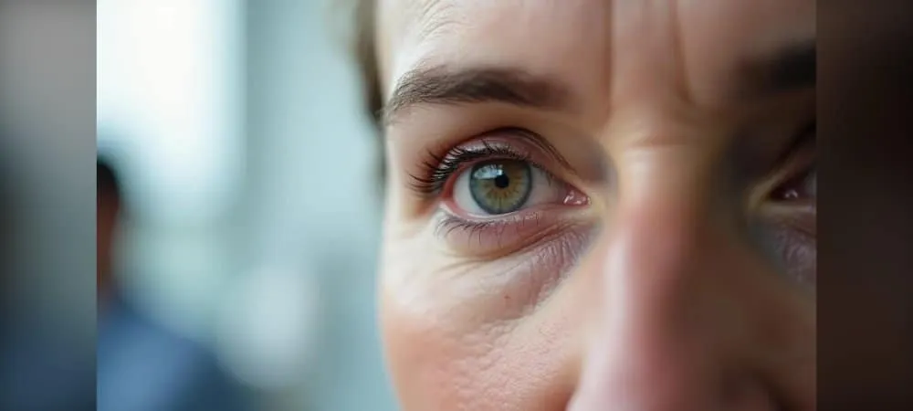
"I cannot see" and "I look tired" complaints of patients who consult ophthalmologists are usually searching for the answer to the question of what ptosis is. If you ask what ptosis means, it is a condition where the upper eyelid is lower than normal. Ptosis in the eye, when it begins to cover the pupil, particularly narrows your upper visual field and makes your daily life difficult. You will find what you are wondering about in this article, such as what ptosis is, why it occurs, who is suitable for ptosis surgery, what ptosis surgery is, and how ptosis treatment is applied.
What is Ptosis and Why Does It Occur?
The condition where your eyelid covers the pupil by more than 1 mm is called ptosis. While the upper eyelid normally covers only about 1-1.5 mm of the colored part of the eye, an increase in this ratio means drooping of the eyelid. To answer the question of what ptosis means more clearly, it is when the eyelid rests lower than its intended position and restricts vision.
Expressed colloquially as "my eyelid has drooped," this condition is different from simple excess skin. If you ask what ptosis in the eye is, it is a functional problem rather than an aesthetic one. It can occur in a single eye or in both eyes simultaneously. Some patients find it difficult to keep their eyelids open; their normal vision is affected or even completely obstructed.
Causes of ptosis: Congenital and acquired factors
The answer to the question of what ptosis is and why it occurs depends on congenital and acquired factors. The cause of congenital ptosis is the underdevelopment of the levator muscle. These cases do not resolve on their own. Ptosis appearing at birth or shortly after birth is unilateral in 75% of cases.
Among the causes of acquired ptosis, weakening of the muscle that opens the eyelid due to aging ranks first. In addition, eyelid trauma due to accidents, excessive eye rubbing, certain medications used, and nerve disorders after head trauma can lead to ptosis. Detachment of the levator muscle tendon from the eyelid due to past eye surgeries or accidents is also among the causes of acquired ptosis.
Neurological diseases and muscle disorders also hold an important place in answering the question of what ptosis in the eye is. In autoimmune diseases such as myasthenia gravis, eyelid drooping is usually one of the first symptoms of the disease and may increase throughout the day. In sympathetic nervous system damage such as Horner's syndrome, pupillary constriction is seen in addition to ptosis.
Eyelid Anatomy and the Levator Muscle
The skin of the upper lid is the thinnest skin of the body and allows the movement of the lid during each blink. The anatomical structures and physiological functions of the eyelids ensure the continuity of the ocular surface. Fully closing, smooth eyelids are essential for a healthy eye and normal vision.
The upper lid is lifted by the levator palpebra superioris muscle, while the height of the lid is adjusted by Müller's muscle. The levator muscle is innervated by the IIIrd cranial nerve and undertakes the task of elevating the eyelid. Müller's muscle, on the other hand, is controlled by sympathetic nerves. The coordinated work of these two muscles maintains the natural position of the eyelid.
Ptosis Signs and Symptoms
Among the signs of ptosis, the most prominent is the drooping or hanging appearance of the eyelid. Restriction in vision or narrowing of the visual field is experienced. This situation becomes more evident especially when looking upward.
Patients may unconsciously tend to constantly raise their eyebrows to open their visual field. This situation leads to deep wrinkles in the forehead region. Children may acquire the habit of looking by lifting their chin up or tilting their head backward to see better. Continuous muscle contraction to keep the eyes open causes fatigue and pain around the eyes.
In severe cases, the eyelid can completely obstruct vision by covering the pupil. Early diagnosis is of great importance, especially in children, as this condition increases the risk of amblyopia (lazy eye).
Types of Ptosis and Who Undergoes Surgery?
Ptosis types are classified according to their causes, and this classification determines the answer to the question of what ptosis treatment is. Each type of ptosis has its own unique characteristics and surgical approaches.
Congenital Ptosis
In this type of ptosis, which is present from birth, there is an abnormality in the development of the levator muscle. It occurs as a result of muscle fibers being replaced by fat and connective tissue while in the womb. Just as the muscle cannot lift the lid sufficiently, it cannot relax on downward gaze either, and the lid remains suspended upward.
The most critical risk in this condition is lazy eye. If the drooping lid covers the center of the pupil, the image traveling from that eye to the brain is blocked. Infancy is the most critical stage during which visual pathways develop. The brain begins to ignore the blurred image coming from this obstructed eye, and amblyopia develops. In severe cases closing the pupillary axis, school age is not waited for surgery; the visual axis is opened with early intervention.
Among the symptoms of congenital ptosis seen in children, head-tilting or chin-up posture stands out. Children may tilt their head backward or lift their chin up to see better. In the long term, this posture can lead to neck and spine problems.
Aponeurotic Ptosis: Age-Related Drooping
The most common cause of adult ptosis cases is aponeurotic ptosis. It is not that the levator muscle loses its strength, but rather that the tendon-like structure where the muscle attaches to the eyelid loosens, thins, or completely detaches with age.
As we age, gravity and loss of tissue elasticity cause the tendon to shift from its place. The patient can contract the muscle, but because the connection in between is broken, the lid does not rise. Long-term hard contact lens use or a severe eye-rubbing habit can cause early detachment of this tendon even at young ages. Past intraocular surgeries such as cataract or glaucoma can also lay the groundwork for this type of ptosis.
In this type, the strength of the levator muscle is usually preserved. When patients raise their gaze upward, the lid can move, but the starting point is lower. The upper eyelid crease appears higher than normal or erased.
Neurogenic and Myogenic Ptosis
In neurogenic ptosis, damage or pressure on the nerve pathways that provide movement of the eyelid is responsible. In the case of third nerve palsy, it is accompanied by severe lid drooping, outward and downward deviation of the eye, and sometimes pupil dilation. In Horner's syndrome, a mild ptosis is seen along with constriction of the pupil on the same side and decreased sweating on the face.
In myogenic ptosis, even if the nerve signal arrives, the muscle itself cannot respond to this signal. Variable ptosis is seen in myasthenia gravis disease. The patient's lid is normal when waking up in the morning, but as the day progresses and the muscle fatigues, the lid begins to drop.
Ptosis Surgery Candidates: For Whom Is It Necessary?
The answer to the question of what ptosis surgery is determines the surgery candidates. The decision for surgery is generally made based on function and appearance. If the lid covers the pupil and narrows the visual field or if the person experiences significant difficulty in daily life, surgical correction comes to the agenda.
Treatment is necessary to improve the patient's quality of life in conditions creating severe visual impairment. Treatment gains urgency, especially when the upper visual field is obstructed. Some patients may have to constantly tilt their heads backward or raise their eyebrows to compensate for this visual obstruction.
Cosmetic concerns can also be effective in the treatment decision. For individuals experiencing social or psychological distress in particular, this is an important source of motivation. However, in neuromuscular diseases, the priority may be the medical management of the underlying disease.
How is Ptosis Surgery Performed?
Preoperative Evaluation and Diagnosis
The answer to what ptosis surgery is shaped by milimetric measurements made before the surgery. The surgeon first measures the distance between the light reflex from the pupil and the upper eyelid margin. This measurement determines the degree of drooping. The total movement capacity of the levator muscle is also checked. A muscle working distance of 12 mm and above is considered excellent, and below that is considered weak.
The Hering's Law Test is definitely applied. Manual elevation of one eyelid can change the signal coming from the brain and cause the other eyelid to drop. This test is of critical importance to prevent experiencing a surprise drooping after surgery. Bell's phenomenon, eye movements, and visual acuity are also evaluated.
Levator Muscle Repair Technique
This technique is preferred in patients with levator function above 4 mm. The levator muscle is reached through an incision made in the natural crease line of the eyelid. The loosened muscle is shortened or advanced from where it attaches to the lid and sutured again. The tendon structure of the muscle is fixed to the tarsal plate, which is the skeleton of the lid.
In this method, since the lid's own muscle is used, the most natural gaze expression is achieved. Since the surgical scar remains within the natural crease line of the eyelid, it is not noticeable from the outside. Age-related lid droops have relatively better and more predictable results than congenital lid droops.
Müller's Muscle Resection (Müllerectomy)
It offers a scarless solution in patients with mild drooping and good levator muscle strength. The surgery is performed from the inner surface of the eyelid. There are no skin incisions or suture scars. Müller's muscle, which is the secondary lifting muscle in the lid, and the tissue over it are shortened to some extent.
Following the resection of Müller's muscle, the change in lid position is a dynamic process rather than a mechanical one. The advantage of this method is that it is not understood that surgery has been performed when looked at from the outside. The recovery process is much faster compared to levator surgery.
Frontalis Sling Surgery
This method is applied in cases where levator function is below 5 mm. In severe conditions closing the visual axis, frontalis sling surgery is performed using special silicone materials. The eyelid is connected to the frontalis muscle on the forehead with the help of a sling. The sling is created using synthetic material or tissue taken from the patient's own leg.
Two lid incisions, two eyebrow incisions, and a forehead incision are made. The sling material is first passed through the lid incisions, then through the eyebrow and forehead incisions. Both ends are joined at the forehead and the lid height is adjusted. When the patient raises their eyebrow, the eyelid is also lifted upward via the sling.
Surgery Duration and Anesthesia Method
Ptosis surgery is usually performed under local anesthesia. Since the patient is awake during the surgery, the surgeon can precisely adjust the ideal height of the lid. General anesthesia is used in children or patients with whom communication cannot be established.
The procedure takes about 30-45 minutes for a single eye and 60-90 minutes for both eyes. The surgery is a procedure from which patients are discharged on the same day. Since patients are under local anesthesia, no pain is felt during the procedure.
Recovery and Care After Ptosis Surgery
The recovery process after ptosis surgery directly affects the success of the surgery. Although the surgery is generally a procedure with same-day discharge, there are many points to pay attention to afterward.
First Week: Edema and Bruising Management
Immediately after the surgery, your eyelid is covered with a bandage. The first control is performed on the 1st and 5th days postoperatively. After the effect of anesthesia wears off, a mild pain and feeling of tightness may occur in the region. Pain relievers recommended by your doctor alleviate this discomfort.
The first 48-72 hours are the period when the surgical area is most sensitive. During this process, swelling and bruises reach their maximum level. Therefore, swelling increases for the first 2 days and begins to gradually decrease starting from the 3rd day. It is completely normal to see edema and mild bruising on the eyelids during the first 3 days after surgery.
Cold compress application is the basic rule of recovery. Throughout the first 48-72 hours, you need to apply cold compresses for 10-15 minutes every hour. Ice should not touch the skin directly; it should be applied over a clean gauze pad. Keeping your head elevated, especially using two pillows while sleeping, helps reduce edema with the help of gravity.
Resting by lying down for the first 2-3 days is strongly recommended. You should avoid heavy lifting, bending over, and activities that will raise blood pressure. Since coughing and sneezing can damage the stitches, you need to be careful.
Removal of Stitches and Recovery Process
Stitches are removed by the surgeon on the 7th day after surgery. In some cases, they can be removed between 6-10 days depending on the recovery process. Because the stitches used are very fine, the removal procedure is painless.
70% of the swelling subsides within the first week. In the second week, bruising largely passes and reaches a level that can be camouflaged with makeup or sunglasses. Bruising of the eye can typically last 2-4 weeks.
Patients can generally return to work within 15 days. If you have a home-office work environment, there is no problem, but if you have a face-to-face work environment, it is appropriate to use 7-10 days of leave. After the first month is completed, the eyelid level settles into place at a rate of 90%.
Dry Eye and Lagophthalmos Condition
A technical condition seen in the early period after ptosis surgery is lagophthalmos, which is the state of the eyelid not closing completely. When we strengthen the muscle lifting the eyelid upward, it may remain open by 1-2 mm during sleep until the lid gets used to its new position. This condition usually improves within 2-4 weeks with the dispersion of edema and the muscle gaining flexibility.
Intensive eye drops should be used during the day to keep the surface of the eye moist. When going to bed at night, more viscous protective gels are applied. Watering of the eye, sensitivity to light, and blurred vision can be seen in the first few weeks after surgery. Regular use of medications is definitely necessary.
Time to Return to Daily Activities
The first 3 days are a rest period. You should avoid straining your eyes, reading books, and using screens. After a few days, you can start activities such as light walking.
You should not use contact lenses for at least 2 weeks. Eye makeup can be started after the 2nd week. You should not swim for a few weeks. Pool, sea, and heavy sports are allowed after the 1st month. You should limit alcohol and tobacco products. It is important to use a hat and sunglasses to protect against the sun.
Frequently Asked Questions About Ptosis Surgery
Is ptosis surgery painful?
Because the surgery is performed under local anesthesia, you do not feel pain during the procedure. You are awake, but the area is completely numbed. A mild tightness or ache may occur in the first few days after surgery. This discomfort is easily controlled with simple pain relievers your doctor will prescribe. Overall, it is a process that passes quite easily for patients.
Is the surgery result permanent?
The results of successfully performed ptosis surgery are generally permanent for life. Re-drooping of the lid is a rare condition. However, ptosis may recur in some people. Sagging may occur in the lid skin years later with the decrease in skin elasticity as a natural result of aging. This condition is a different process from muscle drooping. In case of recurrent ptosis, revision surgery can be performed. It is possible to achieve success with a second or third surgery.
How much does ptosis surgery cost?
For patients without insurance, eyelid drooping surgery prices in private hospitals range between 35,000 TL and 90,000 TL. The price varies depending on whether it is performed on a single eye or both eyes, and whether muscle shortening or sling placement is applied. For current price information, creating a treatment plan specific to the patient after an expert doctor examination is the most reliable method.
What are the risks of surgery?
As with any surgical procedure, there are some risks in ptosis surgery. Immediately after surgery, the height and shape of the eyelid may be asymmetrical. Your doctor may have under-corrected or over-corrected the condition. This situation will improve over time. Other complications include bleeding from the wound, surgical site infection, corneal damage, incomplete or abnormal closure of the eyelids, and recurrent ptosis. Dry eyes and temporary blurred vision can also be seen. These risks are minimized in operations performed by an expert oculoplastic surgeon.
Is non-surgical ptosis treatment possible?
There is usually no permanent non-surgical solution for age-related or congenital ptosis. Ptosis related to diseases such as Myasthenia Gravis can be tried to be controlled with medications. In mild cases, hyaluronic acid filler injection can provide temporary improvement. A ptosis crutch method, an apparatus attached to the eyeglass frame, offers a temporary solution by keeping the lid up. However, the only permanent treatment for true eyelid drooping is surgery.
Does SGK cover ptosis surgery?
Ptosis surgery is covered by SGK (Social Security Institution) if it obstructs vision and involves a medical necessity. Basic expenses of medical ptosis cases affecting the visual field are covered. However, eyelid surgeries performed solely for aesthetic purposes are outside this scope. Supplementary health insurance prevents additional expenses out of the patient's pocket by paying the difference fee requested by private hospitals.
In Summary, Ptosis Surgery
Eyelid drooping is a serious condition that negatively affects your daily life. When not treated in a timely manner, vision problems and aesthetic concerns arise. Ptosis surgery yields very successful results when applied by an experienced ophthalmologist. Detailed preoperative examination and correct technique selection facilitate the recovery process.
If you notice eyelid drooping that restricts your visual field, you should consult an ophthalmology specialist immediately. Early diagnosis is of critical importance, especially in children, to prevent lazy eye. Together with your doctor, you can create the most suitable treatment plan for yourself and achieve healthy vision.
Get detailed information about ptosis surgery. What is eyelid drooping, why does it occur, who is surgery performed on, and what are the treatment methods?