What Is PSMA PET? Which Diseases Is It Used For?
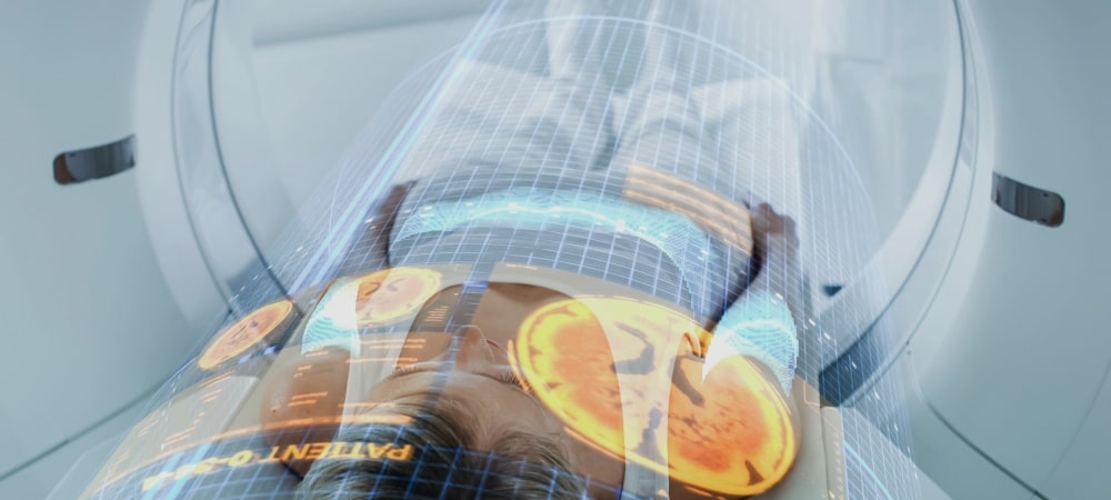
Prostate cancer is one of the most common types of cancer among men worldwide and stands out as a major health problem. Several methods may be used to diagnose prostate cancer, including blood tests, biopsies and imaging scans. However, these may still be unable to precisely determine where the cancer is hiding or how aggressive it is. This is where PSMA PET technology comes in: using the gallium-68 isotope, it can visualize even small clusters of cancer cells with precision. Thanks to this technology, prostate cancer can be diagnosed at an early stage and treated quickly.
What Is PSMA?
PSMA is the abbreviation of prostate-specific membrane antigen. This protein is found on the surface of prostate cells and is present at levels up to 1000 times higher, particularly in cancerous prostate cells, compared with normal cells. Its marked presence in prostate cancer cells makes it an important target for the diagnosis and treatment of prostate cancer. In diagnostic imaging, PSMA is used as a target for positron emission tomography (PET) scans. This property allows prostate cancer within the body to be visualized with a very high degree of precision. PSMA PET imaging offers superior sensitivity and accuracy compared with conventional methods and helps detect the presence and spread of prostate cancer in the body. This imaging technique is particularly useful for showing lesions suspected to be prostate cancer so that biopsies can be taken from the correct areas, for detecting recurrent or metastatic prostate cancer, and for assessing response to treatment.
Beyond its diagnostic use, PSMA also holds promise for the treatment of prostate cancer. Various targeted therapies that act on PSMA are used in the treatment of prostate cancer. Directly targeting prostate cancer cells is more effective and causes far fewer side effects.
In Which Diseases Is PSMA PET Preferred?
PSMA PET imaging is preferred in the diagnosis and follow-up of various cancers, especially diseases related to the prostate. The main areas where PSMA PET is widely used are:
Prostate Cancer: PSMA PET is highly effective in detecting primary and recurrent prostate cancer. It enables detailed imaging of prostate cancer cells and helps accurately stage the disease and determine its location. This allows informed decisions about treatment strategies such as surgery, radiotherapy or targeted therapy.
Biochemical Recurrence: After initial treatment for prostate cancer, some patients may experience an increase in prostate-specific antigen (PSA) levels, which means biochemical recurrence has occurred. PSMA PET is valuable in determining the location and extent of the recurrent disease and guides salvage treatment decisions.
Metastatic Disease: PSMA PET plays an important role in detecting the spread (metastasis) of prostate cancer to other parts of the body, particularly lymph nodes, bones or distant organs. It enables comprehensive imaging of metastatic lesions, helps with treatment planning and helps monitor the response to treatment.
Assessment of Treatment Response: PSMA PET is used to assess response to treatment in patients receiving therapy for prostate cancer. By visualizing changes over time in PSMA expression and tumor burden, it evaluates treatment effectiveness and adjusts treatment approaches accordingly.
Beyond prostate cancer, there are new studies investigating the effectiveness of PSMA PET in other cancers that express PSMA, including renal cell carcinoma, glioblastoma, and certain breast and colorectal cancers. Although initial findings are positive, further studies are needed to determine its effectiveness definitively in these settings.
Which Substance Is Used in PSMA PET Imaging?
In PSMA PET imaging, a radioisotope is used to target and visualize the prostate cell surface antigen (PSMA) found in prostate cancer cells. The most commonly used radioisotope in PSMA PET imaging is 68Ga. The gallium-68 isotope is preferred because of its nuclear properties, which are suitable for PSMA PET imaging. Gallium-68 emits rays that are detected by PET scanners, allowing visualization of PSMA-bearing tissues in the body.
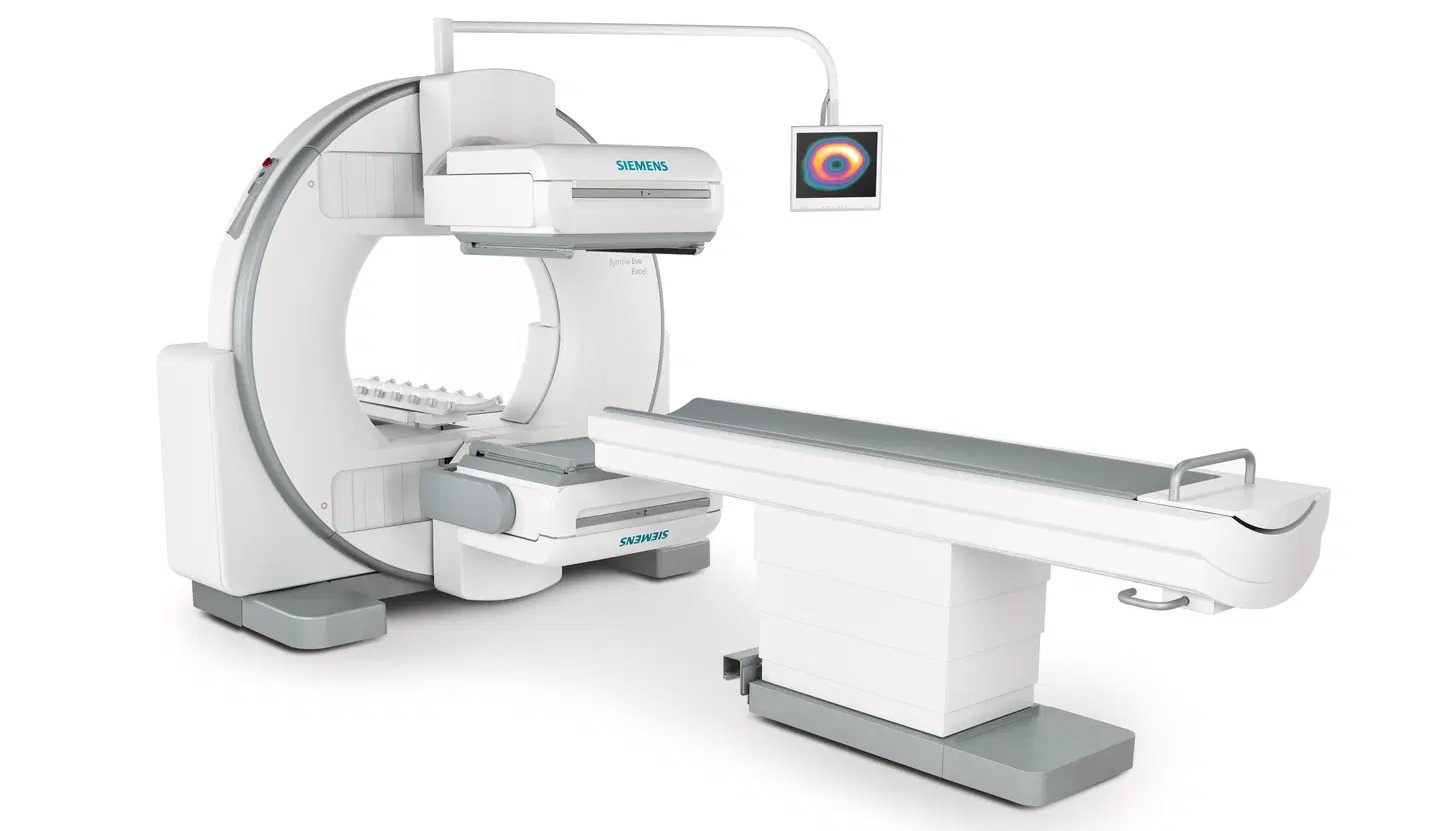
The radioisotope is administered to the patient intravenously and, through the bloodstream, targets cells bearing PSMA, including prostate cancer cells. As the radioisotope accumulates in tumor cells, it emits gamma rays that interact with electrons in the surrounding tissue. These gamma rays are detected by the gallium-68 PET scanner and used to create detailed images of the distribution and intensity of PSMA-bearing tissues. This helps with the diagnosis, staging and monitoring of prostate cancer.
What Is the Preparation Process Before a PSMA PET Scan?
Before a PSMA PET scan, there are specific preparations that patients should follow to ensure the accuracy and effectiveness of the imaging procedure. The preparations may vary slightly depending on the specific instructions provided by the healthcare professional or imaging facility.
Patients do not need to fast for this examination.
Good hydration before the scan is also important. Drinking plenty of water helps the radioisotope distribute better in the body and improves the quality of the PET images. Patients should inform their healthcare professional of all medications they use, including prescription drugs, over-the-counter medications and supplements.
In some cases, patients may need to follow specific bowel preparation instructions to allow better imaging of the pelvic region. These may include following a liquid diet or taking a laxative.
Finally, patients are advised to wear comfortable clothing without metal objects or jewelry that could interfere with the PET scanner. These may include belts, buckles and watches.
What Should Be Considered After a PSMA PET Scan?
After a PSMA PET scan, post-procedure instructions should be followed to ensure patients' well-being and to best interpret the imaging results. Some important points to consider after a Ga 68 PSMA PET scan:
Hydration: Drinking plenty of fluids after the scan is very important to flush the radioactive tracer from the body. This can help reduce radiation exposure to surrounding tissues and organs.
Rest: Some patients may feel tired or weak after the scan, especially if sedation was used during the procedure. If needed, it is important to rest for the remainder of the day and avoid strenuous activities.
Medications: Patients should continue taking any medications that were temporarily stopped before the scan, as directed by their healthcare professionals.
Follow-up Appointments: Depending on the reason for the PSMA PET scan and its findings, follow-up appointments with healthcare professionals may be needed to discuss any further referral or treatment recommendations.
Symptom Monitoring: Patients should be aware of any unusual symptoms or side effects after the scan and report them to their healthcare professional immediately. These may include allergic reactions or excessive bleeding or bruising at the injection site.
Diet: Unless otherwise instructed by healthcare professionals, patients may return to their normal diet after the scan.
Radiation Safety: Patients who have received a radioactive tracer should take measures to minimize exposure, especially pregnant women and young children. This may include avoiding close contact for a certain period, using separate eating and bathing utensils, and following specific waste guidelines for bodily fluids.
Frequently Asked Questions
PSMA PET scans are performed at specialized hospitals and imaging centers. These facilities have advanced PET imaging equipment and experienced healthcare professionals. Scans are carried out in nuclear medicine departments and are performed under appropriate medical supervision according to patient needs. During a PSMA PET scan, a radioactive tracer is injected into the patient through a vein. After the tracer has distributed throughout the body, images of PSMA-containing cells in the body are obtained using a PET scanner. These images are used to diagnose, stage, restage and assess treatment response in prostate cancer and other malignancies that express PSMA. A PSMA PET-CT scan takes approximately 1 to 2 hours. This time includes the time required to inject the radioactive tracer into the body, for its distribution and for acquiring the images. However, the process may vary depending on the patient's individual condition and the specific requirements for the scan. Gallium-68 (68Ga) is a positron-emitting isotope commonly used especially in PET scans. It has a short half-life of approximately 68 minutes, which makes it suitable for imaging procedures requiring rapid decay. Radioisotopes labeled with 68Ga are widely used in imaging various diseases, including cancer and neurological conditions. PSMA PET scans are covered by SGK at hospitals contracted with SGK, provided the criteria specified by SGK are met. Understanding cancer with PET imaging involves analyzing the distribution and intensity of radioisotope uptake in the body. Cancer cells show higher receptor expression, which causes greater uptake of radioisotopes. PET scans visualize this increased uptake as areas of higher radioactivity and indicate the presence and extent of cancerous lesions. Healthcare professionals can diagnose cancer and determine its stage by evaluating the size, location and metabolic activity of these lesions. In addition, PET imaging helps differentiate between benign and malignant tumors, guides treatment decisions and improves patient outcomes. PET scans are performed to diagnose, monitor and evaluate treatment of medical conditions such as cancer, heart and neurological diseases, or a wide range of diseases whose diagnosis has not yet been established. The scans can detect abnormalities by visualizing the condition of cells or tissues, assess disease progression and monitor response to treatment. They provide valuable information about the functioning and health of organs and tissues in the body. PET scan results can usually be available on the day of the scan or the following working day. However, the exact time they will be available may vary depending on factors such as the complexity of the case and the need for additional analysis by nuclear medicine specialists. In PET images, cancer is identified by detecting areas of high metabolic activity. Because cancer cells take up more glucose, these areas appear as bright spots on the image. These bright areas help distinguish cancerous lesions from benign abnormalities when compared with normal tissues. You may consult your doctor to learn more about PSMA PET scanning.