What is PET-CT?
PET-CT, an advanced imaging method in which two separate devices are combined into a single unit, plays an active role especially in oncology, as well as in cardiac and neurological disorders. Thanks to its low radiation dose and high-speed scanning of the entire body from head to toe, it is used in the diagnosis of cancer, treatment planning, and post-treatment evaluation.
PET-CT is an advanced imaging method formed by combining positron emission tomography (PET) and computed tomography (CT).
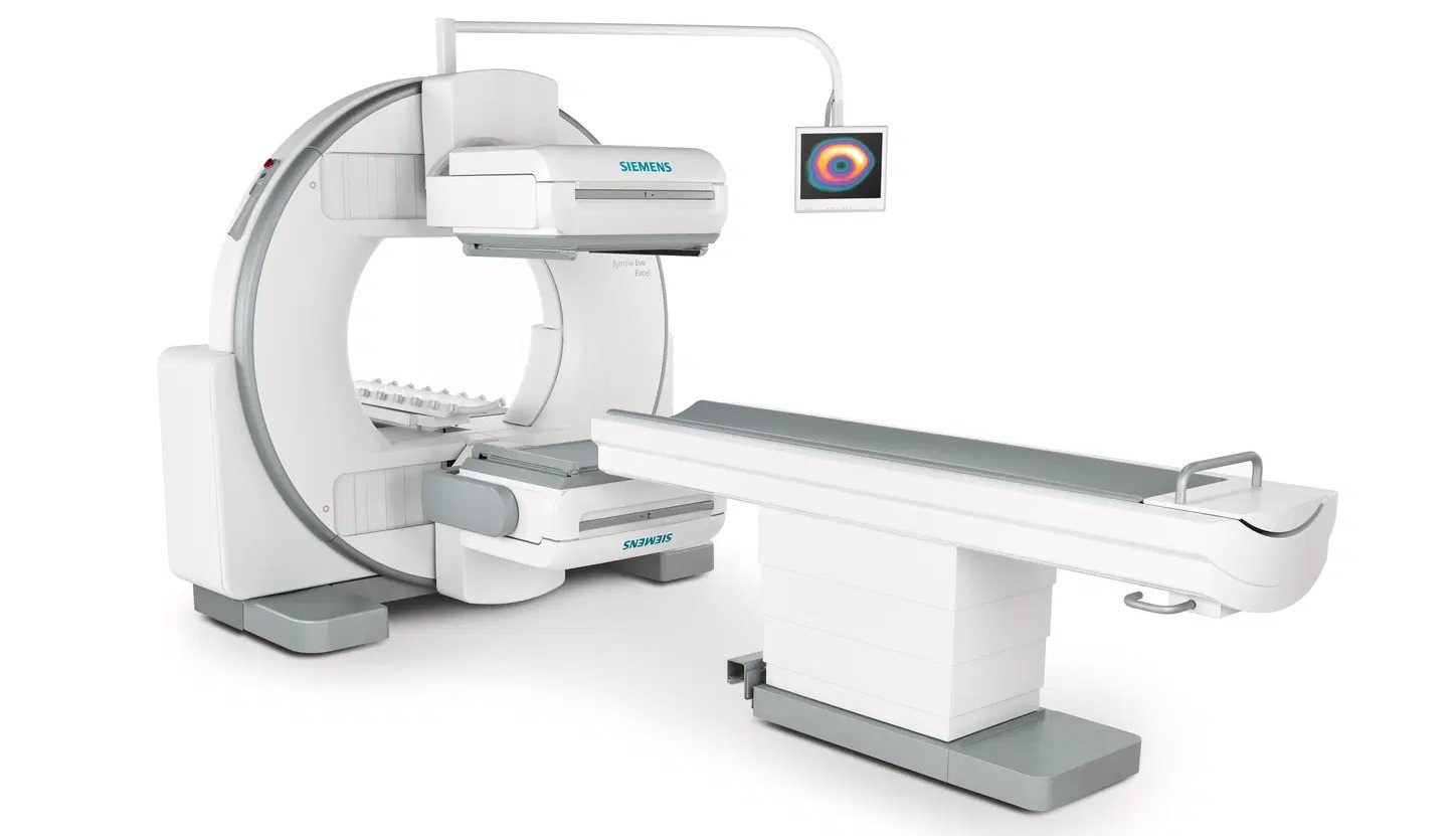
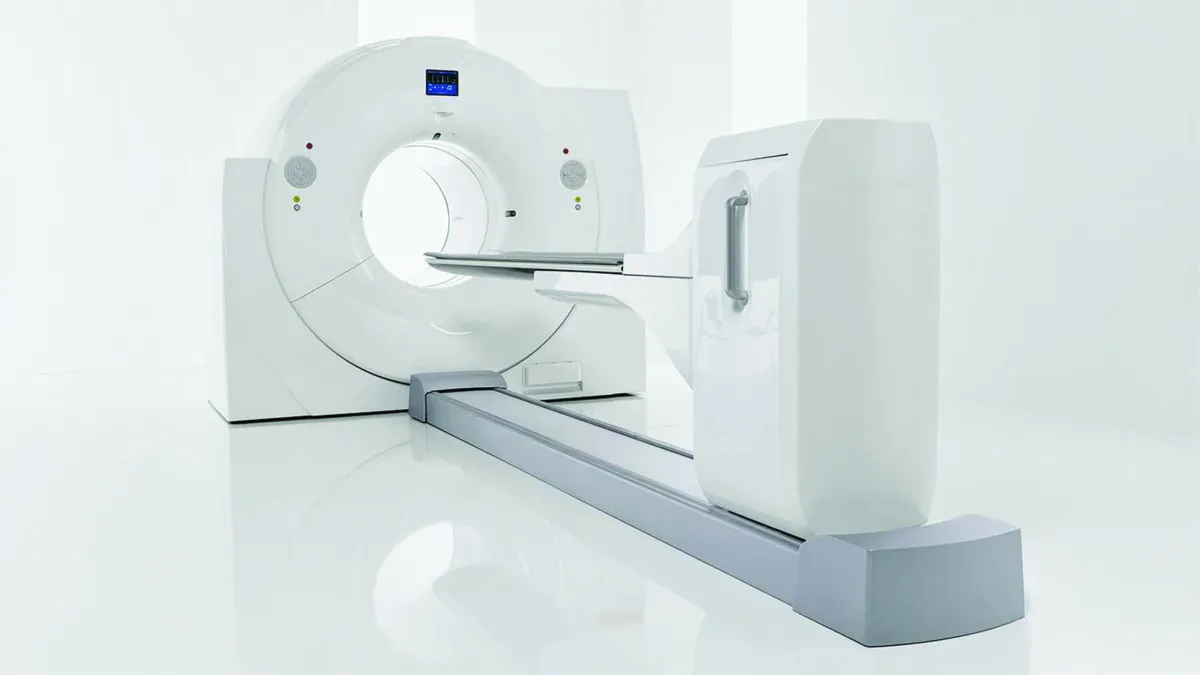
The PET-CT device;
Cancer
Undiagnosed disorders
Infections
Heart diseases
Brain disorders
Differentiation of Alzheimer's and dementia
serves as a guide for early diagnosis and treatment planning in cases such as these.
PET-CT in Oncology
In oncological diseases, PET-CT is used for the early diagnosis of cancer, treatment planning, staging, detection of recurrences, identification of metastases, planning of radiotherapy, and evaluation of treatment success.
The cancer types in which PET-CT is most effective are:
Lung Cancer
Breast Cancer
Thyroid Cancer
Head and Neck Cancers
Colorectal Cancers
Esophageal Cancer
Stomach Cancer
Gynecological Cancers
Bladder Cancer
Prostate Cancer
Lymphoma
Melanoma
PET-CT in Heart Diseases
PET-CT plays a role in identifying areas with reduced blood flow caused by blockages in the coronary arteries. Identifying these areas is important in determining whether angioplasty or coronary artery bypass surgery would be beneficial.
PET-CT in Brain Diseases
PET-CT technology can be used to detect brain tumors and seizures, and in the diagnosis of Alzheimer's and dementia.
What are the Advantages of PET-CT?
Generally, doctors describe and diagnose diseases by overlaying the results of separately performed PET and CT scans. However, for this, the patient must be positioned in exactly the same way for both separate scans. Since achieving this is not always easy, the accuracy of diagnostic information may be compromised.
The PET-CT device, on the other hand, allows doctors to perform both scans quickly in a single session without having to move the patient. Thus, the metabolic data obtained by PET and the anatomical data provided by CT are acquired simultaneously from a single device, providing more detailed and accurate information than other imaging methods.
Benefits for Our Patients
High Resolution
The PET-CT device at our hospital serves our patients with the highest-resolution images in the sector, with a 400x400 matrix reconstruction and 95 mm3. Thus, even the smallest details that cannot be seen with other devices are detected, contributing to the diagnosis and treatment process and helping to prevent unnecessary surgical procedures and biopsies.
Wider Gantry
Designed with overweight and obese patients in mind, our device has a gantry that is 24 percent wider than those of other manufacturers. The larger space is also an advantage for people with claustrophobia.
Low Radiation, High Speed
Our PET-CT device, which operates with a lower radiation dose, increases patient comfort while scanning the entire body with precision at maximum speed, detecting the location of abnormalities such as cancerous cells.
Diagnosis with Less Radioactive Material
Compared with other devices, scans can be performed to obtain high-resolution images using a lower dose of radioactive material administered through a vein.
How Does PET-CT Work?
The steps followed during the scan are as follows:
Fasting for 6 hours is required before the PET-CT scan.
The patient's blood sugar is measured. For the scan, blood sugar must be below the required limit.
A radioactively labeled sugar molecule, which is absorbed by cells, is injected into the body through a vein.
Approximately 1 hour is allowed for the drug to be retained in the cells.
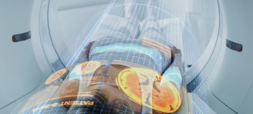
The patient is placed in the PET-CT device and imaging begins.
Detailed 3-dimensional images of the body are taken with the help of X-ray.
Sugar molecules accumulated in abnormal structures or cancerous cells are easily distinguished thanks to their bright images.
The scan takes 20-25 minutes, depending on the area to be imaged.
Process After PET-CT
The radiation received during PET-CT is excreted in urine. For this reason, drinking plenty of water, especially on the day of the scan, and flushing the toilet several times after urination are recommended.
On the other hand, although the radiation received during the procedure does not pose a risk to those around you, it is recommended to avoid close, prolonged contact for 12 hours with young children and pregnant women, who belong to the radiation-sensitive risk group.