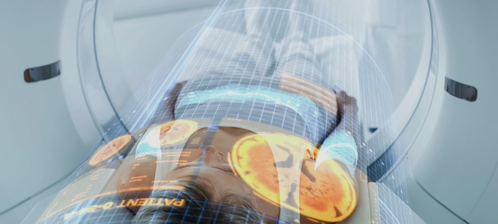
To the question “What is nuclear medicine?” it can be answered as “a branch of science that encompasses the use of radioactive substances for the purpose of diagnosing and treating diseases.” Radioactive substances can be used by labeling samples such as blood taken from a person outside the body, or by administering them directly into the patient’s body through routes such as oral, vascular or respiratory. In this way, physiological images of organs and systems are obtained and their functions are evaluated. The techniques used in diagnosis and treatment are painless, reliable procedures that allow diseases to be observed in advance compared to many other methods. They are procedures frequently preferred especially in the diagnosis and treatment of thyroid diseases, heart and kidney diseases, inflammatory diseases and cancer.
What Is Nuclear Medicine?
The foundation of nuclear medicine is based on the “tracer principle” adopted in the early 1920s. Nuclear medicine can be defined as “the examination and use of radioactive compounds for the purpose of visualizing, diagnosing and treating human diseases.” It is a branch of medicine that can work on a multidisciplinary principle together with sciences such as medicine, pharmacy, physics, biology, nuclear physics, radiology, radiopharmacy and radiotherapy. Nuclear treatments applied in clinics are performed by doctors who have received medical training and specialized in the field of Nuclear Medicine.
Radiopharmaceuticals are radioactive drugs that contain a radionuclide (an atomic nucleus that emits radioactive radiation), can be used for diagnostic and therapeutic purposes, and do not cause a physiological change in the person when administered. These drugs are used in the imaging procedures collectively known as scintigraphy, in blood and urine samples, or directly in the living body. The use of radiopharmaceuticals makes it possible to obtain data that cannot be provided by anatomical methods, and thus makes the treatment of many diseases easier.
What Are Nuclear Medicine Scans Used For?
Nuclear medicine scans are a medical imaging method used to evaluate the functions of organs and tissues in the body. Imaging is obtained through the radiation emitted by radioactive substances within the body. These scans are non-invasive, painless and reliable procedures aimed at the heart, lungs, oncological, neurological, orthopedic and kidney conditions. These scans:
Monitoring of DNA proliferation (the process encompassing the growth, division, repair and multiplication of cells),
Evaluation of organ function and assessment of blood flow,
Detection of abnormal physiological conditions,
Early diagnosis of forgetfulness and dementia,
Early diagnosis of Alzheimer’s disease,
Diagnosis of stroke,
Evaluation of brain and neck vessel surgeries,
Diagnosis of brain death,
Evaluation following head injuries and trauma,
Planning of epilepsy surgeries,
Detection of hidden bone fractures and bone infections,
Detection of tumor sites,
Staging of tumors,
Determining whether tumors have spread,
Relief of pain in cancerous bones,
Determining obstruction in the urinary tract,
Investigating the presence of infection and urinary leakage in the kidneys,
Diagnosis of blood clots in the lungs (pulmonary embolism),
Evaluation of bypass surgery,
Investigation of the cause of disease in hypertension patients,
Detection of coronary artery diseases,
Goiter diseases and treatment of toxic goiter,
Diagnosis of esophageal and stomach diseases,
For problems related to the gallbladder,
Suspicion of hidden infection,
Evaluation of blood flow and function of transplanted kidneys,
Detection of lymphatic system blockage,
Intestinal bleeding,
Diagnosis of cardiomyopathy and coronary artery diseases,
Follow-up of metastatic cancer,
Examination of tear ducts,
Evaluation of salivary gland functions,
Prostate cancer screening,
Imaging of neuroendocrine tumors (pancreas, liver),
Distinguishing between benign and malignant tumors,
Evaluation of responses to cancer treatment,
It can be used for many purposes, such as making and differentiating dementia and Alzheimer’s diagnoses in neurology.
These applications, which provide many benefits to human health, are frequently preferred for the purpose of diagnosing and treating diseases. These applications are performed by specialist physicians in the field. Those who wish to obtain more detailed information and be examined may apply for a specialist consultation by making an appointment.
Why Do We Need a Nuclear Medicine Department?
Nuclear medicine is an important main branch of science with multidisciplinary working principles. It is an important tool used in the diagnosis and treatment of many diseases. It is a method applied especially in conditions where diagnosis and treatment are important, such as cancer, heart, kidney, brain, thyroid and bone diseases. Anatomical imaging techniques such as ultrasonography and radiography may be insufficient for imaging in the early stages of diseases because they rely on morphological changes. Scintigraphic imaging methods, on the other hand, because they rely on the detection of physiological changes, allow much earlier diagnosis of diseases. The scintigraphic images obtained provide information about the functions and anatomy of the systems in the body. They enable early diagnosis of diseased tissues or any physiological abnormality. These methods offer patients a reliable, easily applicable and painless treatment option. Therefore, they are methods that are needed and widely used all over the world.
How Does Nuclear Medicine Imaging Differ from Other Radiological Tests?
Nuclear medicine imaging methods are performed by injecting radiopharmaceutical drugs into the body. In other radiological tests, they are usually performed using techniques such as X-rays and magnetic resonance imaging applied from outside the body. Nuclear medicine imaging enables the evaluation of the functions of organs in the body through radioactive rays, whereas radiological tests are mostly used for anatomical examinations.
What Are the Risks and Benefits of Nuclear Medicine Imaging?
The use of radioactive substances in diagnostic and therapeutic procedures often causes people to wonder about the answers to questions such as “Is nuclear medicine harmful?” and “What are the side effects of nuclear medicine?” However, the dose adjustments of radioactive substances used for treatment are carefully made by specialists. There is no evidence that radiopharmaceuticals at these doses cause significant discomfort or serious radiation-related damage in humans. The amount of radiation to which the patient is exposed in these diagnostic and therapeutic methods is equal to or less than that of other X-ray films.
Radioactive substances are eliminated from the body through urine and the intestines. The rate at which they are lost from the body varies in direct proportion to their half-life. Pregnant women, babies and children are more likely to be affected by radiation. For this reason, individuals in this group need to stay away for a certain period of time from patients undergoing these treatment procedures, in order to protect themselves from the possible side effects of radioactive substances. Care should also be taken that radioactive substances are not spread through urine. Allergic complications are also among the risks of imaging. However, allergic side effects are negligibly rare.
Nuclear medicine is used in the diagnosis of heart diseases, cancer, cerebrovascular diseases, thyroid diseases and many other diseases. Furthermore, in some cases, it can increase the chance of treatment by enabling early diagnosis of diseases. It can be used directly in the treatment of diseases, and also in monitoring the treatment process to evaluate its effectiveness and observe the course of the disease. These imaging methods are reliable, economical and effective. Therefore, applications carried out correctly by experts ensure that the risks of nuclear medicine imaging are minimized.
What Are the Procedures Used for Diagnosis in the Nuclear Medicine Department?
Nuclear medicine uses radiopharmaceuticals for the diagnosis and detection of diseases, and enables the acquisition of data that cannot be provided by anatomical imaging methods. The diagnostic methods used to evaluate the functions of specific tissues and organs are divided into two:
Non-Imaging Diagnostic Methods: Radiopharmaceutical drugs specific to certain tissues and organs are administered into the body. These drugs reach the target area, after which radioactivity counting is performed. Iodine applications used in the diagnosis of thyroid diseases can be given as an example of non-imaging diagnostic methods. Radioactive iodine accumulates in the thyroid gland; from radioactivity measurements taken from the thyroid gland region, numerical data on the metabolism and function of the gland can be obtained. Another non-imaging diagnostic method is the administration of radiopharmaceuticals to determine the location of tumorous tissues.
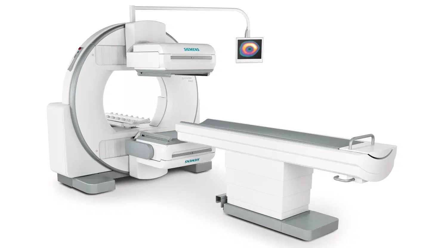
Imaging Diagnostic Methods: In this method, a radiopharmaceutical specific to a certain tissue or organ is administered into the body and, after it reaches the target area, images are obtained using various imaging devices. Depending on the characteristics of the radiopharmaceutical applied, the devices used in imaging diagnostic methods can be listed as gamma cameras, SPECT, PET, SPECT/CT and PET/CT.
PET/CT is a medical imaging device that combines positron emission tomography and computed tomography. The PET/CT device used within Hisar Hospital Intercontinental has the capacity to take detailed images in 40 different slices at the same time. This shows that the device has the ability to obtain high-resolution and detailed images. These devices are frequently used especially in applications aimed at diagnosing cancer, heart and neurological diseases. A 3-dimensional image is obtained with PET/CT scanning. Radioactively labeled drugs are injected into the body, and the drug is allowed to wait for about 1 hour so that it can attach to the cells. The scan duration may last approximately 10–20 minutes.
What Is Radioactive Therapy?
Radioactive substances are low-radiation, short-lived radiopharmaceutical drugs specially produced in the laboratory to be administered to the human body, which do not accumulate in the body and are mostly used in nuclear medicine. These drugs can be used for the diagnosis and treatment of diseases. Radioactive treatment methods can be applied in bone diseases, cancer, kidney, brain and thyroid diseases, and many other medical conditions. Radioactive drugs decrease by spontaneously halving physically, and can also be excreted from the body through urine, sweat and the intestines. The side effects and allergic effects of these drugs are negligibly rare.
For Which Diseases Is Radioactive Therapy Used?
Radioactive therapy is a treatment method used mostly for the treatment of cancer. In order to destroy cancer cells systemically or locally, treatments can be applied with agents that deliver very high doses of radiation specifically to the disease site, using radioactive substances such as Lu-177, Ac-225, Y-90 and I-131. Also, radioactive iodine therapy can be applied to bring under control the disorders that develop due to overactivity of the thyroid gland. It can be used for the treatment of bone marrow disorders and diseases such as rheumatoid arthritis. Radioactive treatment applications are determined according to the type of disease and the patient’s general health status after detailed evaluation by specialist physicians.
How Is Radioactive Therapy Administered?
Patients undergoing radioactive therapy and their companions must be informed in detail about radiation safety before examinations are performed. In nuclear medicine departments, information forms must be distributed to patients and their companions. Pregnant women and children must be prevented from entering radiation areas and areas with contamination risk. Examples of the methods used in radioactive therapy applications include:
Bone Scintigraphy: In this method, a radioactive drug is injected through a vein. Imaging with the gamma camera begins 2–6 hours after the drug is given and lasts about 20–30 minutes. To reduce the dose of radiation received during the procedure, frequent fluid intake is recommended.
Dynamic Renal Scintigraphy: Before this method is started, the patient’s bladder is completely emptied. To perform the test, a radioactive drug is given through a vein and imaging begins immediately. This procedure may last 30–40 minutes.
Thyroid Scintigraphy: Imaging tests with the gamma camera device begin 20–60 minutes after the radioactive drug is administered and last about 10 minutes. Before this procedure, you should consult your doctor and provide information about your general health status and the medications you use. If your physician deems it appropriate, they may ask you to pause the medications you use for a while.
Renal Cortical Scintigraphy: The imaging test, which will last about 10–15 minutes, begins 2–6 hours after the radioactive drug is administered through a vein. It is absolutely necessary not to move during imaging.
Myocardial Perfusion Scintigraphy: For this test, fasting for at least 4 hours and discontinuing some medications are required. Your doctor will give you general information about what you need to do before the procedure. This method is also known as a stress test. You will be asked to walk on a treadmill until you reach at least 85% of your heart rate. The radioactive drug is administered at the peak of the exercise, and the test with the gamma camera begins 30–60 minutes later. All these studies may last 4–5 hours.
What Is PET/CT? Which Diseases Is It Used to Diagnose and Treat?
PET/CT, the combined use of positron emission tomography and computed tomography, is one of the most advanced and powerful ways to image normal and altered physiology in the body. The PET/CT device combines functional and anatomical imaging, and is an imaging technique that plays a role in the response to treatment and in its follow-up. PET/CT is used in the diagnosis and treatment of many diseases. These diseases can be listed as follows:
Head and neck cancers,
Lung cancer,
Lymphoma,
Skin cancers,
Gynecologic cancers,
Gastrointestinal cancers,
Genitourinary cancers.
Another frequently asked question is “What is the difference between PET/CT and PET/BT?” However, these two terms are used to describe the same device. PET/CT is the English abbreviation, while PET/BT is the Turkish abbreviation. PET/BT reports are written by specialist physicians, and you can arrange to speak with your physician by bringing a copy of your PET/BT result.
You too can benefit from nuclear medicine services, a reliable, painless and economical method, in your diagnosis and treatment planning. You can learn about the biological processes of your body through scintigraphy, PET/CT and other nuclear medicine imaging techniques. Our nuclear medicine reports are written by Associate Professor physicians who are experts in their field, and we also have an SGK agreement for nuclear medicine applications. You can contact us and make an appointment for more detailed information.