Myocardial Perfusion Scintigraphy (MPS)
Myocardial perfusion scintigraphy (MPS) is a nuclear imaging method in which blood flow to the heart muscle is evaluated under resting and stress (exercise or pharmacologically induced stress) conditions. In this test, a low dose of a radioactive substance (usually a technetium-99m isotope) administered intravenously reaches the heart muscle, and the distribution of this substance in the heart is monitored with the help of a gamma camera. Thus, it is determined whether the heart is sufficiently perfused, whether there is a vascular occlusion, or to what extent the heart tissue is affected after a previously experienced heart attack.
What is Myocardial Perfusion Scintigraphy?
Myocardial perfusion scintigraphy (MPS) is an advanced nuclear medicine imaging test that shows whether sufficient blood reaches the heart muscle. It is performed to directly evaluate the function of the coronary vessels that meet the heart's oxygen and nutrient needs. Basically, the perfusion levels of the heart tissue are observed; this reveals whether there is any narrowing or blockage in the coronary vessels. During this procedure, a small amount of a radioactive substance is injected into the body. This substance reaches the heart muscle through the bloodstream, and its distribution in different regions of the heart is imaged with a special camera system. Thanks to the images obtained, it can be analyzed in detail which parts of the heart are sufficiently perfused and which are insufficient.
What Are the Indications for Myocardial Perfusion Scintigraphy?
MPS is generally applied to patients with specific complaints and clinical risk conditions. It is a test performed based on the need for diagnosis, not as a symptom of a disease. Myocardial perfusion scintigraphy findings are usually evaluated by comparing stress and rest images. Fixed defects indicate an old infarct, while reversible defects indicate ischemia. The following conditions are among the main reasons for performing MPS:
In individuals who experience chest pain but cannot reach a clear conclusion with ECG and blood tests,
In patients who are at risk of having a heart attack but have not yet had one,
In individuals with known coronary artery disease (CAD), to understand the degree of progression of the disease,
After heart surgery or stenting, to evaluate the effectiveness of the treatment,
As an alternative method in patients who cannot perform an exercise test or whose results are inconclusive,
In cases where cardiac reserve needs to be evaluated before surgical intervention.
Why Is Myocardial Perfusion Scintigraphy Performed?
Myocardial perfusion scintigraphy is performed to evaluate whether the heart muscle receives a sufficient level of oxygen and nutrients. The main purpose of this test is to observe whether the coronary vessels feeding the heart are performing their duties in a healthy way. It is very useful for distinguishing whether these complaints are heart-related in patients who present with symptoms such as chest pain, shortness of breath, and fatigue but cannot reach a clear diagnosis with standard tests. It is also used to see if the disease is progressing in people with known heart disease, to evaluate the effect of the treatments applied, or to predict future risks. MPS is also preferred before a surgical or interventional procedure to understand whether the heart can withstand such an intervention. In cases where an exercise test cannot be performed in some patients, MPS becomes an alternative diagnostic tool by creating stress with drugs.
How Is Myocardial Perfusion Scintigraphy Diagnosed?
Myocardial perfusion scintigraphy is not used to diagnose a disease directly, but to assist in diagnosis by evaluating the perfusion status of the heart muscle. In other words, this test does not define a disease on its own, but it provides a strong basis for the physician to understand the underlying cause of existing symptoms. Myocardial perfusion scintigraphy indications include diagnosis of coronary artery disease, ischemia evaluation, follow-up after revascularization, and differential diagnosis of atypical chest pain. Especially in conditions related to the narrowing or blockage of heart vessels, such as coronary artery disease, if sufficient blood cannot reach the heart muscle from the vessels, this situation can be imaged thanks to MPS.
The data provided by the test contains critical information such as in which region of the heart blood flow has decreased to what extent, and whether this decrease occurs in the resting state or only under stress. Thus, the physician obtains detailed information about the presence, severity, and location of the heart disease. Used as part of the diagnostic process, this test is a guide in planning surgical decisions, stent applications, or drug therapy. Especially in cases where a diagnosis cannot be made with inconclusive exercise tests or classic ECG findings, MPS significantly increases diagnostic accuracy.
Myocardial perfusion scintigraphy results reveal ischemic areas by showing whether blood flow to the heart muscle is sufficient. Exercise myocardial perfusion scintigraphy is a method in which the heart's perfusion under stress is imaged by having the patient exercise. Gated myocardial perfusion scintigraphy evaluates not only perfusion but also functional parameters such as left ventricular wall motion and ejection fraction. Gated SPECT myocardial perfusion scintigraphy provides a three-dimensional imaging of the heart, offering a more detailed and precise analysis.
How Is Myocardial Perfusion Scintigraphy Performed?
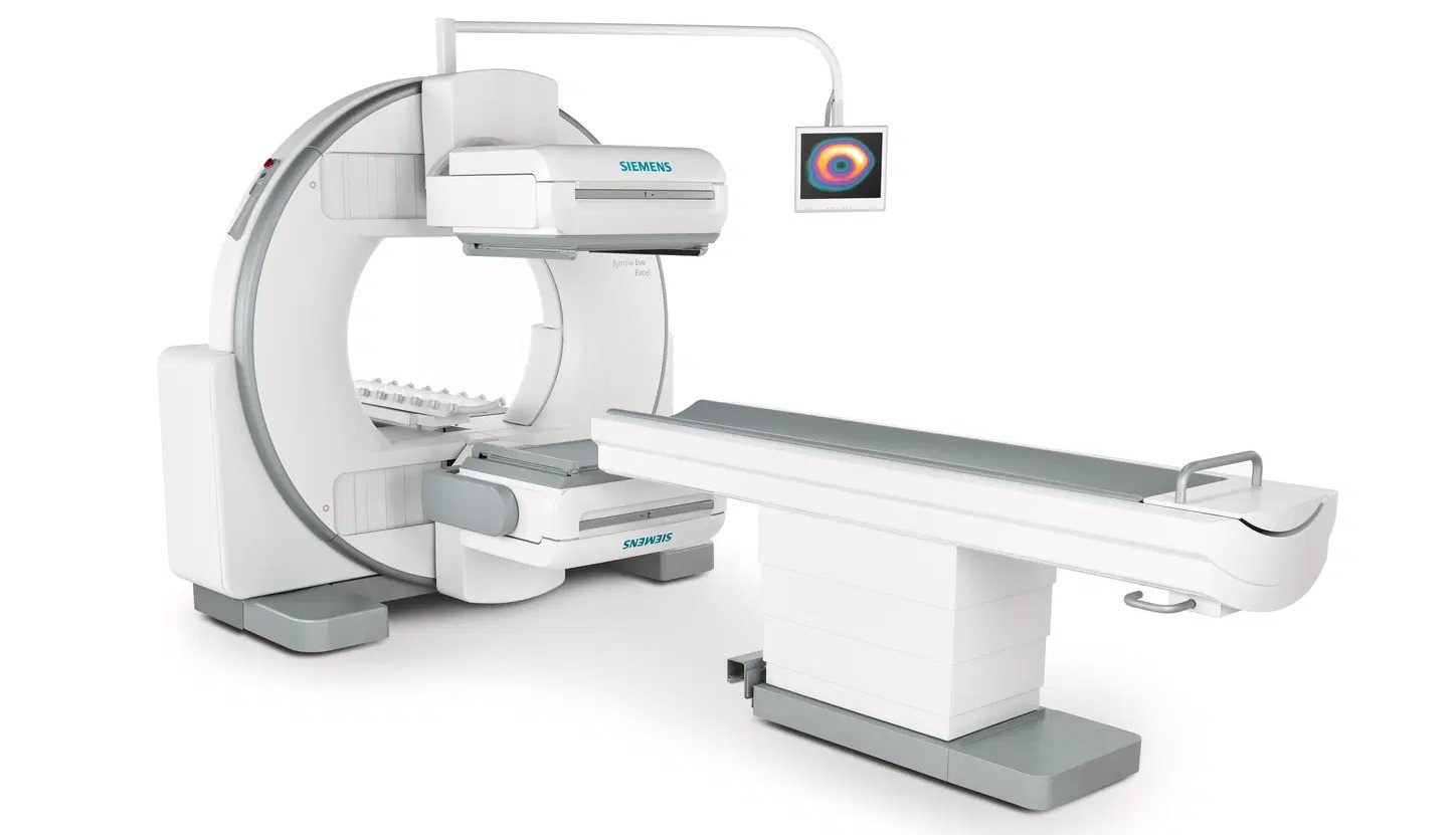
Myocardial perfusion scintigraphy is usually performed in two separate stages: the stress phase and the rest phase. The goal is to see how the heart works both at rest and under strain (exercise) and to analyze the differences between these two states. On the day the test is to be performed, the patient is asked to make certain preparations. Stopping caffeine consumption, temporarily discontinuing some medications, and usually fasting for a few hours are among these preparations. In the first stage, the patient's heart is put under stress. This is usually done with a physical exercise test on a treadmill. However, in patients who are unable to exercise, pharmacological stress is created using a special drug that dilates the heart vessels. At this stage, the heart vessels are forced to work rapidly. During this time, a low dose of a radioactive substance is administered intravenously. This substance follows the flow in the blood to reach the heart muscle, and its distribution in the heart is imaged with a special device called a gamma camera.
The second stage, the rest phase, is performed while the heart is working at its normal rhythm. At this stage, imaging is taken in a similar way by administering the radioactive substance again. The images obtained allow for the comparison of the heart muscle's perfusion under stress and at rest. The entire test can usually be completed within the same day, but some centers may prefer to perform it over two separate days.
During the imaging process, the patient lies on their back on a special table. The device rotates around the patient and takes images of the heart from different angles. There is no feeling of pain or discomfort during the procedure. When the test is completed, the radioactive substance given to the body is excreted naturally over time. The images are analyzed by a nuclear medicine specialist and the results are sent to the physician. In this way, it is clearly revealed how healthy the heart is working and in which regions there is a risk.
Frequently Asked Questions
The amount of radiation received during MPS is generally at an acceptable level and is quite low compared to the diagnostic benefit. The radiation received during an average MPS test is around 10-12 millisieverts. This is a level similar to annual natural radiation exposure. However, it is important that it is not repeated unnecessarily and is performed within medical necessity. Some preparations may be required before the test. Especially caffeinated beverages (tea, coffee, chocolate) should be stopped at least 48 hours before the test. Some medications used may also be temporarily discontinued. The relevant center decides whether fasting is required. The patient should share all current medications, and any diabetes or blood pressure problems with the doctor. It is important for the patient to wear comfortable clothes during the test and to come with appropriate shoes, especially if a stress test is to be performed. If complaints such as dizziness, chest pain, or shortness of breath occur during the exercise test, the test team must be informed. Throughout the process, the patient is under the supervision of expert medical personnel. Although the duration of the test varies according to the center's application protocol, it is usually completed between 2 and 4 hours. Two-day protocols may be applied in some centers. Stress and rest phases can be performed on different days. Serious side effects after MPS are rare. The excretion of the radioactive substance from the body is rapid. Apart from mild headache, fatigue, or slight pain at the injection site, there is usually no significant problem. Allergic reactions are very rare. The area where it is most frequently used is coronary artery disease. It can also be used in many areas such as evaluating myocardial viability after heart attacks, observing the effect of heart valve diseases on the heart, and performing risk assessment before heart surgeries. As long as it does not contain doses much higher than the radiation encountered in daily life, the MPS test is generally safe. However, it should be used with caution in patients during pregnancy or with severe kidney failure. As always, the benefit-risk ratio should be evaluated by the doctor. Results are usually available within 1-3 business days after the completion of the test. However, this period may vary depending on the workload and the operation of the center. The results are reported by an expert nuclear medicine doctor and forwarded to the cardiologist for interpretation. MPS can also be performed in patients who have previously had a stent inserted. This test is quite useful for understanding whether the stent is doing its job sufficiently or if there are new narrowings. However, in this case, the patient's suitability is decided by the doctor. Seeing that there is insufficient perfusion in some regions of the heart as a result of the test may indicate that the risk of heart attack is high in that person. This allows preventive treatments to be planned in a timely manner. It is usually requested by cardiology specialists. However, internal medicine specialists or cardiovascular surgeons can also refer the patient to nuclear medicine departments when they deem it necessary.
For a clear assessment of your heart health, Myocardial Perfusion Scintigraphy (MPS) is one of the most reliable methods. If you have chest pain, shortness of breath, or a suspicion of heart disease, you can apply to our expert Nuclear Medicine Department physicians without delay.
You can make an appointment by calling us or you can easily apply through our online appointment system.