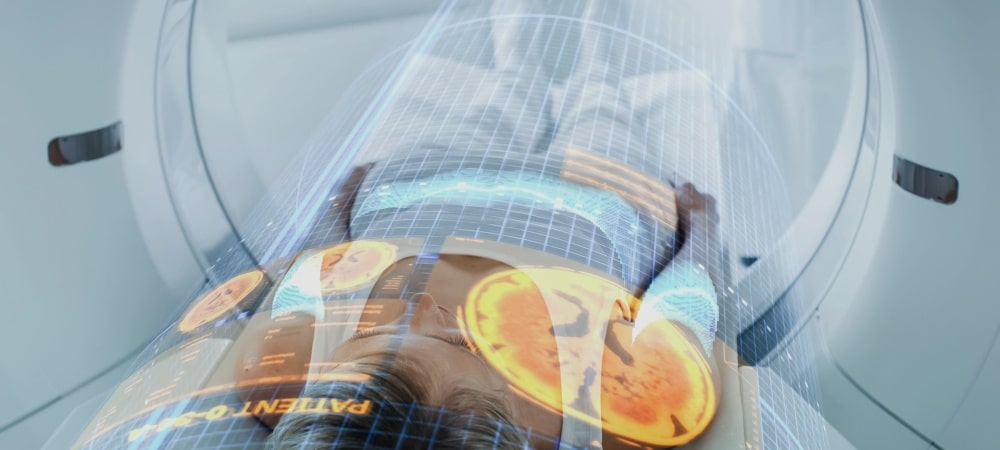
Bone scintigraphy is a nuclear medicine method used in the diagnosis, treatment and follow-up of bone diseases. Thanks to the injection of radioactive tracer substances into the body during bone scintigraphy, abnormal cellular activities can be visualized. Bone scintigraphy is one of the methods that specialists frequently use to identify the factor causing pain, especially in bone pain of unexplained cause. In addition, specialists may resort to bone scanning to check whether cancers such as breast or prostate cancer have spread to the bone.
What Is Bone Scintigraphy?
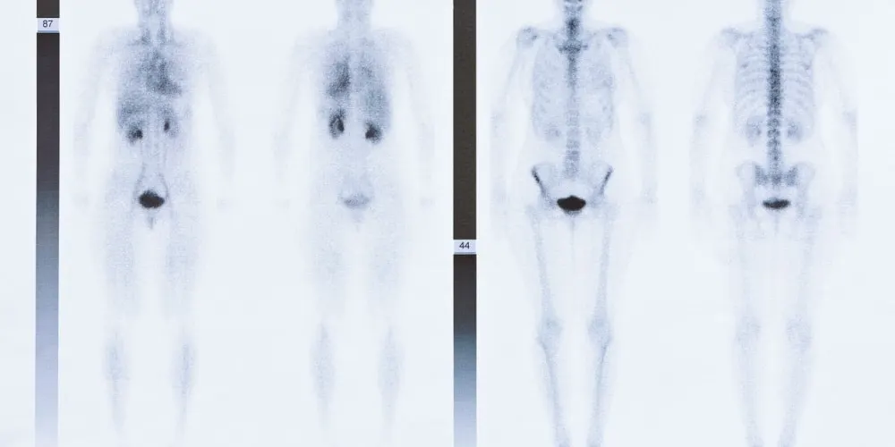
Bone scintigraphy, also called bone scan, is a nuclear medicine method used to diagnose abnormalities occurring in bone tissues, to evaluate whether the applied treatment is working, and for similar purposes. During a bone scan, a radioactive substance is injected into the patient. The injected substance collects in areas where cellular changes are intense, called “hot spots,” and makes these areas stand out on the imaging device. These hot spots may be a sign of many diseases, from infections to trauma, from metastatic cancers to arthritis. Thus, through scintigraphy, fractures, infections or tumors that cannot be detected by classic imaging methods such as X-ray can be seen (1, 2).
Bone scintigraphy also plays an important role in the follow-up of cancer patients. Whole-body bone scintigraphy can be used to determine whether cancer has metastasized to the bone, i.e., spread to the bone, and to evaluate the stage of the disease. Especially since the risk of spreading to the bones is high in cancer types such as breast and prostate cancer, specialists use bone scanning in determining the treatment process (1, 2).
In Which Situations Is Bone Scintigraphy Used?
Bone scintigraphy is an imaging method used in the diagnosis, treatment and follow-up of many health problems, above all bone pain of unexplained cause. In particular, specialists may resort to bone scanning to detect whether cancer cells have spread to the bone, to determine the stage of the cancer before and after cancer treatment, and to evaluate the effectiveness of treatment. In addition, other areas of use of bone scintigraphy are as follows (1):
Detecting bone traumas that cannot be visualized by X-ray,
Finding fractures whose location is difficult to determine,
Determining when the fracture occurred,
Diagnosis of bone infections such as osteomyelitis,
Diagnosing joint or prosthesis infections,
Finding the source of bone pain that cannot be determined by X-ray or other methods
Diagnosis, treatment and follow-up of rheumatic diseases such as arthritis and osteoarthritis,
Paget’s disease, characterized by thickening and softening of the bones,
Avascular necrosis, i.e., a health problem characterized by death of bone tissue as a result of decreased blood flow,
Benign bone tumors,
Cancers that start in the bone,
Cancers that have spread to the bone from different tissues and organs, such as the breast and prostate,
Fibrous dysplasia (abnormal development in bone tissue).
Whole-body bone scintigraphy can help visualize damage or structural changes throughout the entire skeleton. In this way, after bone scintigraphy, specialists can plan the treatment process individually for the person.
How Is Bone Scintigraphy Performed?
For bone scintigraphy, patients usually do not need a special preparation before the procedure, but it is important for patients to inform their doctors of the medications they use regularly. In addition, metal-containing jewelry such as necklaces, earrings and bracelets, or accessories such as belts, must be removed during the procedure. Furthermore, if the patient has medical devices such as a joint implant, dental implant or pacemaker, it is important to inform the doctors of this before the bone scan.
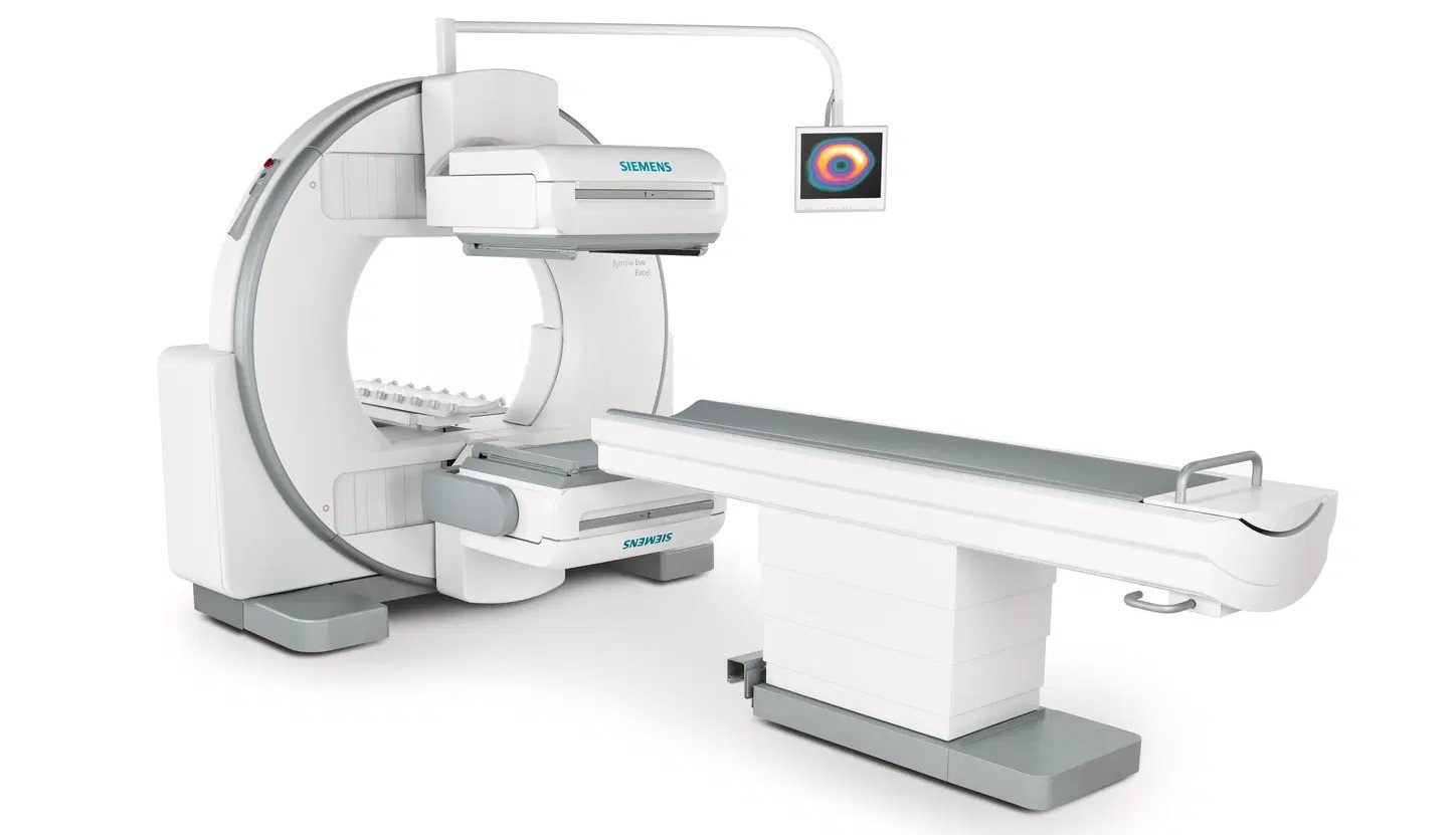
During the bone scintigraphy procedure, the nuclear medicine technicians first inject the radioactive substance into the patient. Although the bone scan itself usually takes 30 to 60 minutes, the patient may need to wait one to four hours at the healthcare facility for the drug in the bloodstream to reach the bones (3). After the waiting period for the drug to reach the bones ends, patients are taken into the examination room and asked to lie supine on the examination table. At this stage, a large camera moving over the body begins to take images. The camera detects the hot spots where the radioactive substance has collected. During the scanning process, to obtain clearer images, specialists may ask patients to hold their breath for a few seconds. In some cases, specialists may use an imaging method called single photon emission computed tomography (SPECT) to visualize the deeper parts of the bones (4).
In Which Diseases Is Bone Scintigraphy Applied?
Bone scintigraphy plays an important role in the diagnosis and follow-up of many health problems, including cancer. Some of the health problems for which bone scanning is applied are as follows (5, 6):
Primary bone cancers and bone metastases: Cancer cells can metastasize to bones as they spread throughout the body, regardless of the tissue or organ of origin. Especially in advanced stages of cancer types such as breast, prostate, lung and kidney cancer, bone metastases may be seen. Bone scanning can be used in the diagnosis, treatment and follow-up of both cancers originating in the bone and cancers that have spread to the bone from other organs.
Osteoporosis: Also known as bone thinning, osteoporosis is a health problem characterized by decreased bone density and an increased risk of fractures in the bones, which especially affects women during menopause. In patients with osteoporosis, bone fractures are usually seen in the vertebrae, i.e., the spinal bones. Other imaging methods may not clearly show fractures in the vertebrae. Bone scanning helps to locate fractures and to evaluate whether the cause of bone pain is a bone fracture.
Paget’s disease: Paget’s disease is a health problem presented by bone destruction and abnormal bone structure, whose cause is not precisely known, and which may affect a single bone or the entire skeletal system (7).
Bone infections (osteomyelitis): Bone infections can cause swelling and inflammation in the bones. Bone scintigraphy can detect osteomyelitis, characterized by these features, at an early stage, and the stage of the disease can be evaluated.
Arthritis and other joint diseases: Bone scintigraphy is also widely used in the diagnosis, treatment and follow-up of inflammatory joint diseases such as rheumatoid arthritis.
Bone scintigraphy is an imaging method used especially in patients with occult or small fractures.
When Are Bone Scintigraphy Results Available?
A bone scintigraphy report generally cannot be obtained immediately. After the scanning procedure, the images obtained are examined by a radiologist who is an expert in the field, and abnormalities in metabolism are analyzed in detail. Interpreting the scan results and delivering them to the patient may usually take 1 to 2 weeks. Specialists inform patients after scintigraphy about when the results can be obtained. If specialists detect suspicious areas as a result of the scan, they may request additional tests in order to plan the treatment process (3).
Frequently Asked Questions
Bone scintigraphy is usually requested to determine whether cancer has spread to the bones. In addition, bone scanning can be performed to determine metastases, as well as to stage the cancer before and after treatment and to evaluate the response to treatment. It can also be used in the diagnosis and treatment of health problems such as bone traumas that cannot be detected by X-ray, hard-to-see fractures, bone infections (osteomyelitis), unexplained bone pain and Paget’s disease. Bone scintigraphy is an important medical imaging method used to detect diseases and abnormalities in the bones. Bone scanning provides important information to specialists in the diagnosis, treatment and follow-up of both cancers originating from the bone and cancers spreading to the bone from other tissues and organs. In addition, bone scanning can be used to evaluate fractures that are difficult to detect with other imaging methods, bone infections, rheumatoid arthritis and metabolic bone diseases such as Paget’s disease. Specialists can also use bone scintigraphy to understand the cause of bone pain whose reason cannot be determined. Bone scintigraphy is generally considered a safe imaging method. Although a radioactive substance is used to examine abnormalities in the bones during the procedure, the dose used is quite low. Therefore, patients can return to their daily lives shortly after the procedure. However, specialists may recommend that patients drink plenty of water for 48 hours after the procedure in order to accelerate the excretion of the radioactive substance from the body. For most people, bone scintigraphy does not pose a serious risk. In addition, the risk of radiation spreading to the patient’s surroundings is also quite low. However, since radiation may cause unwanted effects in pregnant or breastfeeding women, bone scanning is not recommended for these individuals (3). During bone scintigraphy, while the radioactive substance circulates in the patient’s body, patients generally do not feel anything. The only discomfort that may be felt during the scanning procedure is the discomfort of the injection that the radiology technician uses when injecting the radioactive substance through a vein. Bone scintigraphy is one of the medical procedures with virtually no side effects. However, as with every medical procedure, patients may experience some mild effects after the procedure. During the procedure, slight bruising or swelling may occur at the injection site. This usually resolves on its own within a few days. In rare cases, some patients may develop swelling and pain in the arm due to leakage of the radioactive substance outside the vein. Bone scintigraphy is not recommended for pregnant women, since the radioactive substance used during bone scanning may harm the developing baby. The frequency of bone scintigraphy may vary depending on the purpose of the scan and the general health status of the patient. For example, some specialists suggest that women whose bone density is found to be healthy in the first scan may wait 15 years for a new scan, while women with severe bone density loss at the onset of osteopenia, the stage preceding osteoporosis, should have a bone scan once a year. Bone scintigraphy plays an important role in the diagnosis and treatment of many health problems, above all cancer. If symptoms such as bone pain of unexplained cause are present, it is useful to consult a specialist doctor to get information about bone scanning.