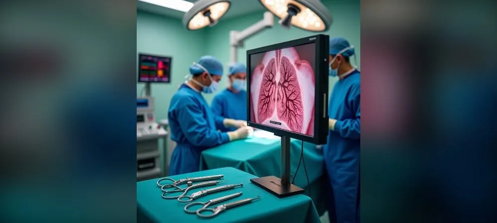
What is Segmentectomy?
Segmentectomy is the removal of only the independent unit containing the diseased section instead of an entire lobe of the lung. This surgical method preserves the patient's respiratory capacity by protecting healthy lung tissue to the maximum extent. Compared to classical lobectomy, this procedure requires much higher technical skill and allows for the preservation of 15-20% more of the patient's postoperative respiratory capacity.
Lung Anatomy and Segmental Structure
Although the lungs appear to be large structures composed of lobes when viewed from the outside, each lobe is internally composed of bronchopulmonary segments. These segments function as independent units of the lung, and each has its own airway and artery. This unit of the lung, where a segmental bronchus distributes, is called the bronchopulmonary segment.
There are three fundamental characteristics that define the structure of each segment: independence, functionality, and protection. The lung can be thought of as an apartment building, and the segments function like independent apartments within that building. If there is a problem in one of the apartments, it is possible to isolate only that apartment instead of demolishing the entire building or an entire floor.
The right lung is divided into exactly ten independent segments. The left lung has a different structure due to the placement of the heart and generally consists of eight main segments. Each segment has its own segmental bronchus and pulmonary artery. Venous and lymphatic drainage occurs along the edges of the segments.
Differences Between Segmentectomy and Lobectomy
Lobectomy is the removal of an entire lobe of the lung. Segmentectomy, on the other hand, involves the removal of only one or several anatomical segments. This difference is important because more lung tissue is preserved compared to lobectomy.
Segmentectomy is preferred especially for small tumors or in patients with limited lung capacity. It is applied to stage I patients who are suitable for lobectomy and to patients who are not suitable for lobectomy due to limited cardiopulmonary reserve. Thanks to this anatomical independence, if the surgeon can separate the segmental vessels and bronchi with millimeter precision, they can remove only the relevant unit without damaging the neighboring segments.
Organ-Preserving Surgical Approach
In surgery, the philosophy of "the more tissue we remove, the safer it is" has now been replaced by the vision of "destroy the disease but preserve the organ's function to the maximum extent." Segmentectomy can be defined as a precision adjustment operation.
Lung tissue cannot regenerate itself volumetrically. Therefore, segmentectomy is vital not for growing tissue, but for not losing existing capacity. This minimally invasive method, which preserves the maximum amount of lung tissue in cancer surgery, provides patients with less pain, shorter hospital stays, and lower complication rates.
In Which Cases is Segmentectomy Performed?
Segmentectomy surgery in lung surgery is not suitable for every patient. Certain conditions must be met. These conditions are determined based on the patient's tumor characteristics, respiratory capacity, and general health status.
Early-Stage Lung Cancer
In non-small cell lung cancer, tumors with a diameter of less than 2 cm are ideal candidates for segmentectomy. For tumors of this size, segmentectomy offers oncological results equivalent to lobectomy. Anatomical segmentectomy is preferred, especially for peripherally located and small-diameter tumors.
Nodules defined as "subsolid" on tomography images fall into this category. These lesions generally tend to have a slower progression. Therefore, removing a large portion of the lung does not provide an additional survival advantage to the patient.
In Patients with Limited Respiratory Function
Even if the tumor is operable, whether the patient can tolerate the surgery is a separate issue. Segmentectomy is a life-saving option for patients with low respiratory functions due to severe COPD or emphysema. Advanced age is also one of the factors that limit respiratory reserve.
Performing a lobectomy in these patients increases the risk of postoperative dependence on an oxygen device. Segmentectomy not only saves patients from cancer but also preserves their quality of life. In patients with borderline respiratory reserve, an organ-preserving approach becomes a necessity.
Segmentectomy in Lung Metastases
In tumors that spread to the lung from organs such as the colon, kidney, or breast, the surgical goal is different. In these cases, the target is the guest tumor, not the lung itself. Certain criteria are sought in metastasis surgery:
The primary cancer must be under control
There should be no metastasis in any organ other than the lung
The patient's respiratory functions must be at a sufficient level for surgery
Sufficient lung tissue must remain for a healthy life after surgery
Metastases are usually multiple, and further surgery may be required in the coming years. Therefore, segmentectomy is preferred over lobectomy in the initial intervention. Thus, tissue reserve is preserved for possible future surgeries.
Ground-Glass Nodules (GGO)
Nodules showing a ground-glass appearance on tomography require special evaluation. According to the JCOG guidelines, lobectomy is recommended for GGO lesions larger than 3 cm. However, segmentectomy is applied for lesions between 2-3 cm.
The consolidation-to-tumor ratio, called the C/T ratio, is decisive in the surgical decision. If the C/T ratio is less than 0.5, wedge resection is sufficient for GGOs smaller than 2 cm. However, if the C/T ratio is between 0.5-1.0, segmentectomy is recommended even for lesions smaller than 2 cm.
In part-solid GGOs, if the solid component is larger than 5 mm, direct surgical resection is planned. In pure GGO lesions, regular follow-up is preferred instead of emergency surgery.
How is Segmentectomy Surgery Performed?
A comprehensive planning process begins before every segmentectomy surgery. This stage directly affects the success of the surgery.
Preoperative Preparation and Evaluation
The extent of the disease is determined by thoracic computed tomography. The metabolic activity of the tumor is evaluated using the PET-CT imaging method. Distant organ spread is checked with brain MRI. Respiratory function tests are performed to measure the patient's capacity to tolerate the surgery.
The DLCO test is particularly critical. It measures how efficiently the lungs can transfer oxygen taken from the air into the blood. Heart health is evaluated with echocardiography. The decision for suitability for surgery is made by a multidisciplinary team. The oncology council evaluates imaging and function tests specifically for the patient.
Before the surgery, 3D CT slices are uploaded to the navigation system to create a roadmap. A marking method is used to locate lesions, especially those located in the S1 segment.
VATS and Robotic Segmentectomy Techniques
In the video-assisted thoracic surgery method, small incisions are made in the chest wall. The procedure is performed by entering through 1-2 small holes using state-of-the-art 4K cameras. The image quality is excellent, but the instruments used by the surgeon are straight. Closed surgery takes an average of 2-2.5 hours.
Robotic surgery is performed with the da Vinci system. The surgeon manages the robot from a console. Thanks to the 3D and 10x magnification imaging system, vascular and nerve structures are protected down to the finest detail. Robotic arms offer 540-degree range of motion. The tremor filtering feature reduces millimeter-scale vascular injuries to almost zero. Robotic lung surgery, including the preparation phase, is completed in an average of 1.5 to 3 hours.
Segmental Vascular and Bronchial Dissection
The surgery begins with the patient transitioning to single-lung ventilation with double-lumen intubation. The first step is to open the mediastinal pleura to reveal the anatomical structures. In S1 segmentectomy, the apical segmental vein is dissected and divided. Then, the apical segmental artery is dissected.
Before bronchial dissection, the inflation-deflation method is used to check that the correct segment bronchus is being dissected. The most commonly used method is clamping the bronchus with an endostapler and ventilating the lung.
Boundary Determination with Indocyanine Green
After cutting the artery of the segment, indocyanine green is administered through the patient's vascular access. When special infrared cameras are turned on, healthy segments that continue to be perfused begin to glow bright green. The segment whose blood supply has been cut off remains dark. With this method, the surgeon sees the boundary line on the screen with millimeter precision.
Surgery Duration and Technical Details
The duration of the surgery varies depending on the patient's anatomical structure, the size of the tumor, and the scope of the procedure to be performed. Lung cancer surgery usually lasts between 2 and 4 hours, depending on the surgical method used. Since tissue trauma is less compared to open surgeries, the operation process proceeds in a more controlled manner.
Post-Segmentectomy Recovery Process
The real journey begins when the surgery ends. The recovery process is as important as the surgery itself, and with the right steps, this process passes quite comfortably.
Hospital Stay and Follow-up
The hospital stay after segmentectomy varies between 3-7 days on average. In surgeries performed with closed methods, this period is usually completed within 2-3 days. The first night after surgery is spent in intensive care or a close follow-up room for precautionary purposes.
Blood pressure, pulse, and respiratory values are monitored instantaneously for the first 24 hours after surgery. If everything is fine the next morning, the patient is transferred to the ward. In modern surgery, immobility is the greatest enemy. Therefore, the patient is stood up and walked the day after the surgery, or sometimes even on the evening of the same day.
Early movement accelerates blood circulation, reduces the risk of clots, and ensures the lung opens to full capacity again. There is no need to worry about pain control. Thanks to nerve blocks, epidural catheters, and new-generation painkillers applied during and after the surgery, the process is quite comfortable.
Chest Drain Management
A chest tube is inserted to drain the fluid and air accumulated in the surgical area. This tube is the greatest helper in recovery. The drain is removed when the air leak in the lung stops and the incoming fluid decreases.
In current practices, digital drainage systems are used. These devices measure the air leak per millimeter and monitor the internal pressure of the lung instantaneously. Instead of classic water-sealed bottles, this technology shortens the hospital stay by 1-2 days.
The decision to remove the drain is made with clear criteria. The air leak must stop completely, and the 24-hour fluid amount must fall below 250 ml. In patients who have undergone surgery with closed methods, the drain is usually removed within 2-3 days, and the patient is discharged.
Respiratory Exercises and Physiotherapy
The best medicine after surgery is a deep breath. Respiratory exercises are vital to prevent the air sacs in the lower parts of the lungs from closing. Patients are given a breathing exercise device containing balls, called a triflo, and are asked to work with it frequently.
Exercise is performed with the triflo device every hour. The goal is not to lift the balls up, but to fill the lungs slowly and in a controlled manner to their maximum capacity. Assisted coughing techniques are taught to clear the phlegm accumulated in the surgical field.
Walking helps the lower parts of the lungs to ventilate thanks to gravity. Every step taken in the corridor is a rehabilitation act that increases lung capacity. Respiratory exercises and physiotherapy accelerate the recovery process and can help lung functions return to normal.
Return to Daily Life Calendar
After discharge, it is necessary to walk regularly, continue breathing exercises, and eat a balanced diet. Heavy lifting should be avoided. Light activities can usually be performed within 2-4 weeks.
The time to return to daily life and work life in the full sense can take 4-8 weeks. However, this period varies depending on age, general health status, and the size of the surgery. The recovery process is usually completed between 4-8 weeks.
Segmentectomy Risks and Success Rates
It is as important to know the risks and long-term results of the surgery as it is to understand the recovery process. Although segmentectomy is a safe surgical method, it carries certain complication risks.
Possible Complications
The morbidity rate after segmentectomy surgery is around 37.78%. The mortality rate is quite low and has been determined as 1.11%. These figures show that the general safety profile of the surgery is high.
The most common complication is prolonged air leak, which is seen in 17.7% of patients. Air leak usually resolves on its own but may prolong the drain removal time. In addition to this, infection, bleeding, and temporary low lung function may be seen.
The 30-day mortality after surgery was recorded as 1.11%, and the 90-day mortality as 3.33%. These risks are further reduced with closed surgeries performed by experienced centers and physicians. The patient's age, accompanying diseases, and the surgical technique used are factors that affect complication rates.
Oncological Success Criteria
For segmentectomy surgery to be successful in cancer treatment, three basic criteria are sought. Sufficient surgical margin, systematic lymph node removal, and the absence of microscopic spread pathways constitute these criteria.
Surgical margin width is the most important parameter that prevents local recurrence. The accepted golden rule is: the surgical margin must be equal to the tumor diameter or at least 2 cm. For example, if a 1.5 cm tumor is being removed, the healthy tissue margin must also be at least 1.5-2 cm.
Local recurrence can result from insufficient surgical margin, undetected metastasis in the same lobe, or lymph node spread. Therefore, the correct evaluation of lymph nodes during surgery affects long-term success.
Long-Term Survival Results
The five-year survival rate in segmentectomy surgeries performed for early-stage lung cancer is 70.87%. The average survival time in stage I patients has been calculated as 146 months.
When segmentectomy and lobectomy were compared, similar results were obtained. This proves that segmentectomy is an oncologically safe option in selected patients. Survival rates increase significantly in surgeries performed at an early stage.
Chemotherapy or immunotherapy can be planned according to postoperative pathology results. Long-term success depends on the stage of the tumor, its histological type, and the quality of the surgery.
In Summary, Segmentectomy
Segmentectomy has an important place in modern lung surgery. All things considered, this method both treats the disease and preserves your respiratory capacity to the maximum extent. Especially in early-stage lung cancer, it offers results equivalent to lobectomy while leaving more lung tissue behind.
Before making a decision about the surgery, you should consult an experienced thoracic surgeon. Your doctor will make an evaluation specific to your condition and determine the most appropriate treatment option for you. Thanks to closed surgical methods, the recovery process progresses quite quickly, and you can return to your normal life within a few weeks.
Frequently Asked Questions
What is segmentectomy and how does it differ from lobectomy?
Segmentectomy is a surgical method where only the diseased segment of the lung is removed. While the entire lobe of the lung is removed in lobectomy, only the affected section is removed in segmentectomy. In this way, more healthy lung tissue is preserved, and respiratory capacity is better maintained.
In which patients is segmentectomy applied?
Segmentectomy is preferred especially in early-stage lung cancer patients. Also, in patients with limited respiratory capacity, advanced age, or additional health problems, segmentectomy may be a safer option than lobectomy.
Is the surgery performed with a closed method?
Yes, segmentectomy is usually performed with minimally invasive methods such as VATS (Video-Assisted Thoracoscopic Surgery) or robotic surgery. These methods provide smaller incisions, less pain, and faster recovery.
How long does the postoperative recovery process take?
When closed surgical techniques are used, most patients are discharged from the hospital within 3-5 days. Full recovery usually occurs within 3-6 weeks, and patients can return to their normal lives. However, this period may vary depending on the person's general health status.
How much is my respiratory capacity affected after segmentectomy?
Segmentectomy preserves more lung tissue than lobectomy, so its effect on respiratory functions is less. Most patients can preserve a large part of their preoperative capacity and continue their daily activities comfortably.
What are the risks of the surgery?
Like every surgical procedure, segmentectomy has risks: bleeding, infection, air leak, and respiratory complications are some of them. However, when performed with minimally invasive techniques, complication rates are quite low. An experienced thoracic surgeon minimizes these risks.
What kind of preparation is required before the surgery?
Detailed imaging tests (CT, PET-CT), respiratory function tests, and general health evaluation are performed before the surgery. Smokers are asked to quit smoking at least 2-4 weeks before the surgery. Your doctor will give you specific preparation instructions.
How are follow-ups performed after segmentectomy?
Regular follow-up after surgery is very important. Follow-up examinations and imaging tests are performed every 3-4 months in the first year, and then once or twice a year. These follow-ups are necessary to check whether the disease has recurred and to evaluate lung functions.