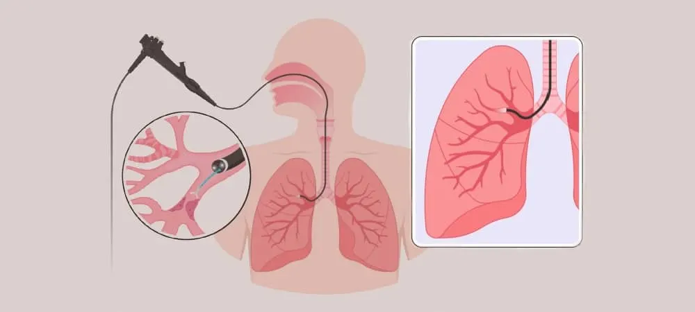
Endobronchial ultrasound (EBUS) is an interventional procedure performed with a device developed by attaching an ultrasound probe to the tip of a fiberoptic bronchoscope equipped with a video camera. It is used as an advanced diagnostic method that allows for the precise visualization of both the internal structure of the lungs (the inner surfaces of the bronchi) and the adjacent surrounding structures (lymph nodes, tumoral formations, vascular structures) via ultrasound. The procedure is preferably performed under general anesthesia. It can also be performed with local anesthesia and sedation in suitable patients who can tolerate the procedure. The device is inserted through the mouth into the trachea and advanced into the bronchi. During the procedure, while the inner surfaces of the bronchi are directly visualized, structures outside the bronchi are also detected via ultrasound, their boundaries are determined, and the physician is simultaneously enabled to safely obtain tissue samples (biopsies) for diagnosis from the correct location. The collected samples are sent for pathological examination and, in case of suspected infection, to the microbiology laboratory. The procedure takes an average of half an hour to 1 hour, depending on the extent of the disease and the number of samples to be taken. The patient can be discharged on the same day after the procedure.
This method is highly valuable in the diagnosis of diseases characterized by lymph node enlargement, such as sarcoidosis, tuberculosis, and lymphoma, as well as mediastinal (occurring in the chest cavity) masses (benign/malignant), especially lung cancer, or in detecting cases where cancers located in other organs of the body have spread (metastasized) to the lymph nodes of the lungs. With EBUS, it is possible to diagnose diseases more accurately, quickly, and safely using a minimally invasive method—meaning with the least possible intervention to the body—without the need for a surgical procedure like mediastinoscopy.
In the diagnosis of which diseases is EBUS used?
Endobronchial ultrasound (EBUS) is a targeted diagnostic method preferred especially in cases requiring detailed examination of the lymph nodes within the chest cavity and structures around the lungs. In this way, both serious diseases such as cancer can be diagnosed, and ambiguous situations can be clarified. Some of the disease groups where EBUS is most frequently used are as follows:
Lung Cancer: EBUS is one of the most reliable methods in the diagnosis and staging of lung cancer. It plays a critical role, especially in understanding whether the cancer is limited to the lung or has spread to the surrounding lymph nodes. This information is decisive for shaping the treatment. Being able to take a biopsy without the need for surgery increases patient comfort while also increasing the accuracy of the diagnosis. EBUS is an advanced approach used to clarify the stage of the cancer and to prepare a personalized treatment plan (surgery? /chemotherapy, radiotherapy?).
Sarcoidosis: Sarcoidosis is an inflammatory disease that causes the formation of abnormal cell clusters in the lungs and lymph nodes, but it is not always easy to understand what these clusters are caused by. EBUS is used to clarify the reason behind lymph nodes that appear enlarged radiologically. Thanks to the opportunity to take direct samples, the disease can be verified at the cellular level rather than just based on imaging.
Tuberculosis: Tuberculosis, an infectious disease, does not always present with classic symptoms. In some cases, it can progress silently, especially in the intrathoracic lymph nodes, which makes diagnosis difficult. EBUS makes it possible to diagnose the disease at a microscopic level by taking direct samples from these nodes. Thus, treatment is started by clearly proving the presence of the disease rather than just looking at an image. It makes a significant contribution to the differential diagnosis of diseases such as tuberculosis and cancer, especially in cases of unexplained lymph node enlargement seen in the lungs.
Lymphoma: Lymphoma is a type of cancer belonging to the lymphatic system and can also develop in the lymph nodes located within the chest cavity. Since surgical intervention in these areas can be difficult and risky, EBUS is preferred both for diagnosis and for determining the subtype of the disease.
What is the recovery process like after the EBUS procedure?
The recovery process after EBUS is generally fast and uneventful. The patient is kept under observation for a few hours after the procedure. If a biopsy has been taken, there may be temporary complaints such as a mild sore throat, hoarseness, or a dry cough. These symptoms usually disappear on their own within 1–2 days. Rarely, a mild fever or blood in the sputum may be seen. Since the pathological examination of the samples taken may take a few days, it is necessary to wait for this process to be completed for a definitive diagnosis. It is recommended that the patient consume plenty of fluids after the procedure and avoid heavy physical activities for a few days.
What are the differences between EBUS and bronchoscopy?
Both procedures are performed with devices called bronchoscopes and allow for the examination of the respiratory tract, but they contain significant differences in terms of their intended use and the level of information they provide. Bronchoscopy is used to directly visualize the inner surface of the trachea and bronchi. It is used for the visual detection of problems such as tumors, inflammation, or bleeding. If there are pathological images, superficial tissue samples can be taken from the inner lining of the bronchi (mucosa). The EBUS procedure, on the other hand, allows for the evaluation of deeper areas that are not visible to the eye. Thanks to the ultrasound probe at the tip of the bronchoscope, lymph nodes, tissues, and vascular structures outside the bronchial wall are visualized. The most important difference is the ability to safely obtain biopsies from deep tissues directly while performing ultrasound simultaneously.
Who is the EBUS procedure suitable for?
The EBUS procedure is recommended especially for individuals who have unexplained enlargements, masses, or irregularities detected in the lymph nodes located within the chest cavity or in the tissues around the lungs. The goal of this method is to safely obtain information that cannot be obtained with classic methods by taking tissue samples from hard-to-reach areas.
How is lung cancer diagnosed with the EBUS procedure?
Lung cancer rarely shows symptoms in the early stages. For this reason, the disease may have reached a certain stage by the time it is diagnosed. The EBUS device is a critical tool that allows both the confirmation of the diagnosis and the determination of the stage of the disease. During the procedure, the surrounding lymph nodes are visualized in detail with an ultrasound probe from inside the trachea via a bronchoscope. If suspicious lymph nodes have been detected in PET-CT or CT imaging, these areas are directly targeted and biopsied under ultrasound guidance. Thanks to this “targeted biopsy” method, the probability of making a diagnosis with a sample taken from the correct spot increases significantly. EBUS not only makes a diagnosis but also helps determine the stage of the disease, which is necessary for the treatment plan. Determining whether there is a spread to the lymph nodes directly affects the decision for surgery or chemotherapy planning. In this way, the treatment process is created with clearer data.
Frequently Asked Questions
The EBUS procedure usually lasts between 30 and 60 minutes. The duration may vary depending on factors such as the number of regions imaged, the need for a biopsy, and the patient's general condition. When the preparation of the patient before the procedure, the administration of medication, and the short observation period after the procedure are added, the total time spent in the hospital can reach a few hours. The procedure is usually planned so that the patient is discharged on the same day. The procedure both speeds up the diagnosis and does not interrupt daily life for a long time. If general anesthesia is administered, the patient feels nothing, sleeps, and wakes up when the procedure is finished. If a decision for local anesthesia is made, mild sedation is usually given. The patient is not completely put to sleep. Since the throat is numbed, no serious discomfort is felt during the advancement of the tube. Sometimes a feeling of mild pressure or a short-term cough may be seen, but this is temporary and is usually well-tolerated in patients who are compliant with the procedure. EBUS is a procedure with a high safety profile when performed by experienced teams. Since it does not require surgical intervention and can make a diagnosis without damaging external tissues, it carries fewer risks compared to classic methods. The rate of complications during the procedure is quite low. Rare side effects include throat irritation, slightly bloody sputum, or a temporary cough after the procedure. More serious complications (such as infection, air leakage) are extremely rare and can usually be prevented by evaluations performed before the procedure. Therefore, when appropriate patient selection is made, EBUS is a reliable and effective diagnostic method for both the patient and the physician. The recovery process after the EBUS procedure is generally fast, but there are some important points the patient should pay attention to. A feeling of stinging in the throat, mild hoarseness, or a dry cough may occur for a few hours after the procedure. Such symptoms are usually temporary and disappear on their own within a few days. In order not to irritate the throat, it is recommended to avoid very hot or very cold drinks and to limit spicy foods within the first 24 hours. Not smoking after the EBUS procedure positively affects not only throat irritation but also the course of any underlying lung diseases. Biopsy samples taken during EBUS bronchoscopy are sent to the laboratory and subjected to a detailed pathological examination. This process is not limited to the examination of cellular structures. If necessary, the samples are evaluated with special staining techniques or molecular-level analyses. This makes the process technically sensitive and time-consuming. In general, biopsy results are available within a few weeks, but in some cases, this period may be extended by a few more days. If infectious diseases are included in our preliminary diagnoses, microbiological examinations are also performed.
Early diagnosis is very important for your lung health.
Our chest diseases specialists offer modern, safe, and fast solutions in diagnosis and staging procedures with EBUS.
Contact Hisar Hospital Chest Diseases to make your appointment or to get detailed information.