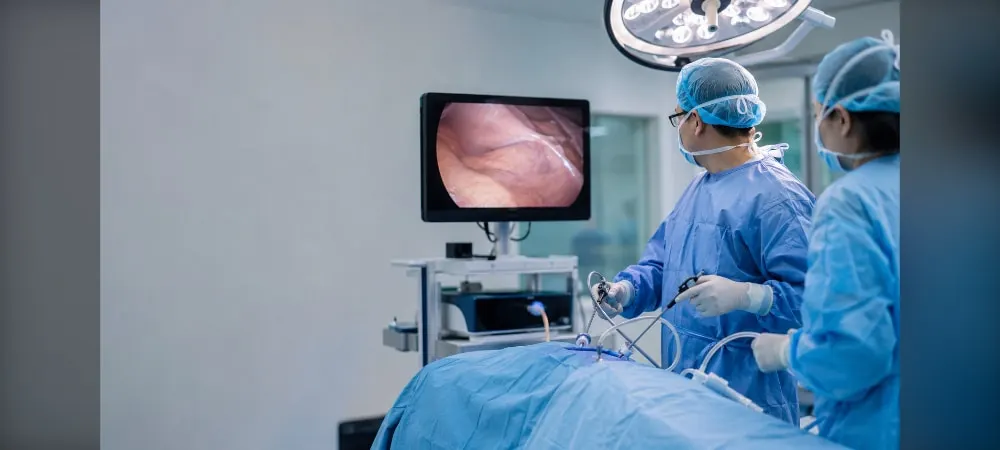
When thoracic surgery is mentioned, large incisions and long recovery times usually come to mind. However, VATS is a modern approach that changes this perception. Known as video-assisted thoracic surgery, this minimally invasive method provides access to the thoracic cavity through small incisions. The surgeon treats conditions such as lung cancer, pneumothorax, and pleural effusion using a camera and specialized instruments. In this article, you will find answers to the questions of why VATS surgery is performed, how VATS surgery is performed, and what the advantages of VATS surgery are.
What is VATS (Video-Assisted Thoracic Surgery)?
Definition and Key Features of VATS
VATS is a minimally invasive surgical method used to access the thoracic cavity and treat diseases within this area. Also known as video-assisted thoracoscopic surgery, this procedure involves the surgeon inserting a camera and specialized surgical instruments into the chest cavity through small incisions.
During the procedure, which is generally performed under general anesthesia, 1 to 3 small incisions are made in the chest wall. One of these incisions is for inserting a camera used to view the inside of the thoracic cavity. Surgical instruments are inserted through the other incisions.
The camera provides a high-definition view of the chest cavity, allowing the surgeon to perform direct interventions. The surgeon performs surgical interventions on the lung tissue, lymph nodes, or other intrathoracic structures by monitoring the images on a monitor. Thus, the operation area can be evaluated in real time.
VATS as a Minimally Invasive Surgical Method
VATS stands out among minimally invasive surgical techniques. It is a method of performing surgery through small incisions rather than making large incisions on the body. It is a thoracic surgery applied using specialized instruments to minimize trauma.
Unlike thoracotomy, which is known as the traditional method, the VATS technique does not require entering between two ribs via open surgery. There is no need to spread the ribs apart to perform procedures in the chest cavity as in open surgery. It is possible to reach the lung through small incisions between the ribs.
The camera images inside are projected onto the screen in the operating room. Depending on the case, the area where the chest cavity must be entered and intervened is also determined via the camera. This increases the success rate of the surgery and provides the opportunity for direct intervention to the problem.
Differences from Traditional Surgery
VATS is a preferred method instead of traditional thoracotomy and greatly shortens the recovery process of patients. It has many advantages compared to traditional open surgery.
Due to smaller incisions, the surgical cuts are quite small, which means less scarring. Small incisions significantly reduce postoperative pain. Patients can return to their daily lives much faster compared to open surgeries.
Patients who undergo VATS can usually be discharged within a few days. The risk of surgical complications such as bleeding and infection is lower. High-definition cameras provide the surgeon with the opportunity for more precise intervention.
The surgery is performed without stretching or breaking the ribs. Minimal damage is inflicted on the muscles supporting respiration. Allowing the surgeon to perform complex operations without performing an open surgery is one of the most important advantages of VATS. It improves the quality of life of patients by offering an effective solution even in complex surgical interventions.
How is VATS Surgery Performed?
Preoperative Preparation Process
Before the surgery, your doctor will inform you about what preparations need to be made. First, tests such as blood tests, chest X-rays, and ECG are performed. Computed tomography, urine tests, and special functionality tests are also among the primary tests requested from the patient. At the end of these tests, if the patient is evaluated as suitable, they are taken for surgery.
If you are using blood-thinning medications, your doctor will ask you to discontinue them. It is recommended not to smoke and to perform light daily exercises. Food intake must be paused 12 hours before the operation. Paying attention to these warnings reduces the risk of complications during the operation and helps complete the surgery successfully.
Steps During Surgery
The patient is placed under general anesthesia and a sterile surgical environment is ensured. In the VATS method, double-lumen intubation is performed on the patient. The surgeon makes one or small-sized incisions on the chest wall of the patient in the lateral decubitus position.
A camera entry site is opened in the area where the surgery will be performed, namely the rib cage, and this opening is 1-1.5 cm in length. The thoracic cavity is entered with a camera through the opened entry site. First, the area where the surgery will be performed is evaluated with the aid of the camera. Following this evaluation, an idea is gained about where the specially made surgical instruments enabling the surgery will enter the thoracic cavity.
Depending on the case, entry sites can be opened either single or double. After the entry sites are determined, specially manufactured surgical instruments are placed into the chest through these locations and the operation is performed. A thoracic tube is placed in the chest area to ensure the lungs inflate after surgery and to prevent fluid accumulation. This tube is removed before the patient is discharged once air leakage stops and fluid drainage decreases.
Use of Camera and Surgical Instruments
In the VATS procedure, every corner of the inside of the rib cage is visualized with the help of a camera, magnified, and projected onto the monitor. The surgeon at the bedside and the entire team perform the surgery by watching these images. Images captured with the help of a 10mm camera advanced into the chest cavity are projected onto screens.
Classically, VATS is usually performed through 3 incisions (ports). Later on, major surgeries such as lung cancer began to be performed comfortably and safely through 2 incisions. One port is used for the camera and the second port for the surgical instruments. In the Single-Port VATS method, the surgery is performed with only a single small incision. This incision is generally desired to be 2 cm. The chest cavity is entered by making only a single small incision (2-4 cm).
Surgery Duration and Procedure Details
The duration of the surgery varies depending on the scope of the procedure to be performed. VATS surgery takes approximately 2-3 hours. In general, surgeries can be completed within 1 to 3 hours. A simple biopsy or hyperhidrosis surgery can be completed in 30-45 minutes. More complex procedures, such as the removal of a lung lobe (lobectomy), can take 2-3 hours.
The average surgery duration is 2 hours. The type of surgery, the details of the procedures it involves, and the patient's specific medical condition are the main factors determining how long the surgery will take. Thus, minimally invasive thoracic surgery is applied. Without opening the patient's rib cage, the team places the tumorous lung tissue and lymph nodes inside a special bag (endobag) and removes them out through the 5 cm incision made.
In Which Conditions is VATS Surgery Applied?
Video-assisted thoracic surgery can be applied in almost all diseases concerning the chest region. This method is used for both diagnostic and therapeutic purposes. Surgeons intervene in many different problems in the thoracic cavity with VATS.
Lung Cancer and Pulmonary Nodules
VATS surgery is applied as the first choice in early-stage lung cancers. Especially in non-small cell lung cancers, the closed method can be safely applied. Scientific data show that operations performed with VATS in early-stage cases yield results equivalent to open surgery in terms of cancer control.
When a nodule is noticed in the lung, it is investigated whether it is benign or malignant. In suspicious nodules, the nodule is removed using the VATS method. In this procedure, the nodule is removed along with a certain amount of healthy surrounding tissue. The nodule removed during surgery is immediately sent to pathology. If the result comes back malignant, the surgeon can expand the operation and conclude the cancer treatment right then and there.
It is completely possible to recover from lung cancers caught at an early stage with surgical treatment. The patient group in which we can be most beneficial in lung cancer is patients caught at the small nodule stage. The cancerous lung lobe and lymph node are removed together using the closed method.
Pleural Diseases and Effusion
VATS is the most effective surgical route for draining fluids accumulated between the lung membranes. In case of fluid accumulation in the pleura, cytological examination is sometimes insufficient. By entering the chest cavity with VATS, the pleural membrane is directly observed, and biopsies are taken directly from suspicious areas.
This method has an accuracy rate of over 96.5% in the diagnosis of cancerous fluids. In cases of undiagnosed pleural fluid, VATS is a safe method with a high diagnostic value of 96.5% and a low complication rate. Adequate material sampling is performed, and lesions are macroscopically observed and evaluated.
If it is decided that there is malignant pleural fluid, pleurodesis is performed in the same session so that fluid does not form again. Since talc powder will be powdered everywhere, pleurodesis performed with VATS is more effective than other methods. Procedures for cleaning inflamed tissues and draining fluid accumulations of unknown cause are carried out.
Treatment of Pneumothorax
In cases where the lung collapses due to air leakage, the procedure of removing the air sacs causing the leak is performed with VATS in minutes. Air sacs on the lung surface are closed using a stapling method, and an adhesion procedure is applied to the pleura. Recurrence rates are similar compared to open surgery, but the recovery process is much faster.
The most commonly used method is the closed endoscopic VATS method. A camera is advanced into the chest cavity through a single 2 cm incision. It is the operation method that causes the least harm to the patient. Following this method, pain is less, the surgical incision is much smaller, and the patient's return to normal life is much faster.
Mediastinal Masses and Lymph Node Biopsy
Masses developing in the mediastinum located in the middle of the rib cage require expert evaluation. Thymoma, thymus gland tumors, and mediastinal cysts can be successfully treated with the closed method. A large proportion of mediastinal masses must be surgically removed both to establish a diagnosis and to eliminate compression symptoms.
In the closed method, the operation is performed with a camera and specialized instruments. It is frequently preferred especially for biopsy purposes or the removal of simple cysts. Mediastinal masses are applied with VATS for diagnostic, staging, and therapeutic purposes. Even very large masses can be removed with VATS.
Mediastinal lymph node dissection performed to understand the spread of cancer is one of the areas where the VATS method is most successful. Some deep areas that are difficult to reach in open surgery can be reached more easily with the angled camera systems offered by the closed method. This facilitates the accurate staging of the patient.
What are the Advantages of VATS Surgery?
The advantages of VATS surgery are the most important reasons why patients choose this method. Its many superiorities compared to open surgery have been scientifically proven. These advantages manifest themselves both during and after the surgery.
Smaller Incisions and Less Scarring
While large incisions of 20-25 cm are made in open surgery, a 1 cm camera incision and a 4-5 cm auxiliary incision are sufficient in the VATS method. In some cases, the surgery can also be performed with a single incision. For procedures such as pneumothorax, a single 2 cm incision is sufficient.
Since the surgery results in only a single small scar, it has a cosmetic benefit. Instead of a large surgical scar, a few small dots remain that become almost invisible over time. The small puncture marks remaining on the rib cage become indistinct over time and do not cause cosmetic discomfort.
Postoperative Recovery Process
The recovery process is faster compared to traditional open surgery. Since the ribs are not spread apart and wide muscle incisions are not made, postoperative pain is significantly less. This situation lowers the risk of complications by enabling the patient to breathe and cough more comfortably.
Having less pain ensures that patients mobilize earlier. Patients can get up on the day of the surgery or the next day. It is possible for office workers to return to work within 10 to 14 days. It is recommended that those working in professions requiring heavy lifting rest for about 4 weeks.
The full recovery process can take 1-2 weeks. Postoperative pain is usually minimal, and patients can return to their normal activities faster. 1 month after the operation, the patient can return to all daily activities, including sports, as if they had never had surgery.
Length of Hospital Stay and Return to Daily Life
The length of hospital stay varies depending on the complexity of the surgery. Discharge generally occurs within 2-3 days. In the VATS technique, the hospital stay is usually 2 to 4 days. Patients are generally discharged within 1-3 days.
Patients undergoing single-port VATS lung biopsy or wedge resections can go home 1 to 3 days after surgery. Patients undergoing VATS lobectomy can usually go home 3 to 4 days after surgery. After surgeries performed with minimally invasive methods, patients are generally ready to be discharged within 2-3 days.
Most patients can start their normal daily routines, such as driving and returning to office work, within 3-4 weeks after the operation. Patients are generally discharged within a week. The return period to normal life is shorter.
Reduction of Complication Risks
Scientific studies have shown that minimally invasive thoracic surgeries reduce postoperative pain and the risk of complications. It comes with less bleeding, infection, and other complication risks. Problems such as infection, hemorrhage, and respiratory issues are fewer compared to traditional open surgery.
Since the rib cage is not opened, contact with the external environment is less, which ensures a decrease in the risk of infection. Major surgical traumas can suppress the body's immune response, but VATS minimizes this suppression with less trauma. The immune system is affected less.
Rapid mobilization reduces the risk of lung collapse and blood clots. Having minimal nerve damage also reduces the risk of chronic chest pain.
Frequently Asked Questions
1. What is VATS surgery?
VATS (Video-Assisted Thoracoscopic Surgery) is a minimally invasive surgical method used to operate on organs inside the rib cage using small incisions. It is performed with smaller incisions compared to traditional open surgery, and the procedure is carried out by obtaining visualization with the aid of a camera.
2. How long does VATS surgery take?
The duration of VATS surgery varies depending on the procedure to be performed. While simple procedures take 1-2 hours, more complex surgeries can take 3-4 hours or more. Procedures such as lung lobe resection are usually completed between 2-3 hours.
3. What is the recovery process like after VATS surgery?
The recovery process after VATS surgery is much faster compared to open surgery. The length of hospital stay is usually between 2-5 days. While most patients return to their daily activities within 2-4 weeks, full recovery may take 6-8 weeks. Pain is also significantly less compared to open surgery.
4. What are the risks of VATS surgery?
As with any surgical procedure, VATS has some risks: bleeding, infection, air leakage, nerve damage, and very rarely, the need to convert to open surgery. However, these risks are lower compared to traditional open surgery. When performed by an experienced surgeon, VATS is a safe procedure.
5. For which diseases is VATS surgery performed?
VATS surgery can be used for lung cancer, removal of lung nodules, pneumothorax (collapsed lung), pleural effusion, mediastinal tumors, chest wall diseases, treatment of hyperhidrosis (excessive sweating), and biopsy procedures.
6. What are the differences between VATS and open surgery?
While 1-3 small incisions (1-2 cm) are made in VATS surgery, a large incision of 15-20 cm is made in open surgery. In VATS, there is less pain, a shorter hospital stay, faster recovery, less scarring, and a lower risk of complications. Cosmetic outcomes are also much better in VATS.