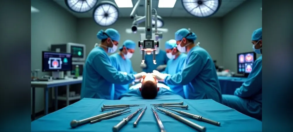
If you are wondering how brain tumor surgery is performed, it is important to first understand what a brain tumor is. A brain tumor occurs when abnormal cells, which arise during cell regeneration in the brain, grow and combine to form masses. These tumors can be benign or malignant; benign ones do not contain cancerous cells and have a low risk of recurrence when removed surgically. Malignant tumors, however, carry the risk of spreading.
Brain tumor surgeries are procedures typically performed by expert neurosurgeons that can last between 2 and 15 hours. In these surgeries, doctors aim to remove the entire tumor from the brain. If the tumor cannot be completely removed, additional treatments such as chemotherapy or radiotherapy may be required. Brain tumors can be treated using different techniques such as craniotomy, microsurgery, and neuroendoscopic brain surgery. In this article, we will share with you all the details you need to know about brain tumor surgery in a clear and understandable way.
What Is Brain Tumor Surgery?
Brain tumor surgery is a surgical procedure aimed at removing or shrinking an abnormal mass of tissue in the brain. This procedure can be performed using different techniques depending on the type, size, and location of the tumor, as well as the patient's general health status. The primary goal of the surgery is to remove the tumor as safely as possible, reduce pressure on brain tissue, and control the progression of the disease.
The difference between benign and malignant tumors
Tumors in the brain are divided into benign and malignant. This distinction significantly affects the treatment approach and outcomes. Benign tumors usually grow slowly and have a low tendency to damage surrounding tissues. Furthermore, recurrence rates after surgical intervention are quite low. In contrast, malignant tumors grow rapidly and are notable for their structure that damages surrounding tissues.
Benign tumors generally do not metastasize, meaning they do not spread to other parts of the body. This makes them easier to remove surgically, and the likelihood of them reforming after removal is usually low. On the other hand, malignant tumors have the ability to metastasize within the brain and therefore require treatment even after surgery.
Meningiomas are mostly benign and respond well to surgical intervention. However, malignant tumors such as glioblastoma multiforme are more aggressive in nature and may be resistant to treatment.
When is brain mass surgery necessary?
Brain tumor surgery is recommended especially when the tumor causes serious symptoms or poses a threat to the patient's health. The presence of a brain tumor does not always require surgical intervention, but in certain cases, surgery may be inevitable.
Common symptoms that may lead to a recommendation for surgery include:
Persistent headaches that do not respond to typical pain relief methods
New seizures or seizures that change in frequency or intensity
Cognitive changes such as memory loss or confusion
Weakness or numbness in the limbs
Changes in vision or hearing
Balance problems or difficulty with coordination
The decision for surgery is usually made after imaging studies such as MRI or CT scans. This decision is made after evaluating the size and location of the tumor and its effect on surrounding brain structures. In some cases, a biopsy may be taken to diagnose the tumor type and determine the most appropriate treatment method.
Does brain tumor surgery affect life expectancy?
Life expectancy after brain tumor surgery varies depending on many factors. The type, location, and stage of the tumor, as well as the patient's age and general health, are some of these factors.
Complete removal of the tumor usually provides the best results. Removing as much of the tumor as possible during surgery while preserving functional capacity reduces recurrence rates and ensures a reduction in tumor burden. However, in some cases, the tumor cannot be completely removed and is surgically debulked.
Specifically, in aggressive tumors like glioblastoma multiforme, life expectancy in untreated patients usually ranges from 6 to 18 months. With modern surgical methods and radiotherapy and chemotherapy applied after total resection of the tumor, this period can be extended beyond 18 months. In contrast, for benign tumors, there is a high probability of long-term or even lifelong non-recurrence with appropriate treatment.
In conclusion, brain tumor surgery is a delicate surgical intervention performed under a microscope according to the location, size, and type of the tumor, which carries vital risks but aims to increase the patient's life expectancy and quality of life. Surgical treatment is usually the first-choice treatment method for brain tumors and has the potential to positively affect life expectancy when applied correctly.
How Is Brain Tumor Surgery Performed?
To treat brain tumors, surgeons use different surgical techniques depending on the location and size of the tumor. With the development of modern technology, many innovative methods have emerged in the field of neurosurgery. Each technique aims to provide the most benefit to the patient with the least risk.
Craniotomy method
Craniotomy is the most commonly applied technique for surgical intervention in brain tumors. In this method, the surgeon gains access to the brain by temporarily removing a portion of the skull bone. During the procedure, the patient is usually put to sleep under general anesthesia. The craniotomy method is used specifically to reach all problems within the skull that require surgery.
The procedure consists of the following stages: First, the scalp is incised according to the location of the tumor. Then, with the help of modern devices and surgical instruments, the relevant part of the skull is opened. After the skull is opened, the surgeon reaches the tumor and attempts to remove it entirely if possible. After the tumor is removed, the skull bone is replaced and fixed with sutures or plates.
Today, thanks to the use of technological devices (pneumatic drill, neuronavigation), the craniotomy procedure can be performed in approximately half an hour.
Closed (endoscopic) brain surgery
Endoscopic brain surgery is a minimally invasive procedure in which a thin camera and surgical instruments are inserted through a small incision made in the skull to reach a specific area in the brain. A special device called an endoscope is used in this method. The endoscope contains a small camera and a light source to magnify and illuminate the surgical field.
This technique has significant advantages:
Tissue damage is minimized thanks to smaller incisions
The patient recovers faster and stays in the hospital for a shorter time
Post-operative pain and discomfort are less
The risk of infection, bleeding, and nerve damage is minimal
Endoscopic surgery is generally effective in removing small tumors located on the brain surface or in the depths. In recent years, it has become possible to reach tumors located at the skull base endoscopically by entering through natural cavities in the nose.
How is brain tumor surgery performed through the nose?
Brain surgery performed through the nose (transsphenoidal surgery) is a preferred method, especially for pituitary tumors and tumors in the front or middle part of the brain. In this approach, the surgeon reaches the tumor by entering through the nostrils without opening the skull.
Surgery performed through the nose has many advantages over classic surgery. While in classic surgery, the skull is lifted and the tumor is reached by advancing through natural cavities in the brain or brain tissue, in surgery performed through the nose, the skull is not opened and the brain tissue is not touched. This provides a more comfortable experience for the patient, reduces the need for dressings, and minimizes side effects.
This surgery usually lasts 45-50 minutes, and patients can stand up 1 day after the surgery and can be discharged 2 days later. While the recovery time in open surgery is 1-1.5 months, patients can return to their normal lives 2 weeks after this surgery.
Microsurgery and ultrasonic aspiration techniques
Microsurgery is an advanced surgical technique used in the treatment of brain tumors. In this method, surgeons work under a microscope, allowing them to intervene in extremely precise and small areas. Microsurgery aims to remove the tumor completely with minimal damage to brain tissue.
Thanks to the microsurgery technique:
Complete surgical removal of the tumor is achieved while protecting healthy brain tissue
Working with high precision becomes possible
The risk of damaging surrounding tissues is reduced
The patient recovers faster and can return to their normal life
The ultrasonic aspirator (CUSA) is a special technique that allows only the tumor tissue to be fragmented using sound waves during surgery, without damaging normal brain tissue and blood vessels. This system allows the tumor to be fragmented and the pieces to be removed by suction using sound waves emitted by an ultrasound probe inserted into the tumor. Thanks to ultrasonic aspiration, many small tumors in the brain can all be removed safely at once.
Pre-operative Preparation Process
Preparing for brain tumor surgery is a critical process that directly affects the success of the operation. The pre-operative preparation phase is a comprehensive journey that starts with making an accurate diagnosis and extends to informing the patient and their relatives.
Diagnostic and imaging methods
Various imaging techniques are used for the accurate diagnosis of a brain tumor. Magnetic resonance imaging (MRI) is considered the gold standard in brain tumor diagnosis. MRI is of vital importance in surgical planning by showing the location and size of the tumor in detail. To support the MRI, contrast material is usually administered intravenously to increase the effectiveness of the imaging.
Computed tomography (CT), on the other hand, helps in evaluating the relationship of the tumor with the bone, as it shows skull bone structures more clearly. Additionally, if there is calcification in the tumor, this is better seen with CT.
Other methods used in the diagnostic phase include:
Positron emission tomography (PET): Shows the metabolic activity of the tumor
Angiography: Examines the vascular structure and blood supply characteristics of the tumor
Electroencephalography (EEG): Measures brain activity
Biopsy: Performed to definitively determine the type of tumor
Thanks to advanced MRI imaging techniques (diffusion, tensor MRI (tractography), functional MRI, perfusion MRI, MR spectroscopy), the extent, type, and relationship of the tumor with important centers in the brain can be evaluated in detail. These techniques play a major role in the surgeon's determination of the surgical strategy.
Neurological examination is also an important part of the diagnostic process. Neurological functions such as mental functions, coordination, speech, vision, hearing, and reflexes are evaluated in detail.
Anesthesia and risk assessment
Before the surgery, comprehensive tests are performed to evaluate the patient's suitability for anesthesia. At this stage, the patient's past medical history, medications used, and allergies are meticulously examined. Suitability for anesthesia, bleeding risk, and possible signs of infection are investigated with blood tests.
A consultation with the anesthesiologist is an important opportunity to discuss anesthesia options and the patient's concerns. During this meeting, detailed information is provided about the anesthesia process and the recovery period.
Before the surgery, the use of blood-thinning medications is reviewed, and if necessary, these medications are discontinued a certain period before the surgery. Additionally, the fasting period begins 8 hours before the surgery.
Informing the patient and their relatives
After the decision for surgery is made, the process of informing the patient and their relatives begins. During this process, how the surgery will be performed, possible risks, and the recovery process are explained in detail. At the same time, information is provided about what needs to be considered after the surgery.
In an important process such as brain tumor surgery, the patient's mental and emotional preparation is as important as their physical preparation. During this period, it is necessary for the patient's relatives not to pressure the patient and to provide only as much support as requested.
Especially making decisions on behalf of the patient or thinking that one has the right to speak is not the right approach in this tense process. It should not be forgotten that every patient experiences the surgical process differently.
The pre-operative preparation process is a comprehensive evaluation in which the location of the tumor, the degree of difficulty of the operation, the surgical technique, the post-operative process, possible risks, costs, and the effects on the patient's quality of life are analyzed in detail. This process forms the basis for a successful surgery and a smooth recovery period.
Risks of Brain Tumor Surgery
As with any surgical procedure, brain tumor surgeries also have some risks. These risks vary depending on the location and size of the tumor, the patient's general health status, and the surgeon's experience. However, these risks can be significantly reduced thanks to modern surgical techniques and expertise.
Risk of infection and bleeding
In brain surgeries, the risk of infection is tried to be minimized with sterile conditions and the use of appropriate antibiotics. Post-operative meningitis or infection in the surgical area may develop. In some cases, cerebrospinal fluid leakage may occur due to problems with the closure of the meninges, which can lead to infection.
The risk of bleeding is present in every brain surgery. Due to the dense vascular structure in the brain, damage to blood vessels may occur during the operation. Especially, damage to a large vessel while reaching the tumor can cause serious bleeding. This situation can lead to post-operative neurological losses or, in rare cases, life-threatening situations.
Nerve damage and speech disorders
If the tumor is close to the speech center of the brain, affecting this area during surgical intervention can cause various speech disorders. These disorders can manifest as the inability to speak at all or understanding what is spoken but speaking nonsensically. In some cases, to preserve speech functions during surgery, the patient is not put to sleep and a technique called "awake craniotomy" is used.
Nerve damage can occur, especially during the removal of tumors close to motor control, balance, or vision centers. Modern microscopic surgical techniques and intraoperative imaging systems help reduce these risks.
Brain edema and memory loss
Fluid accumulation (edema) in the brain may develop after surgery. This condition occurs due to trauma and inflammation caused by the surgical intervention. Brain edema can cause headaches, nausea, vision disturbances, or changes in consciousness. However, it can be brought under control with medical treatment when detected early.
Memory loss is also a complication that can be seen after surgery. Especially in the surgery of tumors located close to the temporal lobe and hippocampus, problems with recording and remembering new information may be experienced. This effect is usually temporary, but in some cases, it may be permanent.
Personality changes and stroke risk
Personality or emotional changes may be seen in some patients after brain tumor surgeries. This condition is usually encountered more frequently in operations performed in the frontal or temporal lobe. Although most of the changes are temporary, rehabilitation and psychological support may be required.
The risk of stroke (paralysis) can occur as a result of damage to brain tissue and its vessels during surgery. This risk is higher, especially in surgeries performed in areas close to the motor center. Detailed imaging performed before surgery and neuromonitoring devices used during surgery help reduce the risk of paralysis.
Things to Consider After Surgery
The period after brain tumor surgery is the most critical stage of the recovery process. The issues to be considered after surgery will ensure that you regain your health and prevent possible complications. Following your doctor's instructions during this process forms the basis for a successful recovery.
Nutrition and lifestyle recommendations
Healthy nutrition in the post-operative period is an important part of the recovery process. Consuming foods rich in protein and vitamins helps your body repair itself. It is recommended to prefer protein-rich foods, especially in the first 3 months. Additionally, getting plenty of fluids is of critical importance during this process.
You need to pay attention to your post-operative diet and stay away from smoking. Especially in the first period, preferring pureed foods in addition to liquid foods helps your digestive system gradually return to its normal function. Furthermore, it is recommended to avoid excessive alcohol consumption for life.
It is important to stay away from caffeinated beverages such as cola, coffee, and tea and to follow an appropriate diet to prevent constipation problems. Keeping your weight under control during this process helps reduce pressure on the brain.
Psychological support and the importance of morale
It is known that up to 90% of patients experience anxiety and depression after brain tumor surgery. Therefore, receiving psychological support will positively affect your recovery process. Especially, keeping your morale high is an important element in your recovery.
It is normal to experience emotional fluctuations in the post-operative period. Do not hesitate to seek professional help to cope with this situation. Spending time with your family members and friends can provide emotional support. Besides this, activities such as keeping a diary, art therapy, or engaging in hobbies can help you express your feelings.
Driving and protection from the sun
It is recommended not to drive for at least 2 months after the operation. During this period, your reflexes and motor skills may be temporarily affected. For your safety, it is important to avoid driving for the duration specified by your doctor.
Protecting yourself from the sun is an issue you should pay attention to both after surgery and throughout your life. In addition to this, you need to keep the surgical area away from water contact during the wound healing process. These precautions accelerate wound healing and reduce the risk of infection.
Things not to do during the recovery process
You should pay attention to the following issues during the recovery process:
Use the medications given to you such as antibiotics, stomach protectors, and painkillers until the boxes are finished, then do not take other medications
Use your anti-seizure medication regularly as specified by your doctor
Avoid sexual activity for at least 2 weeks to 1 month after surgery
Avoid excessive physical activity and heavy lifting
If you notice redness, swelling, or discharge in the surgical area, consult your doctor immediately
You usually need to go to your doctor for your first check-up after 1 month. In this meeting, your doctor will evaluate your recovery and readjust your medications. You can also discuss the time to return to work in this meeting.
In Summary, Brain Tumor Surgery
Brain tumor surgery is a step of vital importance in this life-threatening disease. As we have explained in detail above, this process requires careful preparation, correct technique selection, and a meticulous recovery period. Especially, imaging studies performed before surgery and an accurate diagnosis form the basis for a successful surgical intervention.
Undoubtedly, every surgery has its own unique risks. However, thanks to experienced surgeons and advanced technology, these risks can be significantly reduced. Careful post-operative care and compliance with doctor's instructions are the most important factors that will accelerate the recovery process.
Additionally, receiving psychological support after surgery, adhering to a healthy diet, and gradually increasing physical activities play a major role in the recovery process. As a result, when a brain tumor is diagnosed, it is necessary to determine the most appropriate treatment method by consulting with expert doctors in the field without panicking.
Brain tumor surgeries now provide safer and more successful results thanks to advancements in the medical world. It is possible to overcome this challenging process with the right information, patience, and determination. We wish you healthy days.
Frequently Asked Questions
How long does brain tumor surgery take?
The duration of the surgery varies according to the size, location, and type of the tumor. While simple surgeries take 2-3 hours, complex surgeries can extend up to 8-10 hours. Your surgeon will give an estimated time before the surgery.
How does the post-operative recovery process progress?
The recovery process varies from patient to patient. In general, a hospital stay of 3-7 days is required. Full recovery can take 6-12 weeks. Physical therapy and rehabilitation may be required during this process. Patients generally start to return to their daily activities gradually within 4-8 weeks.
What are the risks of brain tumor surgery?
Like every surgical procedure, brain surgeries also have risks. These include infection, bleeding, neurological damage, seizure activity, brain edema, and anesthesia complications. Your surgeon will explain the possible risks in detail before the surgery.
Can the entire tumor be removed?
This depends on the type of tumor, its location, and its proximity to vital brain regions. While the entire tumor can be removed in some cases, partial resection may be performed in some cases to prevent damage to the surrounding tissue. Surgeons try to balance maximum tumor removal with minimum neurological damage.
Are additional treatments required after surgery?
Depending on the type of tumor and the results of the surgery, radiotherapy, chemotherapy, or targeted therapies may be required. Post-operative pathology reports will determine the exact diagnosis of the tumor and reveal the need for additional treatment.
Which symptoms should I take seriously after surgery?
You should immediately report symptoms such as severe headache, vision disturbances, fever, discharge from the wound site, seizures, difficulty speaking, weakness in the arms or legs, and changes in consciousness to your doctor.
What should I pay attention to before surgery?
Follow all instructions given by your doctor. You will usually be asked to fast for 8-12 hours before the surgery. You should inform your doctor about all the medications you use and get guidance on which ones you should discontinue. Especially blood thinners should be discontinued a certain period before the surgery.
When can I return to normal life after brain tumor surgery?
This varies from person to person and according to the type of tumor. While patients can return to normal activity within 6-8 weeks in simple surgeries, the recovery process may be longer in more complex cases. You should get your doctor's approval to return to activities such as work, sports, and driving.
What is brain tumor surgery and how is it performed? Get information about benign and malignant types of brain tumors, pre-operative preparation, and the post-operative recovery process.