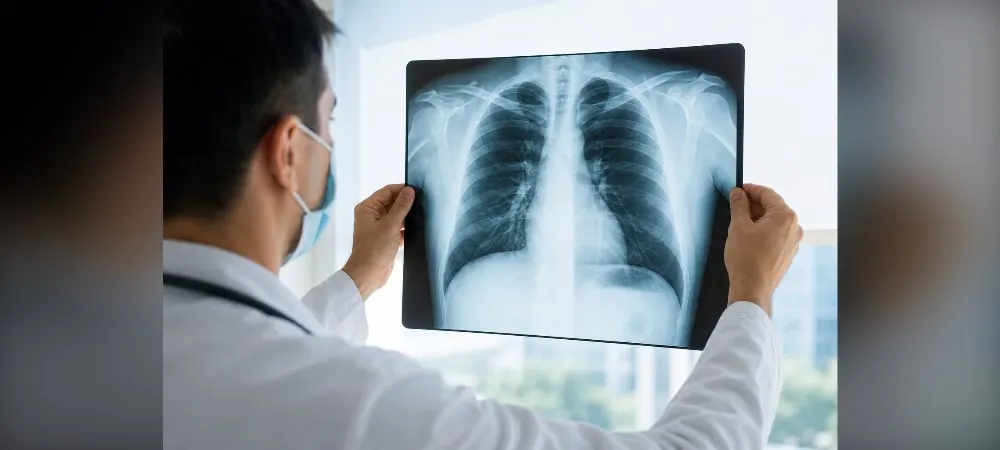
A chest X-ray is one of the primary diagnostic tools doctors use for complaints such as persistent cough, shortness of breath, or chest pain. Thanks to this simple imaging method, many diseases such as lung infections, tumors, fluid accumulation, and rib fractures can be detected in a short time. In this article, you will find the answer to what a chest X-ray is and learn in which situations it is performed. We will also clarify topics such as how a chest X-ray is taken, what a normal chest X-ray looks like, and how healthy chest X-ray results are evaluated.
What is a Chest X-ray?
Definition and basic characteristics of a chest X-ray
Imaging of the chest region using X-rays is called a chest X-ray. Through this method, the structural condition of the lungs, heart shadow, ribs, diaphragm, and mediastinum are examined. This imaging technique, which is the first step in radiological examinations, is widely used due to its low cost and easy accessibility.
A chest X-ray allows for a detailed evaluation of the lung tissue and structures in the chest cavity. Air sacs, bronchi, and the bone structures forming the chest wall are clearly visible on this radiograph. This procedure is painless, non-invasive, and is completed in a few seconds.
The most commonly used method for radiological imaging of the respiratory system is the postero-anterior (PA) chest X-ray. Interpreting an optimally obtained PA chest X-ray is a process that requires discipline and attention. In practice, a single-view PA chest X-ray is usually performed.
The relationship between a chest radiograph and a chest X-ray
A chest radiograph and a chest X-ray mean the same thing medically. Both terms refer to the X-ray imaging method applied to the chest region. The reason they are called by different names is due to the variations in common terminology used by the public.
While the term "radiograph" is generally preferred in more technical and medical literature, the term "X-ray" is more widely known among the public. However, in both cases, the goal is to evaluate the structure, size, air spaces, and signs of infection in the lungs. As a result, there is no difference between a chest radiograph and a chest X-ray.
Working principle of the imaging method
A chest X-ray uses a very small amount of ionizing radiation in addition to X-rays. This creates a black-and-white image. While lung tissue absorbs a very small amount of radiation, the vast majority of the radiation is absorbed by the ribs and spine.
As a result, the spine and ribs appear white on a chest X-ray. Lung tissue, on the other hand, is dark. Dense body parts like bones block X-rays from passing through the body. Softer body tissues like skin and muscles allow X-rays to pass through.
The probability of the radiation absorbed by the body during imaging causing harm is quite low. Since technically inadequate radiographs can lead to incorrect interpretations, the technical adequacy of the radiograph is checked first when evaluating chest X-rays.
In Which Situations Is a Chest X-ray Performed?
Necessity of imaging for respiratory complaints
Doctors request a chest X-ray if a patient presents with complaints of persistent cough, shortness of breath, or chest pain. This imaging plays a critical role, especially in febrile respiratory tract infections. In conditions such as pneumonia and bronchitis, infection areas in the lungs are detected.
Symptoms such as productive cough, wheezing, or rapid breathing are among the complaints that need to be evaluated. For shortness of breath presentations, a posteroanterior chest X-ray is requested to evaluate the thoracic cavity. This examination is also considered necessary in cases of coughing up blood.
Trauma and emergency situations
A chest X-ray is performed on patients suspected of having rib fractures or internal bleeding following chest trauma. It is the most frequently requested radiological imaging method by physicians in the emergency department. This examination is performed in almost every presentation of chest pain and shortness of breath.
A radiograph is used when investigating suspected air or fluid accumulation around the lungs. In sudden chest traumas, respiratory-focused complaints are evaluated. It plays a critical role as the first-line imaging method in detecting post-traumatic damage.
Routine pre-operative checks
It is requested as a routine pre-operative test before surgical operations to confirm the patient's suitability for general anesthesia. It is a frequently used test before heart surgery because the lung condition can affect anesthesia and recovery.
A pre-operative chest X-ray helps evaluate heart size, chest structures, fluid accumulation, infection, and chronic lung problems. It is mandatory if the patient has active complaints such as chest pain, shortness of breath, persistent cough, or signs of heart failure. If the planned surgery involves the heart, lungs, or other major vessels in the chest, it provides a baseline for comparing the post-operative period.
Job entry and periodic health screenings
A chest X-ray is a very important test in job entry health reports. It is one of the fastest methods that helps workplace physicians evaluate the lung health of employees. It is used to assess the current lung health of individuals who will start a new job.
It is requested to be performed in routine job entry health reports for screenings of individuals occupationally exposed to chemical gases and dust. It should be performed at least once a year for people working in factory environments and must be kept by the workplace physician for at least 10 years. It may be directed for those who have been exposed to harmful substances such as asbestos, smokers, or those with respiratory diseases.
Chronic disease follow-up
More than 90% of sarcoidosis cases have abnormalities on a chest X-ray. It has an important place in determining whether the disease is progressing and in forming treatment decisions. Radiological checks must be performed at certain intervals for patients followed up both with and without treatment.
In the early period after heart surgery, especially during the intensive care process, it is one of the most fundamental pillars of patient follow-up. Regular checks are performed on individuals with known heart or lung diseases such as COPD and congestive heart failure.
Diseases Detected by Chest X-ray
Radiology specialists examine the chest cavity, lung tissue, heart structure, and vascular system in detail on a chest X-ray. Infectious diseases, masses, heart problems, and fluid accumulations are detected during this examination.
Lung infections (pneumonia, bronchitis, tuberculosis)
In pneumonia patients, white patchy images appear in the lung tissue. This appearance indicates the inflammatory density in the infection area. In bronchitis cases, thickening in the airways and mucus accumulation are noticed.
Tuberculosis gives characteristic findings on a chest X-ray. Cavity formations and widespread fibrotic changes are seen. Lesions located especially in the upper regions of the lungs raise suspicion for tuberculosis. A chest X-ray plays a critical role in the early diagnosis of the disease.
Evaluation of tumor and mass findings
The first finding of lung cancer often appears as a solitary pulmonary nodule. These nodules are single, round, or oval-shaped lesions smaller than 3 cm. When the edges are smooth, they resemble a coin.
A mass can be of any size, but it is rarely seen on a radiograph before it is over 1 cm. Twenty to 30 percent of lung cancers initially appear as nodules. Due to the different growth rates of different parts of the nodule, lobulation is seen on its edges.
Peripheral tumors account for 42 percent of lung cancers. Centrally located tumors manifest themselves as endobronchial or perihilar masses. Hilar enlargement and collapse due to bronchial obstruction in the periphery of the tumor are common findings.
Heart and vascular structure problems
The structural condition, size, and position of the heart are evaluated on a chest X-ray. Heart enlargement may indicate congestive heart failure or congenital heart defects. Ballooning of the aorta and other major blood vessels is evident on the radiograph.
Congenital or acquired disorders in vascular structures are detected. Vessel wall diseases and pericardial problems are imaged. Hardening of the heart valve or aorta is seen as calcification on the radiograph.
Fluid accumulation and pleural diseases
Fluid accumulation between the lung membranes, called pleural effusion, creates a gray opacity on the radiograph. At least 175 mL of pleural fluid is required for the blunting of the lateral costophrenic sinus. Heart failure, pneumonia, and cancer are the most common causes of pleural effusion.
Blunting of the costophrenic sinuses is a sign of pleural effusion. These fluids compress the lung, leaving no room for it to work, and lead to shortness of breath. Infectious diseases, tuberculosis, and pulmonary embolism cause fluid accumulation. Bilateral pleural effusion is the accumulation of fluid in the membranes of both lungs and is usually seen in diseases such as heart, kidney, or liver failure.
How Is a Chest X-ray Performed?
Preparation process before the procedure
It does not matter whether the patient is fasting or not before a chest X-ray. No special fasting preparation is required. It is requested to remove clothing from the waist up and wear a patient gown. Metal parts such as metal buttons, zippers, necklaces, piercings, and bra wires must be removed as they can distort the image.
Female patients are asked about the possibility of pregnancy before being taken into the imaging room. If you use medical devices such as a pacemaker or heart valve, you must inform your doctor. If there is severe pain or limited mobility, it should be told to the technicians. The patient's identity information and complaint are recorded.
PA and AP imaging positions
A posteroanterior (PA) chest X-ray is always performed standing if possible. The patient rests their chest against the X-ray plate and pulls their shoulders forward to expand the rib cage. The X-ray beam is delivered from back to front at a right angle from a distance of 180 cm. This distance prevents the heart from appearing larger than its actual size on the film. PA imaging is the most standard chest X-ray method.
An anterior-posterior (AP) chest X-ray is mostly used for emergency situations or patients in intensive care. It is preferred for patients who cannot stand. X-rays are taken from the front and delivered toward the back. In an AP radiograph, the heart and mediastinal structures appear larger compared to a PA radiograph. Portable X-ray machines are used for patients who cannot get up from their beds.
Special imaging techniques and types
In a lateral chest X-ray, the patient turns to the side and lifts their arms up. X-rays are delivered from the side from a distance of 100 cm. Lateral decubitus imaging is performed while the patient is lying on their side and is used to detect fluid accumulation in the lung membrane. In apical lordotic imaging, the patient leans backward toward the device, and the lung tips behind the collarbones are imaged.
Expiratory imaging is performed by the patient exhaling completely. It is effective in detecting air trapped in the chest cavity. Oblique views are preferred to highlight rib fractures by giving the patient a slight angle of rotation. Fluoroscopy allows for live monitoring of the movements of the diaphragm muscle.
Imaging duration and patient comfort
A chest X-ray procedure takes about 10 to 15 minutes, including preparations. The imaging process itself is completed in a total of 1 to 3 minutes, starting from when the patient enters the room and takes the appropriate position. Actual X-ray exposure occurs in milliseconds, less than a second.
No pain or suffering is felt during the procedure. To obtain a clear image, one must stand still throughout the imaging. The patient holds their breath for a few seconds. The technician observes and checks that the correct breath is taken and held. Since the imaging time is completed within a few minutes, patient comfort is at a high level.
Normal Chest X-ray and Healthy Results
Healthy chest X-ray features
In the image obtained after the imaging is completed, the lungs are expected to be clear and well-inflated. There are no visible masses, fluid accumulation, or abnormal opacities on a normal chest X-ray. Lung fields appear symmetrical, black, and nodule-free.
In a healthy radiograph, heart size and shape are within normal limits. Diaphragm domes are observed to have a smooth structure and are symmetrical. Costophrenic sinuses show a sharp-angled appearance. The trachea is positioned exactly in the middle of the chest.
The rib cage and spine bones must be intact. Bone structures are assembled in a smooth manner and appear normal. No fluid accumulation or thickening is observed in the pleural regions. In summary, no pathological consolidation, fluid accumulation, or air leakage is observed.
Report retrieval and result evaluation process
A chest X-ray report is usually issued on the same day. However, in some healthcare institutions, this process is completed within 1 to 3 business days. In emergency situations, the report can be prepared in a shorter time.
The radiology specialist examines the images and interprets the findings. The report includes the technique used (PA or AP), the quality of the radiograph, and the separate evaluation of anatomical structures. Detected abnormalities or normal findings are stated in detail.
The prepared report is uploaded to the hospital's information system and made accessible to the doctor. It can also be viewed by the patient via the E-Nabiz system or the hospital's digital platform. The physician makes the final diagnosis by evaluating this report along with the clinical condition and other test results.
Abnormal findings and the necessity for further examination
Abnormal findings include white, patchy infiltrations seen in the lungs in cases of pneumonia. Ground-glass opacities or masses, which are usually bilateral and peripheral, are also noticed. Lung collapse, tumors, or malformations of blood vessels can be cited as examples of abnormal results.
Clinical suspicion or abnormal radiography regarding lung cancer requires further examination. Computed tomography is the primary imaging method. Such findings are supported by further diagnostic methods by the relevant specialist. Sometimes, some diseases may not give a clear finding on a radiograph. Therefore, the interpretation should always be made by a radiology specialist or the relevant branch physician.
Frequently Asked Questions
Does a chest X-ray contain radiation?
Since X-rays are used, it contains a low dose of radiation. However, thanks to modern digital devices, this rate has been minimized. The radiation dose exposed to with a single-view chest X-ray is 0.02 mSv, which is half the radiation exposed to during a 6-hour flight.
It is clinically safe as it is equivalent to the background radiation a person is naturally exposed to from the environment in a few days. It does not pose a health risk unless repeated unnecessarily. It is frequently used as a diagnostic tool, especially in smokers and individuals with respiratory-related ailments.
Is it performed during pregnancy and in children?
Caution should be exercised in pregnant individuals. It is not performed unless there is an acute respiratory problem that threatens the life of the expectant mother or a severe trauma. In case of pregnancy, the doctor must be informed. The radiation level that could harm the baby is above 5 rad (5000 mrad).
In mandatory situations where imaging is vital, the fetus is protected at the maximum level by completely covering the expectant mother's abdomen and pelvic region with special lead aprons. There is no known harm to the baby in the womb from a one-time diagnostic imaging method.
In children, a chest X-ray is performed when clinically necessary for the diagnosis of respiratory tract infections or foreign body aspirations. Since it is known that pediatric patients are more sensitive to radiation than adults, the X-ray dose exposed during imaging is adjusted to the minimum level according to the child's age and weight. Sensitive tissues such as the child's reproductive organs and thyroid gland are placed under absolute protection with the help of lead protective aprons.
When will the results be ready?
Results are usually ready within 1 to 3 business days after the imaging procedure. This time may vary depending on the density of the center where the imaging is performed and the urgency of the situation. In digital systems, the image appears on the screen just a few minutes after the imaging is completed.
It usually takes between 30 minutes and a few hours for the radiology specialist to examine and report this image in detail, depending on the hospital's workload. In emergency situations, the image can be checked instantly by the physician on the screen and interpreted in seconds. In non-emergency situations, the result comes out within 1-2 days.
What is the difference between a chest X-ray and a tomography?
A chest X-ray provides a two-dimensional and fast preliminary assessment. Computed tomography, on the other hand, uses X-rays to create a multi-dimensional, cross-sectional, and detailed map of the rib cage. While a chest X-ray may miss millimetric structures, tomography clearly shows the smallest masses, vascular occlusions, and detailed tissue damage within the lung tissue.
However, this dose is at the 5-7 mSv level in a lung tomography. Therefore, tomography contains higher radiation. It is necessary to avoid having more than 2 tomographies per year, except in cases of absolute medical necessity.
In Summary, Chest X-ray
A chest X-ray is the first and most effective step in the diagnosis of many diseases, from respiratory problems to heart conditions. Being fast, painless, and low-cost, this widely used method is completed in minutes, and results are usually ready within a few days. If you have complaints such as shortness of breath, chest pain, or a persistent cough, your doctor may request this test. The radiation dose is at a minimal level, so you can have it done safely when there is a medical necessity. When it comes to your health, do not neglect the necessary checks by following your doctor's recommendations.